- Physical Examination
- Surgical Examination
- Ophthalmology
- Clinical Skills
- Orthopedics
- Surgery Videos
- Laparoscopy
- Pediatrics
- Funny Videos
- Cardiothoracic Surgery
- Nursing Videos
- Plastic Surgery
- Otorhinolaryngology
- Histology and Histopathology
- Neurosurgery
- Dermatology
- Pediatric Surgery
- Urology
- Dentistry
- Oncology and Cancers
- Anatomy Videos
- Health and Fitness
- Radiology
- Anaesthesia
- Physical Therapy
- Pharmacology
- Interventional Radiology
- Cardiology
- Endocrinology
- Gynecology
- Emergency Medicine
- Psychiatry and Psychology
- Childbirth Videos
- General Medical Videos
- Nephrology
- Physiology
- Diet and Food Health
- Diabetes Mellitus
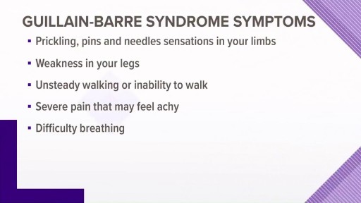
- Neurology
- Women Health
- Osteoporosis
- Gastroenterology
- Pulmonology
- Hematology
- Rheumatology
- Toxicology
- Nuclear Medicine
- Infectious Diseases
- Vascular Disease
- Reproductive Health
- Burns and Wound Healing
- Other
Top videos
Watch that video to learn How to Study The Human Anatomy
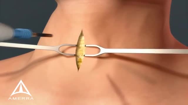
A tracheotomy or a tracheostomy: is simply an opening surgically created through the neck into the trachea (windpipe) to allow direct access to the breathing tube and is commonly done in an operating room under general anesthesia. A tube is usually placed through this opening to provide an airway and to remove secretions from the lungs. Breathing is done through the tracheostomy tube rather than through the nose and mouth. The term “tracheotomy” refers to the incision into the trachea (windpipe) that forms a temporary or permanent opening, which is called a “tracheostomy,” however; the terms are sometimes used interchangeably.
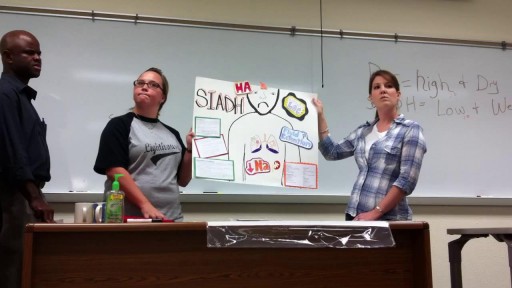
In this condition, the body retains water instead of excreting it normally in urine. This process upsets the body's balance of minerals called electrolytes, especially sodium. Symptoms can vary depending on how rapidly the condition develops. In some cases, nausea and vomiting, headache, confusion, weakness, and fatigue may be experienced. Treatments include fluid restriction and, possibly, medications to adjust electrolyte balance. Underlying conditions also need treatment.

Heparin is an anticoagulant (blood thinner) that prevents the formation of blood clots. Heparin is used to treat and prevent blood clots in the veins, arteries, or lung. It is also used before surgery to reduce the risk of blood clots.

Watch that video to know How to Remove Blackheads From Your Nose
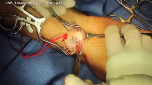
Surgery involves removing the cyst as well as part of the involved joint capsule or tendon sheath, which is considered the root of the ganglion. Even after excision, there is a small chance the ganglion will return. A ganglion cyst at the wrist is removed during a surgical procedure called excision.
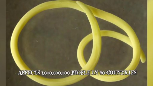
Watch that video of The 10 Most Invasive Parasites in the World
In human anatomy, the radial artery is the main blood vessel, with oxygenated blood, of the lateral aspect of the forearm.

Examination of Varicose Veins

OPAXIO Mechanism of Action
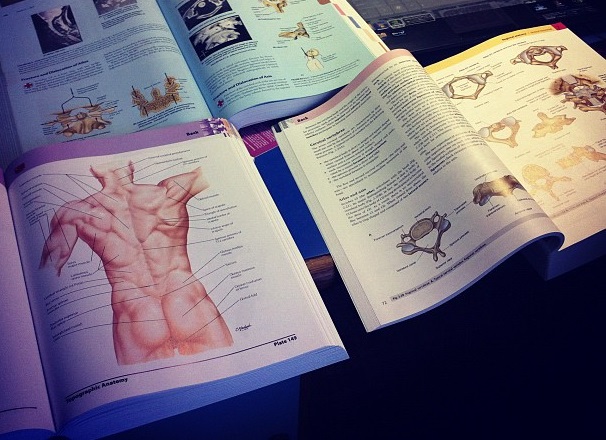
Anatomy of Back Muscles and Spinal Cord
Anatomy of The Peritoneal Cavity
Lymphoma is a cancer that arises from the cells of the lymphatic system. In the brain, this type of cancer is called Primary CNS Lymphoma (PCNSL). Location. Lymphoma occurs most often in the cerebral hemisphere, but may also involve the cerebrospinal fluid, the eyes, or the spinal cord.
Alpha blockers relax certain muscles and help small blood vessels remain open. They work by keeping the hormone norepinephrine (noradrenaline) from tightening the muscles in the walls of smaller arteries and veins, which causes the vessels to remain open and relaxed. This improves blood flow and lowers blood pressure.

Access my FREE Online Membership today → https://www.thenotedanatomist.com
___
Unlock my Premium Tutoring Memberships → https://www.thenotedanatomist.com/premium-memberships
Lifetime Access to Online Anatomy Course
Foundational Q&A Cards Per Video
Notes and Key Takeaways
Downloadable Documents
Flashcards for Each Course
Weekly Group Tutoring Sessions
Direct Tutoring Sessions
___
Discover A Simplified Approach to Master the Complexity of Anatomy with me, Dr. David Morton ... The Noted Anatomist!
This video tutorial discusses an Introduction to Histology (study of tissues):
0:00. Intro
0:35. Hierarchical organization of living matter
1:56. H&E stains
3:00. Epithelium overview (characteristics and classifying scheme)
- 9:12. Simple squamous epithelium
- 11:05. Simple cuboidal epithelium
- 12:20. Simple columnar epithelium
- 13:36. Stratified squamous epithelium
- 15:51. Urinary epithelium (transitional epithelium)
- 16:45. Pseudo-stratified ciliated columnar epithelium (respiratory epithelium)
18:55. Connective tissue overview (characteristics and classifying scheme)
- 21.14. Connective tissue proper (loose CT, dense irregular CT, dense regular CT, adipose tissue)
- 24:50. Cartilage (hyaline cartilage, elastic cartilage, fibrocartilage)
- 26:04. Bone (osteoblasts, osteocytes, osteoclasts, calcium ...)
- 27:34. Blood (RBC, WBC, platelet, plasma)
28:54. Muscle tissue (skeletal muscle, cardiac muscle, smooth muscle)
32:54. Nervous tissue (neurons and glial cells)
36:58. In-a-Nutshell
37:07. Acknowledgements
For a more detailed study of histology go to The Histology Wizard: https://www.youtube.com/channe....l/UCAeLLruy9RkUWaW_r

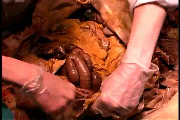
knife spoon and toothbrush removed from stomach
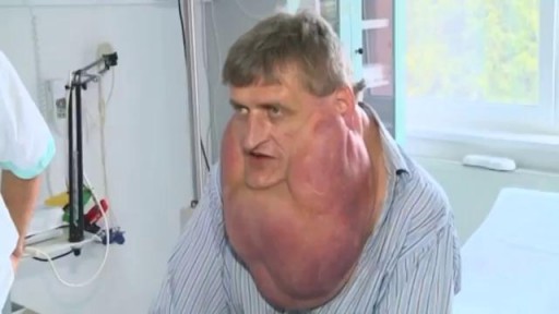
Massive Tumor Removed from Man's Face
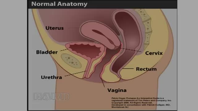
-Rectocele is a relatively common condition in older women and is characterized by the displacement of the rectum through posterior vaginal wall defect(s). The condition is typically caused by damage to the rectovaginal septum incurred during vaginal childbirth and is exacerbated by periodic increases in intraabdominal pressure (e.g., when laughing or coughing) and the effects of gravity. Women with symptomatic rectoceles who are poor surgical candidates may be treated with pessaries, which are structures designed to support the vaginal wall. Pessaries should only be used in conjunction with vaginal
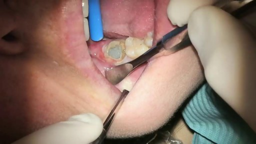
What Is It? Your wisdom teeth (third molars) usually start to erupt (enter your mouth) during the late teen years. Sometimes, there's not enough room for them. They may come into your mouth partially or not at all. Partial eruption of a wisdom tooth can create a flap of gum tissue next to the tooth. The flap can trap bits of food and debris. It can turn into a hotbed for bacteria. It's called pericoronitis if the tissue around the tooth becomes inflamed. Pericoronitis also can occur around a wisdom tooth that is still completely under the gums. Symptoms Symptoms include: Painful, swollen gum tissue in the area of the affected tooth. It can be difficult to bite down comfortably without catching the swollen tissue between your teeth. A bad smell or taste in the mouth Discharge of pus from the gum near the tooth More serious symptoms include: Swollen lymph nodes under your chin (the submandibular nodes) Muscle spasms in the jaw Swelling on the affected side of the face Diagnosis Usually, someone with pericoronitis goes to the dentist, complaining of pain in the area of the back tooth. Pericoronitis is diagnosed during the clinical exam. Your dentist will see inflamed gum tissue in the area of the unerupted or partly erupted wisdom tooth. The gums may be red, swollen or draining fluid or pus. Expected Duration Pericoronitis can be managed with antibiotics and warm salt water rinses. It goes away in about one week. However, it can return. This is likely to happen if the tooth does not completely enter the mouth and food and bacteria keep building up under the gum. Prevention You can help to prevent pericoronitis by brushing any erupting wisdom tooth and flossing around it. This will help make sure that food and bacteria do not build up under the gums. However, sometimes these steps do not work. If pericoronitis returns, you may need to have the flap of gum tissue removed. In some cases, the flap of tissue grows back and the wisdom tooth will need to be extracted. Treatment Pericoronitis can be tricky to treat. That's because the flap of gum tissue won't go away until the wisdom tooth emerges naturally, the tissue is removed or the tooth is removed. Your dentist will clean the area thoroughly by rinsing under the flap with water to remove bits of food and pus. Your dentist also may need to remove damaged tissue. If the area is infected, you'll most likely be given antibiotics. Your dentist will explain how to keep the area clean, which is the best way to prevent the problem from returning. This usually involves brushing and flossing daily and rinsing your mouth with water several times a day. These steps will help to prevent food from getting stuck under the gum flap. In some cases, your dentist may suggest removing the erupting tooth. Or the dentist may want to remove the tooth above it, which bites down on the gum below. If your dentist thinks the tooth may erupt fully into the mouth without problems, he or she may leave it alone. However, if pericoronitis comes back, the tooth may be extracted. Pericoronitis that causes symptoms should be treated as soon as possible. If it is not, the infection can spread to other areas of your mouth. The most severe cases are treated in a hospital. They sometimes require intravenous antibiotics and surgery. When To Call a Professional If you have symptoms of pericoronitis, make an appointment to see your dentist. If your wisdom teeth are coming in, visit your dentist at least twice a year for regular checkups. During those visits, the dentist can check on the progress of your wisdom teeth. Prognosis Pericoronitis does not cause any long-term effects. If the affected tooth is removed or erupts fully into the mouth, the condition cannot return.
