
- Physical Examination
- Surgical Examination
- Ophthalmology
- Clinical Skills
- Orthopedics
- Surgery Videos
- Laparoscopy
- Pediatrics
- Funny Videos
- Cardiothoracic Surgery
- Nursing Videos
- Plastic Surgery
- Otorhinolaryngology
- Histology and Histopathology
- Neurosurgery
- Dermatology
- Pediatric Surgery
- Urology
- Dentistry
- Oncology and Cancers
- Anatomy Videos
- Health and Fitness
- Radiology
- Anaesthesia
- Physical Therapy
- Pharmacology
- Interventional Radiology
- Cardiology
- Endocrinology
- Gynecology
- Emergency Medicine
- Psychiatry and Psychology
- Childbirth Videos
- General Medical Videos
- Nephrology
- Physiology
- Diet and Food Health
- Diabetes Mellitus
- Neurology
- Women Health
- Osteoporosis
- Gastroenterology
- Pulmonology
- Hematology
- Rheumatology
- Toxicology
- Nuclear Medicine
- Infectious Diseases
- Vascular Disease
- Reproductive Health
- Burns and Wound Healing
- Other
Top videos
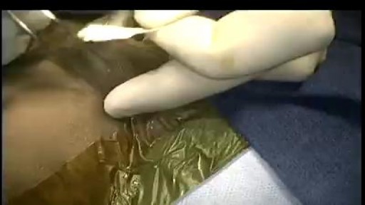

Minimally invasive surgery has been shown to be feasible and safe in pediatric patients since 1975 when laparoscopic surgery was first used to treat a small bowel obstruction. Laparoscopy is an option for surgical repair of inguinal hernias in addition to the traditional open approach.
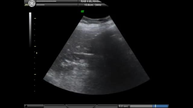
Porcelain gallbladder is a condition characterized by calcium salt deposits in the wall of a chronically inflamed gallbladder. The calcifications can be thin or faintly visible, or may be amorphous, patchy, and thick. The gallbladder is generally large, but its size can vary considerably. Most porcelain gallbladders are associated with gallstones. A plain radiograph generally detects these, but computed tomography (CT) has a higher specificity; therefore, a CT scan is performed to confirm the diagnosis. Due to their high risk of gallbladder carcinoma, all patients with porcelain gallbladder should have an elective cholecystectomy.
NEUROLOGICAL EXAMINATION
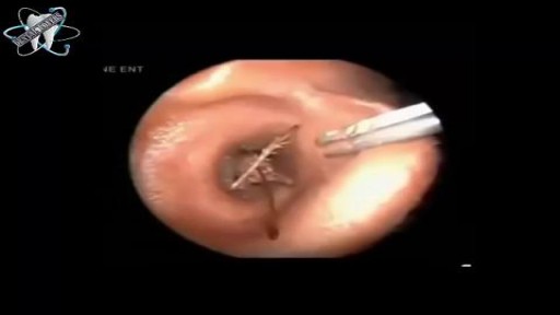
Bugs Removal from Ear Canal
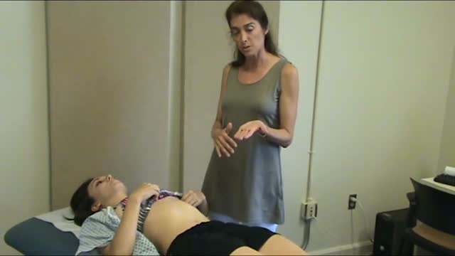
The best way to prepare yourself for your small group exercises is to first watch the cardiac exam video from beginning to end, then proceed through all the explanations of cardiac anatomy and physiology that follow. To go through the material in the recommended sequence, just click the "Next" button in the upper right corner of the screen. But you may also jump to any section using the menu to the left.

Watch Dr. Robert Thomas, of Panorama Orthopedics & Spine Center, perform a Mako Knee replacement. He narrates each step of the process.

Eric knew he needed help when an old knee injury began worsening over the course of time and was significantly affecting his quality of life. That’s when he turned to his hometown orthopedic experts at Mayo Clinic Health System in Mankato, who recommended a total knee replacement. After overcoming some initial fears, Eric decided it was time to have the operation — a fuller and more active life with his family depended on it.
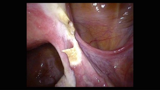
40 years old patient, Parity 3, wanted to have a sterilization. The surgery was perfomed laparoscopically with coagulation technique. This video is not edited and presented in full length.

Watch that video to know if you have Trypophobia
Learn a simple way to tell if you might be suffering from an undiagnosed autoimmune condition. Examples of autoimmune conditions include: • alopecia areata
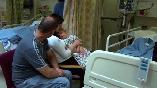
A Documentary on Uterine Transplantation. Dr. Edwin Ramirez pioneers this new breakthrough medical procedure, destined to change the world.

http://www.landging.com/breast-enlargement-animation.html
Breast enlargement product animation designed for TV shopping.
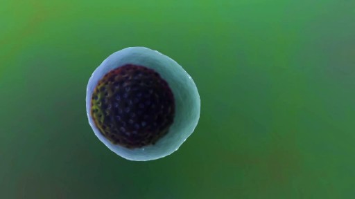
Myeloma is a cancer of the plasma cell, a type of white blood cell that is part of the immune system. This animation explains how a plasma cell can become cancerous and turn into a myeloma cell.
Pulmonary edema is usually caused by a heart condition. Other causes include pneumonia, exposure to certain toxins and drugs, and being at high elevations. Depending on the cause, pulmonary edema symptoms may appear suddenly or develop over time. Mild to extreme breathing difficulty can occur. Cough, chest pain, and fatigue are other symptoms. Treatment generally includes supplemental oxygen and medications.
A video showing amniotomy
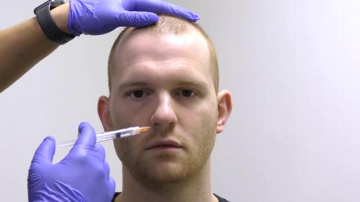
This video demonstrates how a broken nose is fixed using only local anesthesia and without sedation. Of course, this can also be performed while asleep.
Hepatitis and chronic alcohol abuse are frequent causes. Liver damage caused by cirrhosis can't be undone, but further damage can be limited. Initially patients may experience fatigue, weakness, and weight loss. During later stages, patients may develop jaundice (yellowing of the skin), gastrointestinal bleeding, abdominal swelling, and confusion. Treatments focus on the underlying cause. In advanced cases, a liver transplant may be needed.

Percutaneous transluminal coronary angioplasty (PTCA) is a minimally invasive procedure to open up blocked coronary arteries, allowing blood to circulate unobstructed to the heart muscle.
