- Physical Examination
- Surgical Examination
- Ophthalmology
- Clinical Skills
- Orthopedics
- Surgery Videos
- Laparoscopy
- Pediatrics
- Funny Videos
- Cardiothoracic Surgery
- Nursing Videos
- Plastic Surgery
- Otorhinolaryngology
- Histology and Histopathology
- Neurosurgery
- Dermatology
- Pediatric Surgery
- Urology
- Dentistry
- Oncology and Cancers
- Anatomy Videos
- Health and Fitness
- Radiology
- Anaesthesia
- Physical Therapy
- Pharmacology
- Interventional Radiology
- Cardiology
- Endocrinology
- Gynecology
- Emergency Medicine
- Psychiatry and Psychology
- Childbirth Videos
- General Medical Videos
- Nephrology
- Physiology
- Diet and Food Health
- Diabetes Mellitus
- Neurology
- Women Health
- Osteoporosis
- Gastroenterology
- Pulmonology
- Hematology
- Rheumatology
- Toxicology
- Nuclear Medicine
- Infectious Diseases
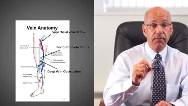
- Vascular Disease
- Reproductive Health
- Burns and Wound Healing
- Other
Top videos
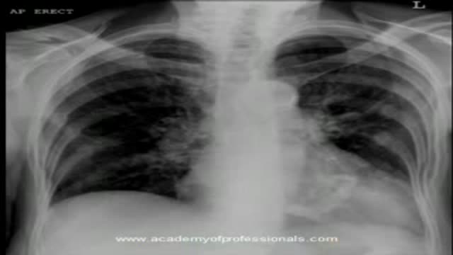
The video will shed light on mitral valve calcification. Please see disclaimer on my website. www.academyofprofessionals.com

An animation showing the Pelvic Inflammatory Disease (PID)
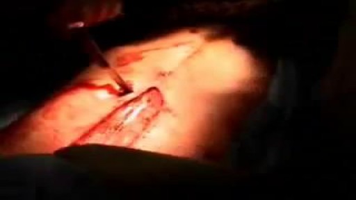
Watch that video of a Knife Stabbed Inside Chest Removing Surgery
secret about human

Breast Reduction Surgery video Operation مركز افارا لجراحات التجميل الخدود تكبير الشفايف
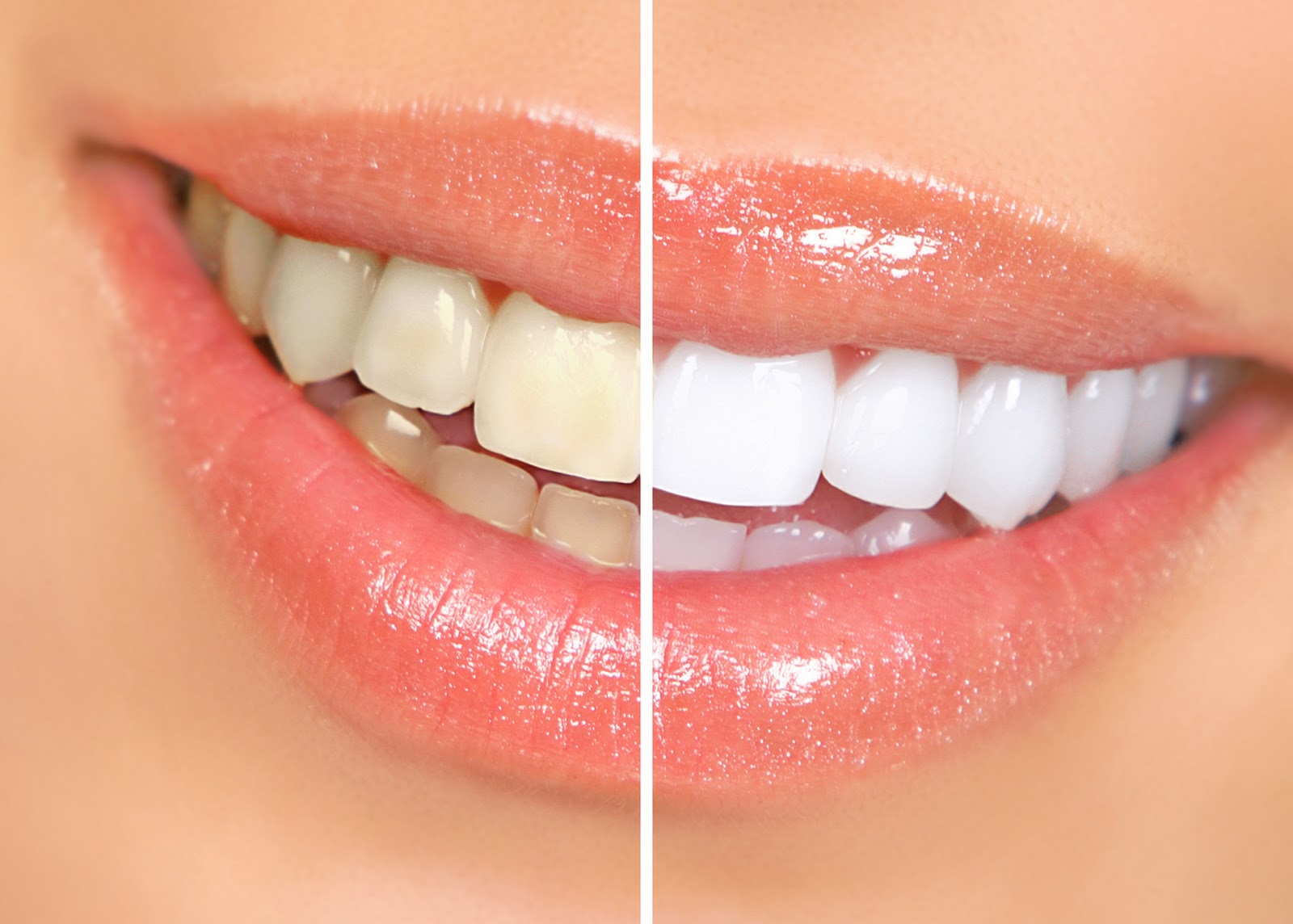
Watch that video to know the Natural Ways to Whiten Teeth at Home
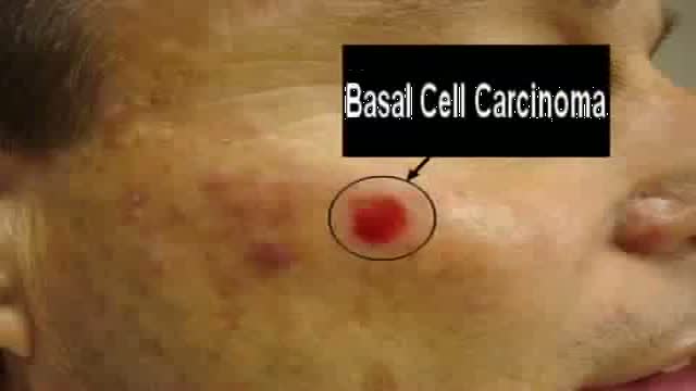
Graphic video demonstrating the reconstruction of a facial cheek defect following the removal of a common skin cancer in a challenging location near the lower eyelid. Visit us @ www.skincancercentre.com.

This clip shows the basic steps of inserting V.T> tubes. This surgery is performed for the treatment of OME resistent to medical TTT.
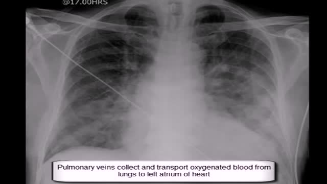
Expand Section. Pulmonary edema is often caused by congestive heart failure. When the heart is not able to pump efficiently, blood can back up into the veins that take blood through the lungs. As the pressure in these blood vessels increases, fluid is pushed into the air spaces (alveoli) in the lungs.

HD Cataract Surgery Video

Normal sperm densities range from 15 million to greater than 200 million sperm per milliliter of semen. You are considered to have a low sperm count if you have fewer than 15 million sperm per milliliter or less than 39 million sperm total per ejaculate.
Anatomy of The Shoulder and Arm Muscles
Anatomy of The Superficial Neck

In the Dialysis Unit you have an opportunity to provide Dialysis care for a variety of patients, including those with End-Stage Chronic Kidney disease and acutely ill patients requiring dialysis and plasmapheresis.
The Chronic Dialysis Nurse focuses on patients receiving Hemodialysis, Peritoneal Dialysis, or Home Hemodialysis. Our patients range in age from newborns to young adults. The Hemodialysis patient receives their dialysis treatment in the clinic 3-5 times a week. The Peritoneal Dialysis and Home Hemodialysis treatments are provided in the patient’s home once the parent/caregiver is trained to operate the machine. They are followed monthly in clinic. The patient receiving Chronic Dialysis is supported by a multidisciplinary team that consists of a physician, nurses, social worker, nutritionist, pharmacist, child-life therapist, teacher, and counselor. The group works together to meet the medical and emotional needs of the patient and caregiver. Care is specialized to meet the needs of each individual patient.
The Acute Dialysis Nurse focuses on acute dialysis related therapies such as: Continuous Renal Replacement Therapy (CRRT); therapeutic plasmapheresis; or acute peritoneal dialysis. The acute dialysis team works with the multi-disciplinary inpatient nephrology team to provide acute dialysis services to the critically ill ICU patients. The work environment is highly technical and fast-paced.
The Dialysis Unit operates on 12hr shifts 7a – 7p; 7 days a week. Night call is required and shared by the nurses. We provide a detailed orientation plan to the nurse to become proficient in providing hemodialysis, peritoneal dialysis, continuous renal replacement therapy and plasmapheresis. Previous experience in dialysis or pediatrics is not required.
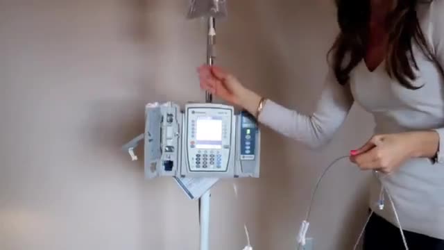
How to use an IV pump..
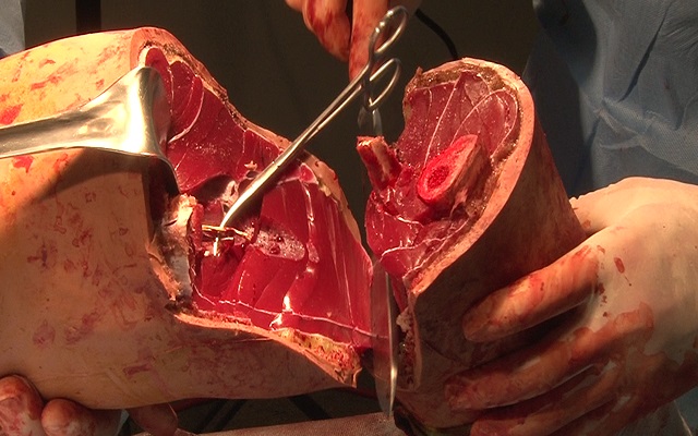
Watch that Above Knee Amputation Surgery video
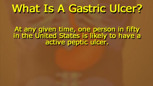
What Causes Ulcers? No single cause has been found for ulcers. However, it is now clear that an ulcer is the end result of an imbalance between digestive fluids in the stomach and duodenum. Most ulcers are caused by an infection with a type of bacteria called Helicobacter pylori (H. pylori). Factors that can increase your risk for ulcers include: Use of painkillers called nonsteroidal anti-inflammatory drugs (NSAIDs), such as aspirin, naproxen (Aleve, Anaprox, Naprosyn, and others), ibuprofen (Motrin, Advil, some types of Midol, and others), and many others available by prescription; even safety-coated aspirin and aspirin in powered form can frequently cause ulcers. Excess acid production from gastrinomas, tumors of the acid producing cells of the stomach that increases acid output (seen in Zollinger-Ellison syndrome) Excessive drinking of alcohol Smoking or chewing tobacco Serious illness Radiation treatment to the area What Are the Symptoms of an Ulcer? An ulcer may or may not have symptoms. When symptoms occur, they may include: A gnawing or burning pain in the middle or upper stomach between meals or at night Bloating Heartburn Nausea or vomiting In severe cases, symptoms can include: Dark or black stool (due to bleeding) Vomiting blood (that can look like "coffee-grounds") Weight loss Severe pain in the mid to upper abdomen
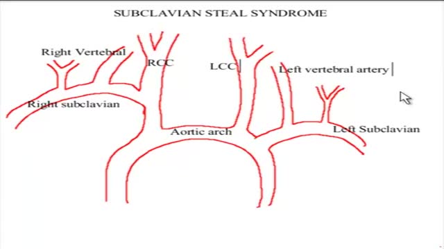
The term subclavian steal describes retrograde blood flow in the vertebral artery associated with proximal ipsilateral subclavian artery stenosis or occlusion, usually in the setting of subclavian artery occlusion or stenosis proximal to the origin of the vertebral artery. Alternatively, innominate artery disease has also been associated with retrograde flow in the ipsilateral vertebral artery, particularly where the subclavian artery origin is involved. Subclavian steal is frequently asymptomatic and may be discovered incidentally on ultrasound or angiographic examination for other indications, or it may be prompted by a clinical examination finding of reduced unilateral upper limb pulse or blood pressure. In some cases, patients may develop upper limb ischemic symptoms due to reduced arterial flow in the setting of subclavian artery occlusion, or they may develop neurologic symptoms due to posterior circulation ischemia associated with exercise of the ipsilateral arm.[1] Treatment has traditionally consisted of open subclavian artery revascularization, typically via carotid-subclavian bypass or subclavian artery transposition, which are generally durable procedures. Newer, less invasive options include endovascular intervention with recanalization as appropriate and angioplasty and stenting if required. The clinical relevance of subclavian steal was described in 1961 by Reivich, Holling and Roberts; however, the recognition of retrograde vertebral artery flow dates back another 100 years to Harrison and Smyth. Some papers, including a previous version of this article, advocate restricting the term subclavian steal to patients with neurologic symptoms only, but this is incorrect in view of the substantial literature using this term to describe the hemodynamic scenario of retrograde vertebral flow and proximal subclavian artery disease.
A leg ulcer is simply a break in the skin of the leg, which allows air and bacteria to get into the underlying tissue. This is usually caused by an injury, often a minor one that breaks the skin. In most people such an injury will heal up without difficulty within a week or two. However, when there is an underlying problem the skin does not heal and the area of breakdown can increase in size. This is a chronic leg ulcer.
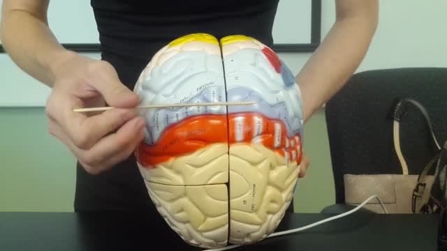
The brain is that part of the CNS contained within the cranial cavity (figure 13.1). It is the control center for many of the body's functions. The brain is much like a complex central computer but with additional functions that no computer can as yet match. Indeed, one goal in computer technology is to make computers that can function more like the human brain. The brain consists of the brainstem, the cerebellum, the diencephalon, and the cerebrum (table 13.1). The brainstem includes the medulla oblongata, pons, midbrain, and reticular formation. The structure of the brain is described in this chapter. Its functions are primarily discussed in chapter 14. Twelve pairs of cranial nerves, which are part of the PNS, arise directly from the brain. Two pairs arise from the cerebrum, nine pairs arise from the brainstem, and one pair arises from the spinal cord.
