
- Physical Examination
- Surgical Examination
- Ophthalmology
- Clinical Skills
- Orthopedics
- Surgery Videos
- Laparoscopy
- Pediatrics
- Funny Videos
- Cardiothoracic Surgery
- Nursing Videos
- Plastic Surgery
- Otorhinolaryngology
- Histology and Histopathology
- Neurosurgery
- Dermatology
- Pediatric Surgery
- Urology
- Dentistry
- Oncology and Cancers
- Anatomy Videos
- Health and Fitness
- Radiology
- Anaesthesia
- Physical Therapy
- Pharmacology
- Interventional Radiology
- Cardiology
- Endocrinology
- Gynecology
- Emergency Medicine
- Psychiatry and Psychology
- Childbirth Videos
- General Medical Videos
- Nephrology
- Physiology
- Diet and Food Health
- Diabetes Mellitus
- Neurology
- Women Health
- Osteoporosis
- Gastroenterology
- Pulmonology
- Hematology
- Rheumatology
- Toxicology
- Nuclear Medicine
- Infectious Diseases
- Vascular Disease
- Reproductive Health
- Burns and Wound Healing
- Other
Top videos

Doctor shares tips on what to expect after a cancer diagnosis

The BMHR uses the same socket (hydroxyapatite-coated metal uncemented cup) and bearing(metal on metal) as the BHR. The modular head component fits onto a hydroxyapatite proximal porous coated cobalt chrome stem. It is an uncemented short stemmed prosthesis. It was invented by Prof. Dereck Mc.Minn a year ago and is performed by very few surgeons the world over. In India it is being done only at the Asian Regional Center for Hip Resurfacing in Chennai.
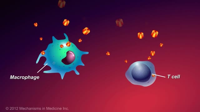
This animation describes what anti-TNF-alpha therapies are, how they work, and how patients with inflammatory bowel disease (IBD) can benefit.
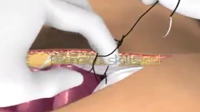
Laparotomy Closure Abdomen Animation
This clip shows the basic steps of inserting V.T> tubes. This surgery is performed for the treatment of OME resistent to medical TTT.
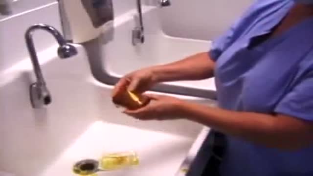
Surgical Scrub How To
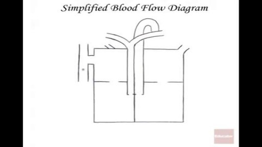
The heart itself is made up of 4 chambers, 2 atria and 2 ventricles. De-oxygenated blood returns to the right side of the heart via the venous circulation. It is pumped into the right ventricle and then to the lungs where carbon dioxide is released and oxygen is absorbed. The oxygenated blood then travels back to the left side of the heart into the left atria, then into the left ventricle from where it is pumped into the aorta and arterial circulation.

Mechanism of Addiction
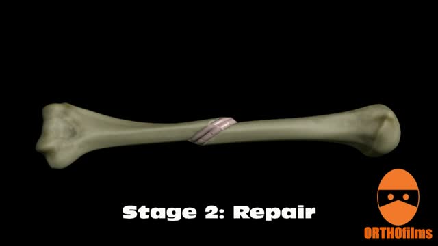
Bone healing can be divided into four stages: inflammation; soft callus formation; hard callus formation; remodeling.
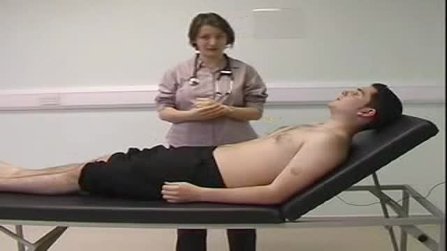
Basic CardioVascular Clinical Exam

Arterial Blood Gas Sampling Technique Video
Large Bowel Epiploica Laparoscopic Resection
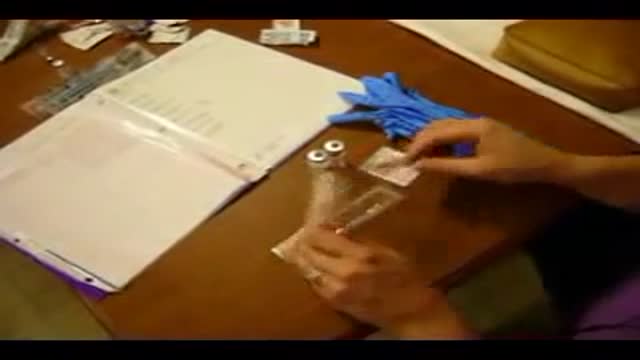
Administering a Subcutaneous Injection
Islam and Medicine

application of intragastric endoscopic baloon for morbid obesity
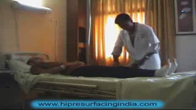
Stephen Jenkins is sharing his experience after underwent the hip resurfacing surgery.

AZT Mechanism of Antiviral Activity
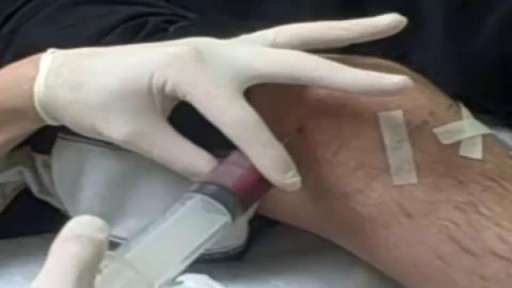
nee joint aspiration and injection are performed to aid in diagnosis and treatment of knee joint diseases. The knee joint is the most common and the easiest joint for the physician to aspirate. One approach involves insertion of a needle 1 cm above and 1 cm lateral to the superior lateral aspect of the patella at a 45-degree angle. Once the needle has been inserted 1 to 1½ inches, aspiration aided by local compression is performed. Local corticosteroid injections can provide significant relief and often ameliorate acute exacerbations of knee osteoarthritis associated with significant effusions. Among the indications for arthrocentesis are crystal-induced arthropathy, hemarthrosis, unexplained joint effusion, and symptomatic relief of a large effusion. Contraindications include bacteremia, inaccessible joints, joint prosthesis, and overlying infection in the soft tissue. Large effusions can recur and may require repeat aspiration. Anti-inflammatory medi
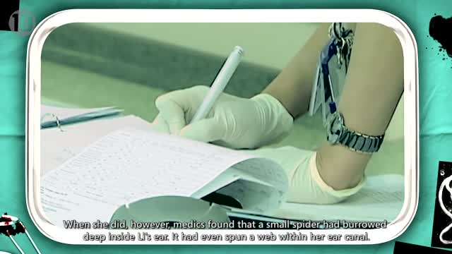
10 Animals Found Living Inside Humans
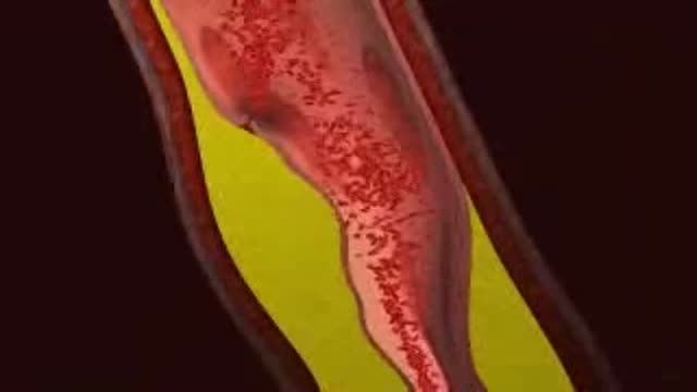
Myocardial infarction (MI), commonly known as a heart attack, is defined pathologically as the irreversible death of myocardial cells caused by ischemia. Clinically, MI is a syndrome that can be recognized by a set of symptoms, chest pain being the hallmark of these symptoms in most cases, supported by biochemical laboratory changes, electrocardiographic (ECG) changes, or findings on imaging modalities able to detect myocardial injury and necrosis. According to the third universal definition of MI, implemented by a joint task force from the European Society of Cardiology (ESC), American College of Cardiology (ACC) Foundation, American Heart Association (AHA), and the World Heart Federation (WHF), MI is diagnosed when either of the following two criteria are met
