- Physical Examination
- Surgical Examination
- Ophthalmology
- Clinical Skills
- Orthopedics
- Surgery Videos
- Laparoscopy
- Pediatrics
- Funny Videos
- Cardiothoracic Surgery
- Nursing Videos
- Plastic Surgery
- Otorhinolaryngology
- Histology and Histopathology
- Neurosurgery
- Dermatology
- Pediatric Surgery
- Urology
- Dentistry
- Oncology and Cancers
- Anatomy Videos
- Health and Fitness
- Radiology
- Anaesthesia
- Physical Therapy
- Pharmacology
- Interventional Radiology
- Cardiology
- Endocrinology
- Gynecology
- Emergency Medicine
- Psychiatry and Psychology
- Childbirth Videos
- General Medical Videos
- Nephrology
- Physiology
- Diet and Food Health
- Diabetes Mellitus
- Neurology
- Women Health
- Osteoporosis
- Gastroenterology
- Pulmonology
- Hematology
- Rheumatology
- Toxicology
- Nuclear Medicine
- Infectious Diseases
- Vascular Disease
- Reproductive Health
- Burns and Wound Healing
- Other
Top videos
Diverticulitis Attack Surgery
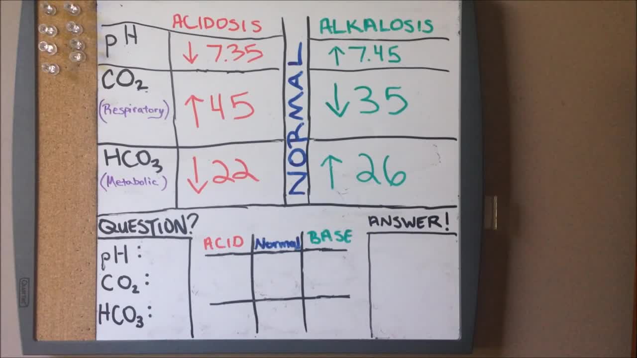
ABGs Made Easy | Arterial Blood Gas | Acid Base Balance: Everything You Need To Know!
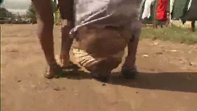
Giant Swollen Leg Elephantiasis Filariasis
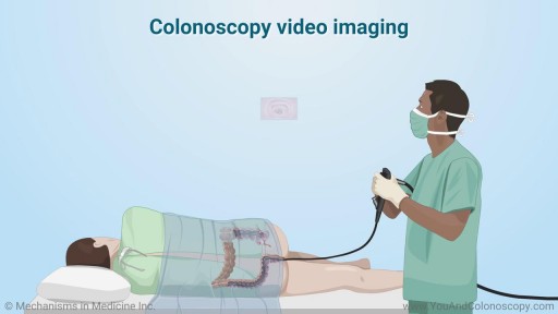
A colonoscope is the special tool used to perform a colonoscopy. It is a thin, flexible, tubular ‘telescope’ with a light and video camera that your doctor carefully guides through your colon in order to see and determine the health of your colon. Watch this animation to learn about the features of the colonoscope, how the colonoscopy procedure is performed and how polyps are removed, and the follow-up care you and your doctor should talk about after your procedure.
A bone marrow biopsy is part of a bone marrow test that takes a sample of your solid bone tissue. This test looks for abnormalities in your blood cells and signs of any diseases. You can request anesthesia or a sedative before the biopsy, and manage any pain afterward with over-the-counter medications.
Craziest Surgeries You'll Never Believe Occurred!
The Distal Femoral Osteotomy System utilizes the same principles of design featured in the Tibial Osteotomy System. Specifically designed femoral osteotomy plates take into account the anatomical differences between the distal femur and proximal tibia.
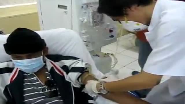
Renal Kidney Hemodialysis
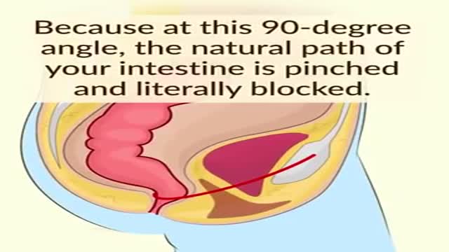
You've been sitting on the toilet incorrectly your whole life
Chronic myelogenous leukemia (CML), also known as chronic myeloid leukemia, is a myeloproliferative disorder characterized by increased proliferation of the granulocytic cell line without the loss of their capacity to differentiate. Consequently, the peripheral blood cell profile shows an increased number of granulocytes and their immature precursors, including occasional blast cells.
CML is one of the few cancers known to be caused by a single, specific genetic mutation. More than 90% of cases result from a cytogenetic aberration known as the Philadelphia chromosome (see Pathophysiology).
CML progresses through 3 phases: chronic, accelerated, and blast. In the chronic phase of disease, mature cells proliferate; in the accelerated phase, additional cytogenetic abnormalities occur; in the blast phase, immature cells rapidly proliferate.[1] Approximately 85% of patients are diagnosed in the chronic phase and then progress to the accelerated and blast phases after 3-5 years. The diagnosis of CML is based on the histopathologic findings in the peripheral blood and the Philadelphia chromosome in bone marrow cells (see Workup).
CML accounts for 20% of all leukemias affecting adults. It typically affects middle-aged individuals. Uncommonly, the disease occurs in younger individuals. Younger patients may present with a more aggressive form of CML, such as in accelerated phase or blast crisis. Uncommonly, CML may appear as a disease of new onset in elderly individuals.
The goals of treatment are to achieve hematologic, cytogenetic, and molecular remission. Although a variety of medications have been used in CML, including myelosuppressive agents and interferon alfa, the tyrosine kinase inhibitor imatinib mesylate is currently the agent of choice, and other drugs in this category are playing increasingly important roles. However, allogeneic bone marrow transplantation is currently the only proven cure for CML.
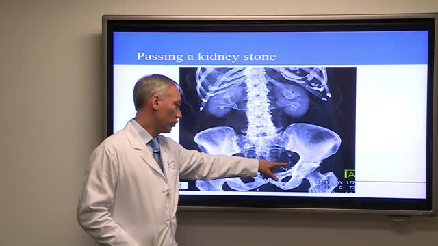
Treatment for kidney stones varies, depending on the type of stone and the cause. Small stones with minimal symptoms Most kidney stones won't require invasive treatment. You may be able to pass a small stone by: Drinking water. Drinking as much as 2 to 3 quarts (1.9 to 2.8 liters) a day may help flush out your urinary system. Unless your doctor tells you otherwise, drink enough fluid — mostly water — to produce clear or nearly clear urine. Pain relievers. Passing a small stone can cause some discomfort. To relieve mild pain, your doctor may recommend pain relievers such as ibuprofen (Advil, Motrin IB, others), acetaminophen (Tylenol, others) or naproxen sodium (Aleve). Medical therapy. Your doctor may give you a medication to help pass your kidney stone. This type of medication, known as an alpha blocker, relaxes the muscles in your ureter, helping you pass the kidney stone more quickly and with less pain. Large stones and those that cause symptoms Kidney stones that can't be treated with conservative measures — either because they're too large to pass on their own or because they cause bleeding, kidney damage or ongoing urinary tract infections — may require more extensive treatment. Procedures may include: Using sound waves to break up stones. For certain kidney stones — depending on size and location — your doctor may recommend a procedure called extracorporeal shock wave lithotripsy (ESWL). ESWL uses sound waves to create strong vibrations (shock waves) that break the stones into tiny pieces that can be passed in your urine. The procedure lasts about 45 to 60 minutes and can cause moderate pain, so you may be under sedation or light anesthesia to make you comfortable. ESWL can cause blood in the urine, bruising on the back or abdomen, bleeding around the kidney and other adjacent organs, and discomfort as the stone fragments pass through the urinary tract. Surgery to remove very large stones in the kidney. A procedure called percutaneous nephrolithotomy (nef-row-lih-THOT-uh-me) involves surgically removing a kidney stone using small telescopes and instruments inserted through a small incision in your back. You will receive general anesthesia during the surgery and be in the hospital for one to two days while you recover. Your doctor may recommend this surgery if ESWL was unsuccessful. Using a scope to remove stones. To remove a smaller stone in your ureter or kidney, your doctor may pass a thin lighted tube (ureteroscope) equipped with a camera through your urethra and bladder to your ureter. Once the stone is located, special tools can snare the stone or break it into pieces that will pass in your urine. Your doctor may then place a small tube (stent) in the ureter to relieve swelling and promote healing. You may need general or local anesthesia during this procedure. Parathyroid gland surgery. Some calcium phosphate stones are caused by overactive parathyroid glands, which are located on the four corners of your thyroid gland, just below your Adam's apple. When these glands produce too much parathyroid hormone (hyperparathyroidism), your calcium levels can become too high and kidney stones may form as a result. Hyperparathyroidism sometimes occurs when a small, benign tumor forms in one of your parathyroid glands or you develop another condition that leads these glands to produce more parathyroid hormone. Removing the growth from the gland stops the formation of kidney stones. Or your doctor may recommend treatment of the condition that's causing your parathyroid gland to overproduce the hormone.
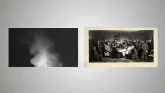
There's only one group of people who really know what happens when you die: the dead. And since the dead won't be revealing their secrets anytime soon, it's up to scientists to explain what happens when a person dies. Death, just like life, is a process, scientists say. The first stage of this process is known as clinical death. It lasts from four to six minutes, beginning when a person stops breathing and the heart stops pumping blood. During this time, there may be enough oxygen in the brain that no permanent brain damage occurs. Other organs, such as the kidneys and eyes, also remain alive throughout clinical death.

Motivation for Medical Students!

TV interview with Adina Nack, Ph.D. about her own cervical HPV experiences, STD research, her new book (Damaged Goods? Women Living with Incurable Sexually Transmitted Diseases), and women's lives after genital warts, HPV and herpes infections. More info is available on STDdatings.com, which is the official STD dating & support site.
Lower Limb Physical Examination
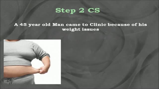
USMLE Step 2 CS - Obesity This is just preview video. To get full access please visit our website : www.usmletutoring.com
ROTIGS medical device by Honolulu inventor Dr. Brad NaPier makes airway intubations easier for medical professionals. For more info, visit www.rotigs.com
Aspirin is a salicylate (sa-LIS-il-ate). It works by reducing substances in the body that cause pain, fever, and inflammation. Aspirin is used to treat pain, and reduce fever or inflammation. It is sometimes used to treat or prevent heart attacks, strokes, and chest pain (angina).

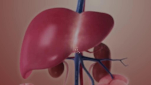
With the help of vitamin K, the liver produces proteins that are important in blood clotting. It is also one of the organs that break down old or damaged blood cells. The liver plays a central role in all metabolic processes in the body. In fat metabolism the liver cells break down fats and produce energy.
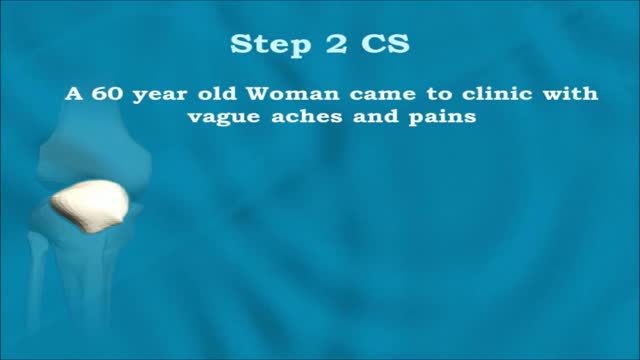
USMLE Step 2 CS - Joint Pain This is just preview video. To get full access please visit our website : www.usmletutoring.com
