Top Videos
Cricothyroidotomy NEJM
Empyema can develop after you have pneumonia. Many different types of bacteria may cause pneumonia, but the two most common are Streptococcus pneumoniae and Staphylococcus aureus. Occasionally, empyema may happen after you've had surgery on your chest. Medical instruments can transfer bacteria into your pleural cavity
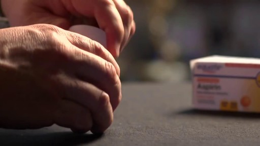
You should not use aspirin if you have a bleeding disorder such as hemophilia, a recent history of stomach or intestinal bleeding, or if you are allergic to an NSAID (non-steroidal anti-inflammatory drug) such as Advil, Motrin, Aleve, Orudis, Indocin, Lodine, Voltaren, Toradol, Mobic, Relafen, Feldene, and others. Do not give this medication to a child or teenager with a fever, flu symptoms, or chicken pox. Salicylates can cause Reye's syndrome, a serious and sometimes fatal condition in children.
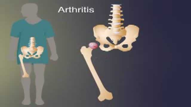
Arthritis of the hip causes severe pain, and sometimes requires surgical treatment, including hip replacement. This animated video show you what hip arthritis is, and how it causes symptoms.
The goal of surgical clipping is to isolate an aneurysm from the normal circulation without blocking off any small perforating arteries nearby. Under general anesthesia, an opening is made in the skull, called a craniotomy. The brain is gently retracted to locate the aneurysm. A small clip is placed across the base, or neck, of the aneurysm to block the normal blood flow from entering. The clip works like a tiny coil-spring clothespin, in which the blades of the clip remain tightly closed until pressure is applied to open the blades. Clips are made of titanium and remain on the artery permanently.
A technique for reducing an inferior shoulder dislocation. watch to learn more
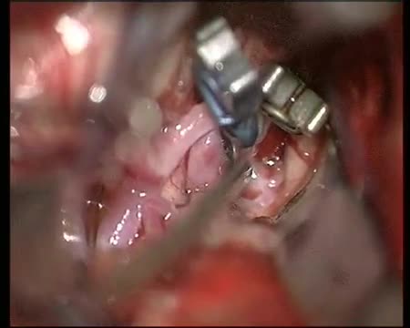
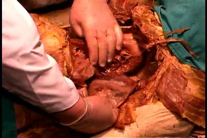
Calcified Brain Abscess complete removal

Ureteral stents are one of the most common devices used by urologists. They are placed with cystoscopic guidance in an operating room setting. Ureteral stents are used to relieve ureteral obstruction, promote ureteral healing following surgery, and to assist with ureteral identification during pelvic surgery. Ureteral stent placement is associated with some degree of morbidity in the majority of patients that ranges from generalized urinary discomfort to urinary tract infection or obstruction. Much of the morbidity is related to the biocompatibility of the materials used to fashion the stent and, to some extent, their design; unfortunately, the ideal stent has yet to be discovered.

Mysterious things happen in nature, and extraordinary birth delivery facts amaze and astound us. And "The baby who didn't know he was born" is one of them; the reason was because his mother didn't break water, so the little one thought was still in the womb. Of course, the amniotic sac was later broken by the doctor, and as soon as this happened the baby began to breath and cry.

LASIK eye surgery is commonly performed laser refractive surgery to correct vision problems. This 3d animation shows how laser-assisted in situ keratomileusis (lasik) can be an alternative to glasses or contact lenses.
It’s one of many vision correction surgeries that work by reshaping your cornea, the clear front part of your eye, so that light focuses on the retina in the back of your eye.
In eyes with normal vision, the cornea bends (refracts) light precisely onto the retina at the back of the eye. But with nearsightedness (myopia), farsightedness (hyperopia) or astigmatism, the light is bent incorrectly, resulting in blurred vision.
During LASIK surgery, a special type of cutting laser is used to precisely change the shape of the dome-shaped clear tissue at the front of your eyes (cornea) to improve vision.
Glasses or contact lenses can correct vision, but reshaping the cornea itself also will provide the necessary refraction.
For more information about medical animation, please visit https://www.amerra.com
Watch more medical animations:
Craniectomy brain surgery - 3D animation: https://youtu.be/1RkseDeYS9g
Accessing an implantable port training - 3D animation: https://youtu.be/xSTpxjyv4O4
Open Suctioning with a Tracheostomy Tube - 3D animation: https://youtu.be/wamB7jpWCiQ
Ventriculostomy Brain Surgery - 3d animation: https://youtu.be/pUy0YDzVNzs
Suctioning the endotracheal tube - medical animation: https://youtu.be/pN6-EYoeh3g
Functional endoscopic sinus surgery (FESS) - 3D animation: https://youtu.be/qKTRyowwaLA
How to insert a nasogastric tube for NG intubation - 3d animation: https://youtu.be/Abf3Gd6AaZQ
Oral airway insertion - oropharyngeal airway technique - 3D animation: https://youtu.be/caxUdNwjt34
Nasotracheal suctioning (NTS) - 3D animation: https://youtu.be/979jWMsF62c
Learn about hemorrhoids with #3d #animation: https://youtu.be/R6NqlMpsiiY
CPR cardiopulmonary resuscitation - 3D animation: https://youtu.be/G87knTZnhks
What are warts (HPV)? - 3D animation: https://youtu.be/guJ1J7rRs1w
How Macular Degeneration Affects Your Vision - 3D animation: https://youtu.be/ozZQIZ_52YY
NeoGraft hair transplant procedure – animation: https://youtu.be/C-eTdH2UPXI
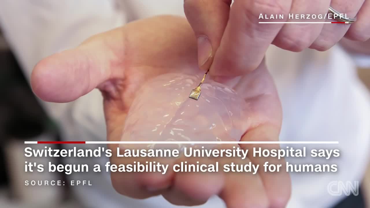
Scientists have developed a wireless brain implant that enabled a paralyzed monkey to walk again.

In this medical video: This 72-year-old patient was unable to resist blinking when we tapped on the glabella. This is the glabellar reflex or Myerson's sign . It is often an early sign of Parkinson's disease, but can also be seen in early dementia as well as other progressive neurologic illness. Note the left (i.e., asymmetrical) hand resting tremor.
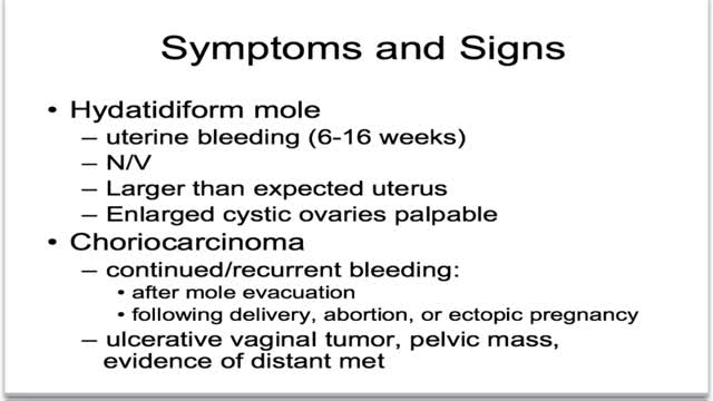
What is gestational trophoblastic disease? Cancer starts when cells in the body begin to grow out of control. Cells in nearly any part of the body can become cancer, and can spread to other areas of the body. To learn more about how cancers start and spread, see What Is Cancer? Gestational trophoblastic (jeh-STAY-shuh-nul troh-fuh-BLAS-tik) disease (GTD) is a group of rare tumors that involve abnormal growth of cells inside a woman's uterus. GTD does not develop from cells of the uterus like cervical cancer or endometrial (uterine lining) cancer do. Instead, these tumors start in the cells that would normally develop into the placenta during pregnancy. (The term gestational refers to pregnancy.) GTD begins in the layer of cells called the trophoblast (troh-fuh-BLAST) that normally surrounds an embryo. (Tropho- means nutrition, and -blast means bud or early developmental cell.) Early in normal development, the cells of the trophoblast form tiny, finger-like projections known as villi. The villi grow into the lining of the uterus. In time, the trophoblast layer develops into the placenta, the organ that protects and nourishes the growing fetus.
NTIS refers to a syndrome found in seriously ill or starving patients with low fT3, usually elevated RT3, normal or low TSH, and if prolonged, low fT4. It is found in a high proportion of patients in the ICU setting, and correlates with a poor prognosis if TT4 is <4ug/dl. The patho-physiology includes suppression of TRH release, reducedT3 and T4 turnover, reduction in liver generation of T3, increased formation of RT3, and tissue specific down-regulation of deiodinases, transporters, and TH receptors. Although long debated, tissue TH levels are definitely reduced, and tissue hypothyroidism is presumably present. This is often not clinically evident because of the brief duration, and reduced but not absent tissue levels of TH. Although recognized for nearly 4 decades, interpretation of the syndrome is contested, because of lack of data. Some observes, totally without data, argue that it is a protective response and should not be treated. Other observers (as in this review) present available data suggesting, but not proving, that thyroid hormone replacement is appropriate, not harmful, and may be beneficial. The best form of treatment (TRH,TSH,or T3+T4) and possible accompanying treatments (GHRH, Cortisol, nutrition, insulin) lack consensus. In this review current data are laid out for reader’s review and judgment.

Grisp Reflex
Preventing Hemodialysis Catheters Problems
Anatomy of The Posterior Thorax
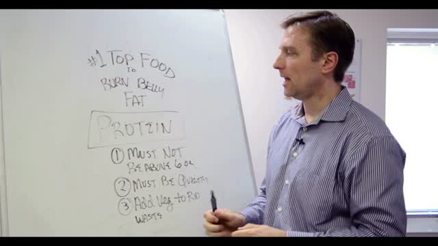
Overweight does not necessarily equal unhealthy. There are actually plenty of overweight people who are in excellent health (1). Conversely, many normal weight people have the metabolic problems associated with obesity (2). That’s because the fat under the skin is actually not that big of a problem (at least not from a health standpoint, it’s more of a cosmetic problem). It’s the fat in the abdominal cavity, the belly fat, that causes the biggest issues (3). If you have a lot of excess fat around your waistline, even if you’re not very heavy, then you should take some steps to get rid of it. Belly fat is usually estimated by measuring the circumference around your waist. This can easily be done at home with a simple tape measure. Anything above 40 inches (102 cm) in men and 35 inches (88 cm) in women, is known as abdominal obesity. There are actually a few proven strategies that have been shown to target the fat in the belly area more than other areas of the body.
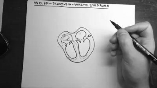
Wolff-Parkinson-White (WPW) syndrome, an extra electrical pathway between your heart's upper and lower chambers causes a rapid heartbeat. The extra pathway is present at birth and fairly rare. The episodes of fast heartbeats usually aren't life-threatening, but serious heart problems can occur. Treatment can stop or prevent episodes of fast heartbeats. A catheter-based procedure (ablation) can permanently correct the heart rhythm problems. Most people with an extra electrical pathway experience no fast heartbeat. This condition, called Wolff-Parkinson-White pattern, is discovered only by chance during a heart exam. Although WPW pattern is often harmless, doctors might recommend further evaluation before children with WPW pattern participate in high-intensity sports.
