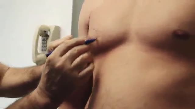
- Physical Examination
- Surgical Examination
- Ophthalmology
- Clinical Skills
- Orthopedics
- Surgery Videos
- Laparoscopy
- Pediatrics
- Funny Videos
- Cardiothoracic Surgery
- Nursing Videos
- Plastic Surgery
- Otorhinolaryngology
- Histology and Histopathology
- Neurosurgery
- Dermatology
- Pediatric Surgery
- Urology
- Dentistry
- Oncology and Cancers
- Anatomy Videos
- Health and Fitness
- Radiology
- Anaesthesia
- Physical Therapy
- Pharmacology
- Interventional Radiology
- Cardiology
- Endocrinology
- Gynecology
- Emergency Medicine
- Psychiatry and Psychology
- Childbirth Videos
- General Medical Videos
- Nephrology
- Physiology
- Diet and Food Health
- Diabetes Mellitus
- Neurology
- Women Health
- Osteoporosis
- Gastroenterology
- Pulmonology
- Hematology
- Rheumatology
- Toxicology
- Nuclear Medicine
- Infectious Diseases
- Vascular Disease
- Reproductive Health
- Burns and Wound Healing
- Other
Top videos
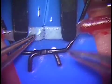
Simple microinstruments and a medical school laboratory microscope were used for anastomosis training. Chicken blood vessels were used as a material for this study. A long segment of blood vessel from the proximal brachial artery to the distal radial artery was used for training. End-to-side anastomosis was practiced first, and the training continued with end-to-end anastomosis of the appropriate segments.
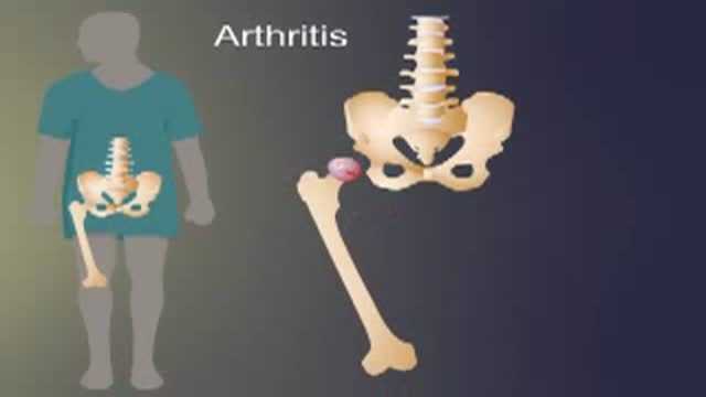
Arthritis of the hip causes severe pain, and sometimes requires surgical treatment, including hip replacement. This animated video show you what hip arthritis is, and how it causes symptoms.
This procedure describes one of the most versatile approaches to the anterior skull base for large tumors of the sinonasal cavity. It may be used with or without a craniofacial resection. The benefits of this approach are: wide access around the tumor; good postoperative cosmesis; & decreased operative & postoperative morbidity. We have used this approach for many bilateral tumors of the nasal & sinus cavities that approach &/or invade the skull base & brain. This video show the resection of a large esthesioneuroblastoma.
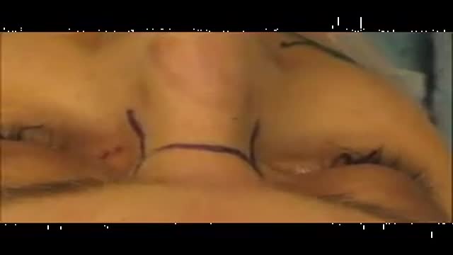
This video was taken 4 days after the surgery. This Patient had a facial rejuvenation procedure performed by Dr. Handal. He was exceptionally pleased with the results. Contact us for a consultation on how our team can help you to look better, (561) 912-9888. https://www.handalplasticsurgery.com
Spinal anesthesia is done in a similar way. But the anesthetic medicine is injected using a much smaller needle, directly into the cerebrospinal fluid that surrounds the spinal cord. The area where the needle will be inserted is first numbed with a local anesthetic. Then the needle is guided into the spinal canal, and the anesthetic is injected. This is usually done without the use of a catheter. Spinal anesthesia numbs the body below and sometimes above the site of the injection. The person may not be able to move his or her legs until the anesthetic wears off.
A filling is a way to restore a tooth damaged by decay back to its normal function and shape. When a dentist gives you a filling, he or she first removes the decayed tooth material, cleans the affected area, and then fills the cleaned out cavity with a filling material. By closing off spaces where bacteria can enter, a filling also helps prevent further decay. Materials used for fillings include gold, porcelain, a composite resin (tooth-colored fillings), and an amalgam (an alloy of mercury, silver, copper, tin and sometimes zinc).
A high definition HD video of Laparoscopic Cholecystectomy surgery
HD Gynecomastia Surgery
This animation describes tools and tests used to diagnose inflammatory bowel disease (IBD), determine IBD type, and predict its probable course and outcome.
Cytomegalovirus is a genus of viruses in the order Herpesvirales, in the family Herpesviridae, in the subfamily Betaherpesvirinae. Humans and monkeys serve as natural hosts.

Thyroid Exam Physical Exam
Anatomy of The Posterior Thorax
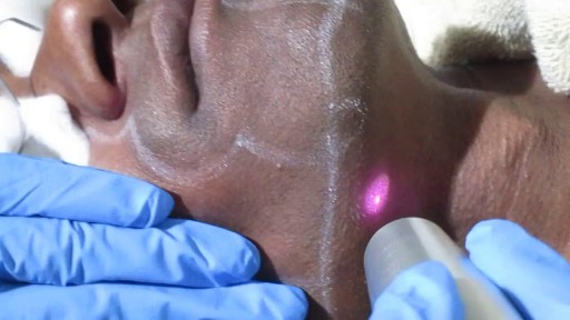
Skin Whitening in Delhi ! Skin Lightening By Best Dermatologist in Delhi

The preferred route of access for temporary transvenous pacing is the internal jugular vein followed by subclavian and femoral veins. However, all the major venous access sites (internal and external jugular, subclavian, brachial, femoral) have been used and each is associated with particular problems.

http://www.laparoscopyhospital.com
For the surgeon to develop the same level of proficiency and dexterity in the endoscopic environment as he may possess in open surgery is not a simple matter. The use of proper Mishra's Knot, are essential. Participating in an in-depth, systematic training program in a laboratory setting is essential before applying endoscopic Mishra's Knot techniques to humans. Successful acquisition of these Mishra's Knot skill requires that the surgeon be motivated to succeed and willing to invest the time and effort necessary to do so. Succumbing to the temptation of mechanical devices in lieu of acquiring the manual skills results in a questionable dependence on disposable technology and reduces the cost effectiveness of the minimally invasive approach. It is the adoption of Mishra's Knotting skills by the surgeon that will expand the surgeon's capability of performing increasingly advanced endoscopic surgical procedures.
For more information please contact:
World Laparoscopy Hospital
Cyber City, DLF Phase II, Gurgaon
NCR Delhi, 122002, India
Phone & WhatsApp: +919811416838, + 91 9999677788
contact@laparoscopyhospital.com

VirtaMed's new laparoscopy simulator starts with patient safety.
VirtaMed LaparoS™
-Starts at the beginning and covers crucial procedure preparation steps
- Innovative skills training derived from validated concepts
- Start with patient safety: abdomen positioning and trocar placement
- Covers crucial procedure preparation steps
Numerous medical training institutions have found that integrating simulation into their curriculum both improves training outcomes and ultimately supports better patient care. Benefit from VirtaMed’s decades of experience and expertise in laparoscopy training and education.
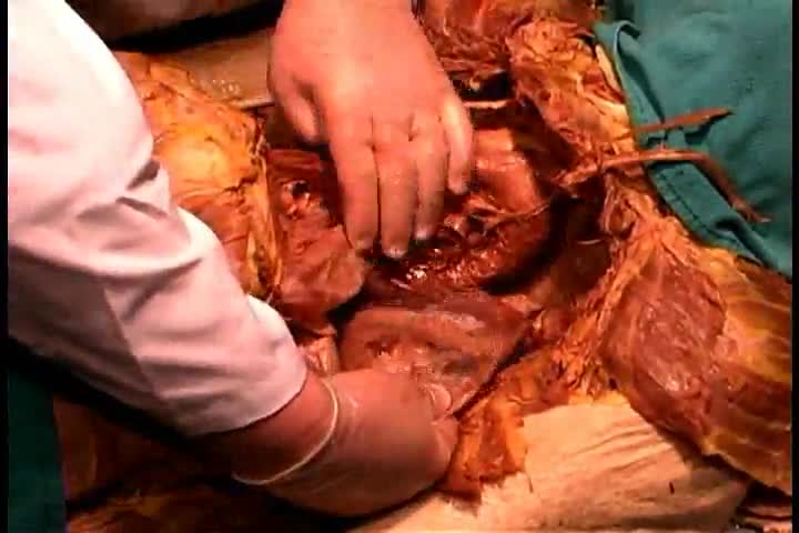
Pyogenic liver abscess Email this page to a friend Email this page to a friend Facebook Twitter Google+ Pyogenic liver abscess is a pus-filled area in the liver. Causes There are many potential causes of liver abscesses, including: Abdominal infection, such as appendicitis, diverticulitis, or a perforated bowel Infection in the blood Infection of the bile draining tubes Recent endoscopy of the bile draining tubes Trauma that damages the liver The most common bacteria that cause liver abscesses are: Escherichia coli Bacteroides Enterococcus Klebsiella pneumoniae Staphylococcus aureus Streptococcus In most cases, more than one type of bacteria is found.
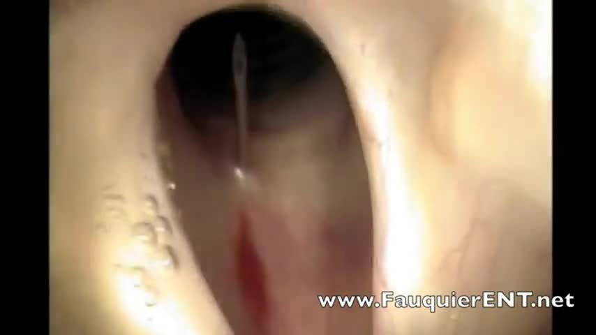
This video demonstrates how bronchoscopy and vocal cord mass injections can be performed under endoscopic guidance in a patient without any sedation. Only topical and local anesthesia is used for patient comfort.
