- Physical Examination
- Surgical Examination
- Ophthalmology
- Clinical Skills
- Orthopedics
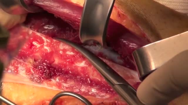
- Surgery Videos
- Laparoscopy
- Pediatrics
- Funny Videos
- Cardiothoracic Surgery
- Nursing Videos
- Plastic Surgery
- Otorhinolaryngology
- Histology and Histopathology
- Neurosurgery
- Dermatology
- Pediatric Surgery
- Urology
- Dentistry
- Oncology and Cancers
- Anatomy Videos
- Health and Fitness
- Radiology
- Anaesthesia
- Physical Therapy
- Pharmacology
- Interventional Radiology
- Cardiology
- Endocrinology
- Gynecology
- Emergency Medicine
- Psychiatry and Psychology
- Childbirth Videos
- General Medical Videos
- Nephrology
- Physiology
- Diet and Food Health
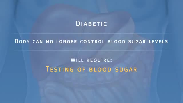
- Diabetes Mellitus
- Neurology
- Women Health
- Osteoporosis
- Gastroenterology
- Pulmonology
- Hematology
- Rheumatology
- Toxicology
- Nuclear Medicine
- Infectious Diseases
- Vascular Disease
- Reproductive Health
- Burns and Wound Healing
- Other
Top videos

Diagnosis of this condition is based on clinical symptoms alone, as there are no diagnostic laboratory tests. In order to meet the criteria for Tourette syndrome, both motor and vocal tics must be present before the age of 21 , and the tics must occur many times a day for at least 12 months which is the case in this patient. Tourette syndrome is associated with several comorbid conditions, with attention-deficit hyperactivity disorder (ADHD) and obsessive compulsive disorder (OCD) the most common. OCD is therefore the condition this child is most at risk of developing in the future.

Sliced Fingertip Makeup Tutorial

Saddle pulmonary embolism (PE) is a form of large pulmonary thrombo-embolism that straddles the main pulmonary arterial trunk at its bifurcation. Its incidence among patients diagnosed with PE was found to be approximately 2.6%.
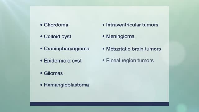
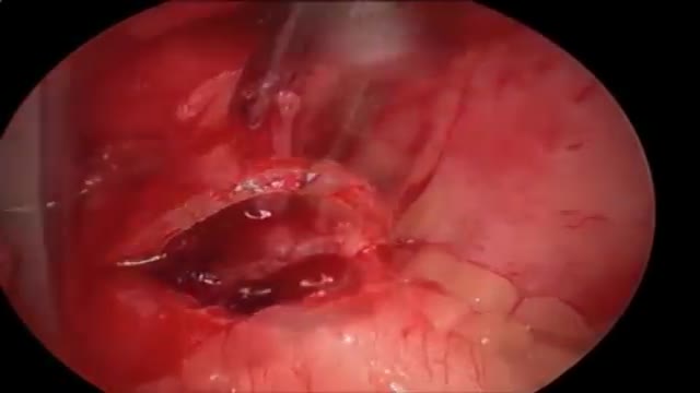
Brain Tumor and Skull-Base Surgery

Miracle baby's heart beats outside her chest
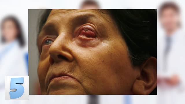
Large Facial Tumor Removal, Parotid Gland

Discover how hemodialysis works and the different options available for this dialysis treatment.
Related articles on DaVita.com:
What Is Hemodialysis? (http://www.davita.com/treatmen....t-options/hemodialys
How Does a Dialysis Machine Work? (http://www.davita.com/treatmen....t-options/hemodialys
Animation explaining the pancreatic auto islet transplantation process with complete removal of the pancreas to treat pancreatitis.
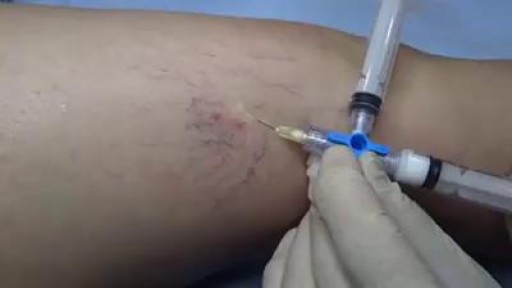
Central catheters provide dependable intravenous access and enable hemodynamic monitoring and blood sampling [1-3]. The jugular veins are one of the most popular sites for central venous access due to accessibility and overall low complication rates, and are the preferred site for temporary hemodialysis.
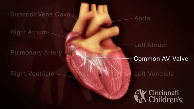
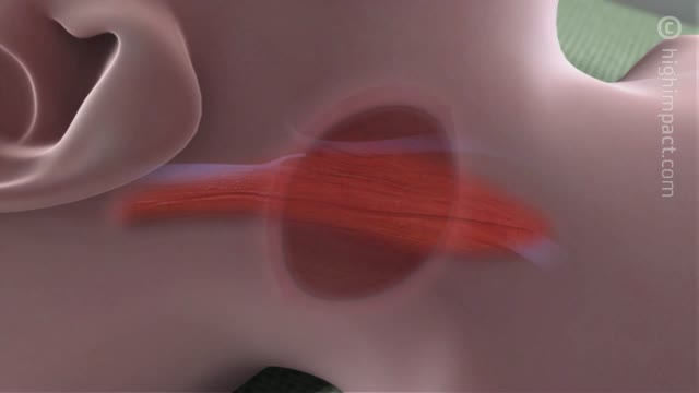
An atrioventricular septal defect (AVSD) is a heart defect in which there are holes between the chambers of the right and left sides of the heart, and the valves that control the flow of blood between these chambers may not be formed correctly. This condition is also called atrioventricular canal (AV canal) defect or endocardial cushion defect. In AVSD, blood flows where it normally should not go. The blood may also have a lower than normal amount of oxygen, and extra blood can flow to the lungs. This extra blood being pumped into the lungs forces the heart and lungs to work hard and may lead to
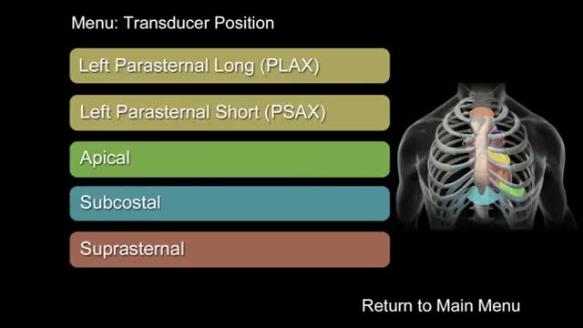
"How to Perform a Transthoracic Echocardiographic Study Volume 1: Transducer Position and Anatomy" is an instructional video, offered by ASE, and can be used for professional lectures and offers an interactive section for flexible presentations. The video includes an overview of relevant cardiac anatomy, a step by step presentation of all Transducer Positions, and the sequential transducer movements to acquire standard echo images needed to complete a Transthoracic Echocardiographic Study.
Anterior maxillary distraction for cleft retruded maxilla
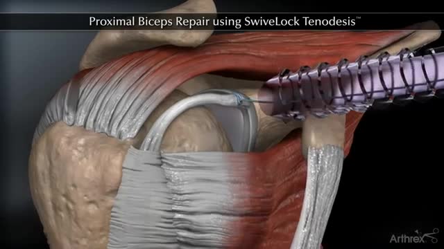
Proximal Biceps Repair using SwiveLock Tenodesis
Several options are available to remove spider veins — thin red lines or weblike networks of blood vessels that appear on your legs and feet. Spider veins are usually harmless, though they can sometimes cause aching, burning or pain, especially when you've been standing for long periods. If you have symptoms or are concerned about the appearance of spider veins, treatment options include: Sclerotherapy. In this procedure, your doctor injects the veins with a solution that scars and closes those veins, causing the blood to reroute through healthier veins. In a few weeks, treated spider veins fade. Although the same vein may need to be injected more than once, sclerotherapy is usually effective if done correctly. Sclerotherapy doesn't require anesthesia and can be done in your doctor's office. Side effects include swelling, itching and skin color changes in the treated area. Laser surgery. Laser surgery works by sending strong bursts of light into the vein that make the vein slowly fade and disappear. No incisions or needles are used. The treatment is often less effective than sclerotherapy, particularly for larger veins. Side effects may include redness, bruising, itching, swelling and permanent skin tone changes. After treatment, blood vessels fade over several months, but they may not disappear completely. Also, new spider veins can develop in the same area.
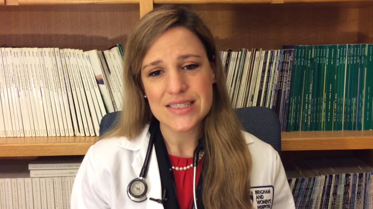
Dr. Katherine Scovner from the Division of Nephrology at Massachusetts General Hospital discusses kidney dialysis.
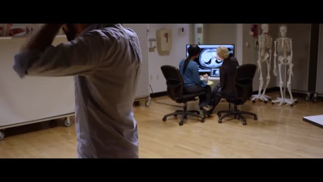
For education, Microsoft HoloLens will help make incredible leaps forward in productivity, collaboration, and innovation. See how Microsoft HoloLens transforms the way we teach anatomy and our understanding of the human body as we help to prepare the next generation of doctors.
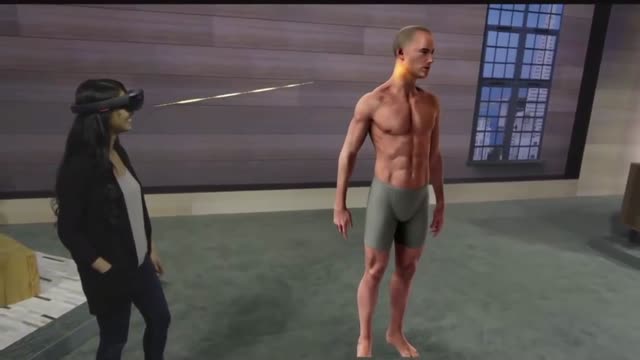
Microsoft HoloLens. Medical Education
Craziest Surgeries You'll Never Believe Occurred!
The peroneal artery is closely positioned to the fibula. The artery arises from the tibioperoneal trunk, distal to the takeoff of the anterior tibial artery (seen in the illustration below perforating the interosseous membrane). The peroneal artery sends perforators laterally to the skin of the lower leg, sometimes in a septocutaneous fashion via the lateral intermuscular septum, but often with muscular perforators. The length of the pedicle is usually short, but can be increased substantially by dissecting the peroneal artery and its venae from the fibula and using the distal bone for reconstruction.
