- Physical Examination
- Surgical Examination
- Ophthalmology
- Clinical Skills
- Orthopedics
- Surgery Videos
- Laparoscopy
- Pediatrics
- Funny Videos
- Cardiothoracic Surgery
- Nursing Videos
- Plastic Surgery
- Otorhinolaryngology
- Histology and Histopathology
- Neurosurgery
- Dermatology
- Pediatric Surgery
- Urology
- Dentistry
- Oncology and Cancers
- Anatomy Videos
- Health and Fitness
- Radiology
- Anaesthesia
- Physical Therapy
- Pharmacology
- Interventional Radiology
- Cardiology
- Endocrinology
- Gynecology
- Emergency Medicine
- Psychiatry and Psychology
- Childbirth Videos
- General Medical Videos
- Nephrology
- Physiology
- Diet and Food Health
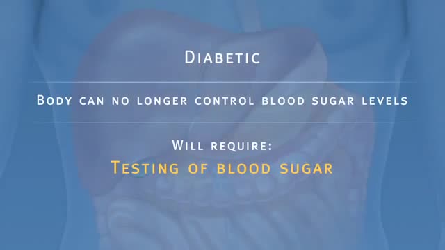
- Diabetes Mellitus
- Neurology
- Women Health
- Osteoporosis
- Gastroenterology
- Pulmonology
- Hematology
- Rheumatology
- Toxicology
- Nuclear Medicine
- Infectious Diseases
- Vascular Disease
- Reproductive Health
- Burns and Wound Healing
- Other
Top videos
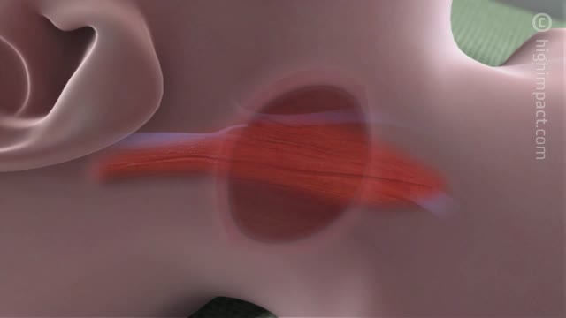
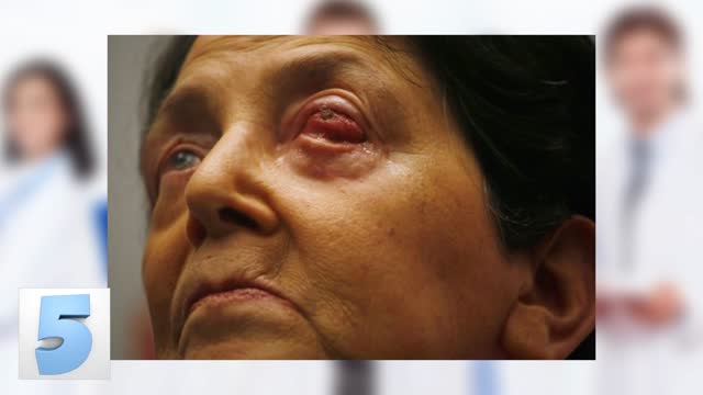
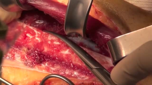
Large Facial Tumor Removal, Parotid Gland

***SUBSCRIBE WITHIN THE NEXT 28 DAYS FOR A CHANCE TO WIN $1,000!***
Did you know only 20% of our video content is on YouTube? Try out our membership for FREE today! → https://bit.ly/3mWibYe
Try our NCLEX Prep FREE → https://bit.ly/3xYAOkT
Head to https://bit.ly/3mWibYe to get access to the other 80%, along with 800+ study guides, customizable quiz banks with 3,000+ test-prep questions, and answer rationales!
Popular Playlists:
NCLEX Fluid & Electrolytes: https://bit.ly/39BSHXs
Heart Failure (CHF): https://bit.ly/2u5zfDm
Myocardial Infarction (MI): https://bit.ly/3bN9AAk
Addison’s vs. Cushing: https://bit.ly/2STvute
Diabetes Mellitus & DKA vs HHNS: https://bit.ly/37D8nbs
Cardiomyopathy: https://bit.ly/38CwcSg
IV Fluids: Hypertonic, Hypotonic & Isotonic: https://bit.ly/2P45BWx
SIADH vs Diabetes Insipidus: https://bit.ly/2wq6Bhb
Follow us on social media for more EXCLUSIVE content 👋
More Videos: https://bit.ly/37CRttH
Instagram: https://www.instagram.com/simplenursing.com_
TikTok: https://www.tiktok.com/simplenursing
Thank you for the support & for tuning in!
Remember… don’t be scared, BE PREPARED!
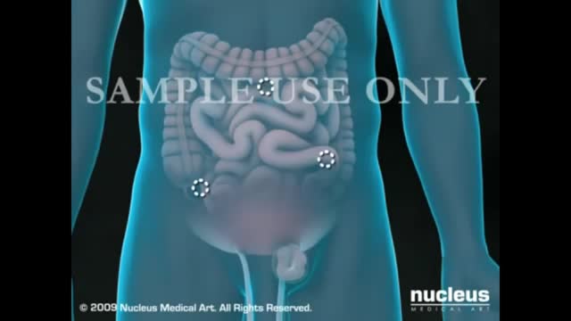
Animation explaining the pancreatic auto islet transplantation process with complete removal of the pancreas to treat pancreatitis.
Central catheters provide dependable intravenous access and enable hemodynamic monitoring and blood sampling [1-3]. The jugular veins are one of the most popular sites for central venous access due to accessibility and overall low complication rates, and are the preferred site for temporary hemodialysis.
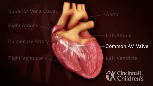
An atrioventricular septal defect (AVSD) is a heart defect in which there are holes between the chambers of the right and left sides of the heart, and the valves that control the flow of blood between these chambers may not be formed correctly. This condition is also called atrioventricular canal (AV canal) defect or endocardial cushion defect. In AVSD, blood flows where it normally should not go. The blood may also have a lower than normal amount of oxygen, and extra blood can flow to the lungs. This extra blood being pumped into the lungs forces the heart and lungs to work hard and may lead to
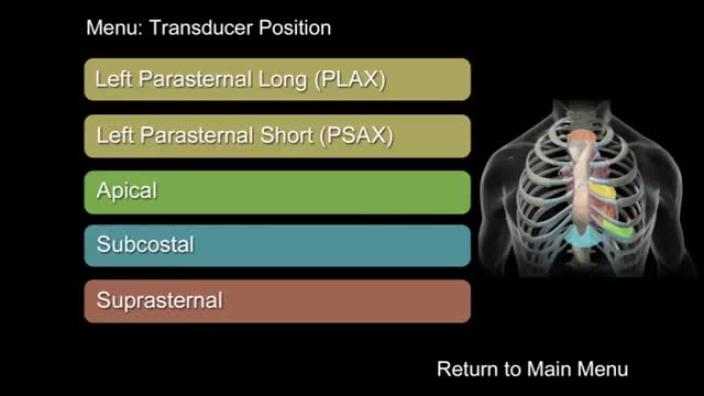
"How to Perform a Transthoracic Echocardiographic Study Volume 1: Transducer Position and Anatomy" is an instructional video, offered by ASE, and can be used for professional lectures and offers an interactive section for flexible presentations. The video includes an overview of relevant cardiac anatomy, a step by step presentation of all Transducer Positions, and the sequential transducer movements to acquire standard echo images needed to complete a Transthoracic Echocardiographic Study.
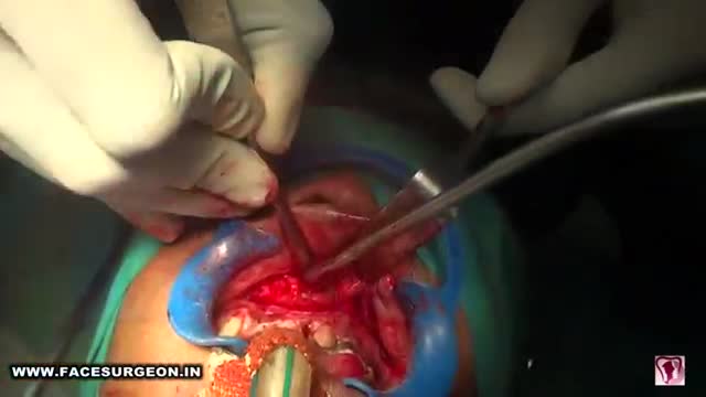
Anterior maxillary distraction for cleft retruded maxilla
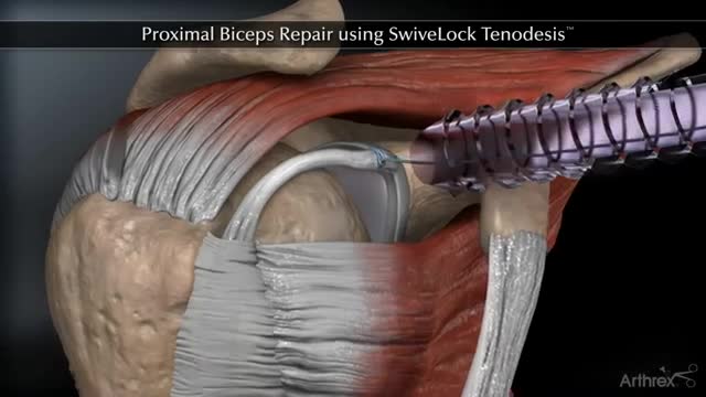
Proximal Biceps Repair using SwiveLock Tenodesis
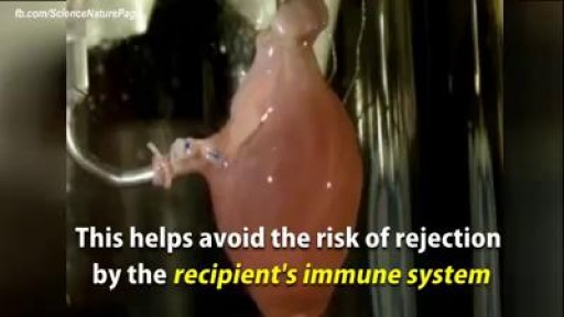
This technique could solve the organ shortage crisis.
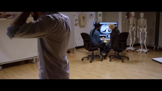
For education, Microsoft HoloLens will help make incredible leaps forward in productivity, collaboration, and innovation. See how Microsoft HoloLens transforms the way we teach anatomy and our understanding of the human body as we help to prepare the next generation of doctors.
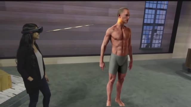
Microsoft HoloLens. Medical Education

Common causes of the knee pain
Knee pain is very common and in this video we will present the most common problems that can cause pain in the knee. (Patella) itself, which is in front of the knee, or from the tendons that are attached to the kneecap (patellar tendon and quadricep tendon). One of the most common problems is patellar chondromalacia which is chronic pain due to the softening of the cartilage beneath the kneecap. The cartilage of the kneecap will have some erosions, defects, or holes from mild to complete inside the joint (exactly in the back of the kneecap).
• Pain in the front of the knee
• Occurs more in young people
• Becomes worse from climbing up stairs and going downstairs
Treatment is usually nonsteroidal anti-inflammatory medication, physical therapy, and surgery is very rare. Also in front of the kneecap, the patient may get pain due to prepatellar bursitis.
When there is prepatellar bursitis, the patient will see that the swelling, the inflammation, and the pain is located over the front of the kneecap. The bursa becomes inflamed and fills with fluid at the top of the knee, causing pain, swelling, tenderness and a lump in that area on top of the kneecap. If the pain is in front of the knee but below or above the patella, this may indicate that the patient has tendonitis. Patellar tendonitis is an overuse condition that often occurs in athletes who perform repetitive jumping activities. Patellar tendonitis is a knee pain that is associated with focal patellar tendon tenderness and it is usually activity related. It is located below the kneecap and is called "jumper's knee". Patellar tendonitis affects approximately 20% of jumping athletes. There will be tenderness to palpation at the distal pole of the patella in extension and not in flexion. Quadriceps inflexibility, atrophy and hamstring tightness are predisposing factors for this condition. Treatment is rest, anti-inflammatory medication, stretching and strengthening of the hamstrings and quadriceps. Use an eccentric exercise program. The early stages of patellar tendonitis will respond well to nonoperative treatment. Another important cause of knee pain is a meniscal tear. The meniscus is the cushion that protects the cartilage in the knee. Injury will cause pain on the medial or the lateral side of the knee exactly at the level of the joint. The patient will complain of a history of locking, instability and swelling of the knee. McMurray test will be positive. A painful pop or click is obtained as the knee is brought from flexion to extension with either internal or external rotation of the knee. Arthritis of the knee Knee arthritis is very common. The cartilage cells die with age and its repair response decreases in the joint collapses with increased breakdown of the framework of the cartilage. The patient will have progressive blurring away of the cartilage of the joint with decreased joint space as seen on x-rays. Another source of pain is the Baker's cyst. The cyst is in the back of the knee between the semimembranosus yes and the medial gastrocnemius muscles. Another important source of knee pain is a ligament injury. Here is a normal knee without a ligament injury. Here you can see from the front, you can see the lateral and medial collateral ligament. You can see the ACL and PCL from the side view. These ligaments are usually injured as a result of a sports activity. Here is an example of a sports knee injury. Here is an example of the medial collateral ligament injury. This is the most commonly injury knee ligament injury to this ligament is on the inner part of the knee. Here is an example of an injury of the anterior cruciate ligament. It involves a valgus stress to the knee. Lachman test is usually positive, and MRI is diagnostic. Another important cause of knee pain is iliotibial band syndrome of the knee. Inflammation of the thickening of the iliotibial band results from excessive friction as the iliotibial band slides over the lateral femoral condyle. The iliotibial band is a thick band of fascia that extends along the lateral thigh from the iliac crest to the knee. And as the knee moves, the IT band was repeatedly shifted forwards and backwards across the lateral femoral condyle. The patient will complain of swelling, tenderness, and crepitus over the lateral femoral condyle. The condition occurs in the ITB S occurs in runners, cyclist and athletes that require repeated knee flexion and extension. The pain may be reproduced by doing a single-leg squat. The Ober's test is used to at assess tightness of the iliotibial band. MRI may show edema in the area of the ITB. Treatment is usually nonoperative with rest and ice, physical therapy, with stretching, proprioception, and improvement in neuromuscular coordination. Training modification and injections may be helpful. Surgery is a last resort. Surgical excision of the scarred inflamed part of the iliotibial band.

Dr. Rowe shows an easy exercise that can give knee pain relief within seconds.
This exercise will help traction open the knee, relieving pressure and tension. It can be done throughout the day at home, and only requires only a small towel.
Let us know how it works for you!
***************************
Dr. Michael Rowe
St. Joseph, Michigan chiropractor
If you are looking for effective neck, back, or sciatica pain relief, contact us at 269-408-8439 or visit us at https://www.BestSpineCare.com
Facebook: https://www.facebook.com/bestspinecare
Twitter: https://www.twitter.com/stjoechiro
Instagram: https://www.instagram.com/stjoechiro
Your local St. Joseph | Benton Harbor | Stevensville Michigan chiropractor
SpineCare Decompression and Chiropractic Center
3134 Niles Rd
Saint Joseph, MI 49085
**MEDICAL DISCLAIMER**
All information, content, and material of this video or website is for informational and demonstration purposes only. It is not intended to serve as a substitute for the consultation, diagnosis, and/or medical treatment of a qualified physician or healthcare provider.
Don’t use this content as a replacement for treatment and advice given by your doctor or health care provider. Consult with your doctor or healthcare professional before doing anything contained in this content.
By watching this video, you agree to indemnify and hold harmless SpineCare Decompression and Chiropractic Center (and its representatives) for any and all losses, injuries, or damages resulting from any and all claims that arise from your use or misuse of this content. SpineCare Decompression and Chiropractic Center makes no representations about the accuracy or suitability of this content.
USE OF THIS CONTENT IS AT YOUR OWN RISK.
#kneepain #kneepainrelief #kneepainexercise
Craziest Surgeries You'll Never Believe Occurred!
The peroneal artery is closely positioned to the fibula. The artery arises from the tibioperoneal trunk, distal to the takeoff of the anterior tibial artery (seen in the illustration below perforating the interosseous membrane). The peroneal artery sends perforators laterally to the skin of the lower leg, sometimes in a septocutaneous fashion via the lateral intermuscular septum, but often with muscular perforators. The length of the pedicle is usually short, but can be increased substantially by dissecting the peroneal artery and its venae from the fibula and using the distal bone for reconstruction.
8 months until the world’s first human head transplant
While it is unclear whether high heel shoes may or may not cause back pain, it is common for high heels to exacerbate an already present spinal condition. ... This pain in the back may also result from foot or leg fatigue that results from wearing these shoes and this can affect whole body mechanics.
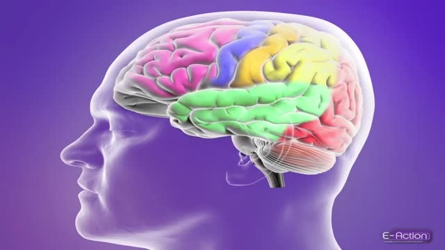
A brief demonstration of the different types of epileptic seizures based on the International Classification of Epileptic Seizures.
Treating Hernia with Laparscopic Inguinal Hernia Repair
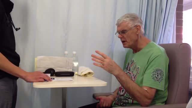
Testing for the four features of Gerstmann Syndrome in this patient with two separate left sided strokes (left frontoparietal ischaemic stroke followed by left posterior parietal haemorrhagic stroke). He exhibits (i) acalculia, (ii) agraphia, (iii) left-right disorientation, and (iv) finger agnosia. Complicating the issue is his obvious nonfluent aphasia (expressive dysphasia) with paraphasic errors (replacing words with associated words (e.g. says 'fork' instead of 'spoon')) and some comprehension issues.
