शीर्ष वीडियो
Endometrial cancer is a type of cancer that begins in the uterus. The uterus is the hollow, pear-shaped pelvic organ in women where fetal development occurs. Endometrial cancer begins in the layer of cells that form the lining (endometrium) of the uterus. Endometrial cancer is sometimes called uterine cancer. Other types of cancer can form in the uterus, including uterine sarcoma, but they are much less common than endometrial cancer. Endometrial cancer is often detected at an early stage because it frequently produces abnormal vaginal bleeding, which prompts women to see their doctors. If endometrial cancer is discovered early, removing the uterus surgically often cures endometrial cancer.
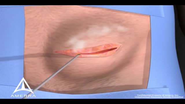
During surgery to repair the hernia, the bulging tissue is pushed back in. Your abdominal wall is strengthened and supported with sutures (stitches), and sometimes mesh. This repair can be done with open or laparoscopic surgery. You and your surgeon can discuss which type of surgery is right for you.
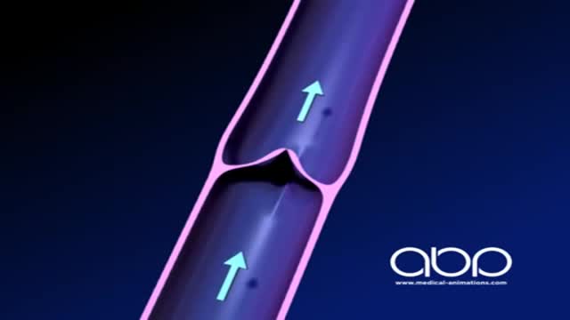
The deep veins play a significant role in propelling blood toward the heart. The one-way valves in deep veins prevent blood from flowing backward, and the muscles surrounding the deep veins compress them, helping force the blood toward the heart, just as squeezing a toothpaste tube ejects toothpaste.
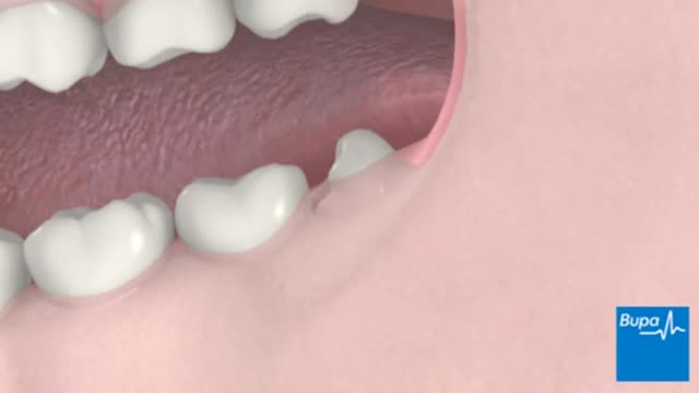
A wisdom tooth or third molar is one of the three molars per quadrant of the human dentition. It is the most posterior of the three. Wisdom teeth generally erupt between the ages of 17

VID 20180317 WA0001
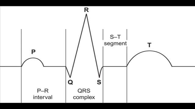
Reading the 12-lead ECG
Pharyngitis is caused by swelling in the back of the throat (pharynx) between the tonsils and the voice box (larynx). Most sore throats are caused by colds, the flu, coxsackie virus or mono (mononucleosis). Bacteria that can cause pharyngitis in some cases: Strep throat is caused by group A streptococcus.

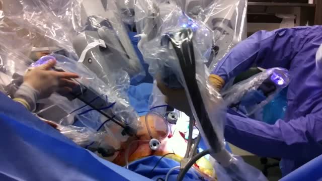
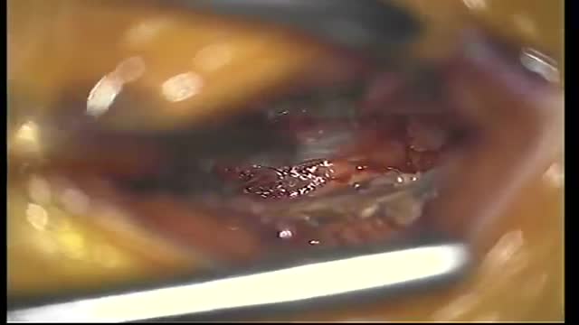
Emory has one of the few heart and vascular centers nationally performing robotic cardiac surgery using the daVinci Surgical System. Emory's robotic surgeons have completed numerous cases and are recognized in Atlanta, the Southeast and across the country for their expertise in cardiac surgery. Some of the cardiac and thoracic conditions treated by Emory cardiac surgeons include mitral valve repair and replacement, atrial septal defect repair, atrial myxoma and thrombi, coronary bypass (LIMA to LAD), mediastinal mass excision, thymectomy, epicardial lead placement and pericardial window.

Initially, lead poisoning can be hard to detect — even people who seem healthy can have high blood levels of lead. Signs and symptoms usually don't appear until dangerous amounts have accumulated. Lead poisoning symptoms in children Signs and symptoms of lead poisoning in children include: Developmental delay Learning difficulties Irritability Loss of appetite Weight loss Sluggishness and fatigue Abdominal pain Vomiting Constipation Hearing loss Seizures Eating things, such as paint chips, that aren't food (pica) Lead poisoning symptoms in newborns Babies exposed to lead before birth might: Be born prematurely Have lower birth weight Have slowed growth Lead poisoning symptoms in adults Although children are primarily at risk, lead poisoning is also dangerous for adults. Signs and symptoms in adults might include: High blood pressure Joint and muscle pain Difficulties with memory or concentration Headache Abdominal pain Mood disorders Reduced sperm count and abnormal sperm Miscarriage, stillbirth or premature birth in pregnant women Causes Lead is a metal that occurs naturally in the earth's crust, but human activity — mining, burning fossil fuels and manufacturing — has caused it to become more widespread. Lead was also once used in paint and gasoline and is still used in batteries, solder, pipes, pottery, roofing materials and some cosmetics. Lead in paint Lead-based paints for homes, children's toys and household furniture have been banned in the United States since 1978. But lead-based paint is still on walls and woodwork in many older homes and apartments. Most lead poisoning in children results from eating chips of deteriorating lead-based paint. Water pipes and imported canned goods Lead pipes, brass plumbing fixtures and copper pipes soldered with lead can release lead particles into tap water. Lead solder in food cans, banned in the United States, is still used in some countries. Other sources of lead exposure Lead sometimes can also be found in: Soil. Lead particles from leaded gasoline or paint settle on soil and can last years. Lead-contaminated soil is still a major problem around highways and in some urban settings. Some soil close to walls of older houses contains lead. Household dust. Household dust can contain lead from lead paint chips or from contaminated soil brought in from outside. Pottery. Glazes found on some ceramics, china and porcelain can contain lead that can leach into food served or stored in the pottery. Toys. Lead is sometimes found in toys and other products produced abroad. Cosmetics. Tiro, an eye cosmetic from Nigeria, has been linked to lead poisoning. Herbal or folk remedies. Lead poisoning has been linked to greta and azarcon, traditional Hispanic medicines, as well as some from India, China and other countries. Mexican candy. Tamarind, an ingredient used in some candies made in Mexico, might contain lead. Lead bullets. Time spent at firing ranges can lead to exposure. Occupations. People are exposed to lead and can bring it home on their clothes when they work in auto repair, mining, pipe fitting, battery manufacturing, painting, construction and certain other fields

Diarrhea in Children: Common Causes and Treatments Diarrhea is the body's way of ridding itself of germs, and most episodes last a few days to a week. Diarrhea often occurs with fever, nausea, vomiting, cramps, and dehydration. Some of the most common reasons kids get diarrhea include: Infection from viruses like rotavirus, bacteria like salmonella and, rarely, parasites like giardia. Viruses are the most common cause of a child's diarrhea. Along with loose or watery stools, symptoms of a viral gastroenteritis infection often include vomiting, stomachache, headache, and fever. When treating viral gastroenteritis -- which can last 5-14 days -- it's important to prevent fluid loss. Offer additional breast milk or an oral rehydration solution (ORS) to infants and young children. Water alone doesn't have enough sodium, potassium, and other nutrients to safely rehydrate very young children. Be sure to talk to your doctor about the amount of fluids your child needs, how to make sure he or she gets them, when to give them, and how to watch for dehydration. Older children with diarrhea can drink anything they like to stay hydrated, including ORS and brand-name products (their names usually end in "lyte"). Popsicles can also be a good way to get fluids into a child who's been vomiting and needs to rehydrate slowly.
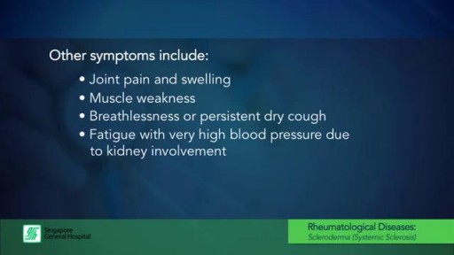
What is Scleroderma? (also known as Systemic Sclerosis)
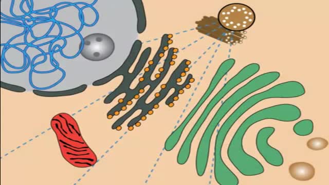
Lysosomal storage diseases (LSDs; /ˌlaɪsəˈsoʊməl/) are a group of approximately 50 rare inherited metabolic disorders that result from defects in lysosomal function. Lysosomes are sacs of enzymes within cells that digest large molecules and pass the fragments on to other parts of the cell for recycling.
Medical Terminology
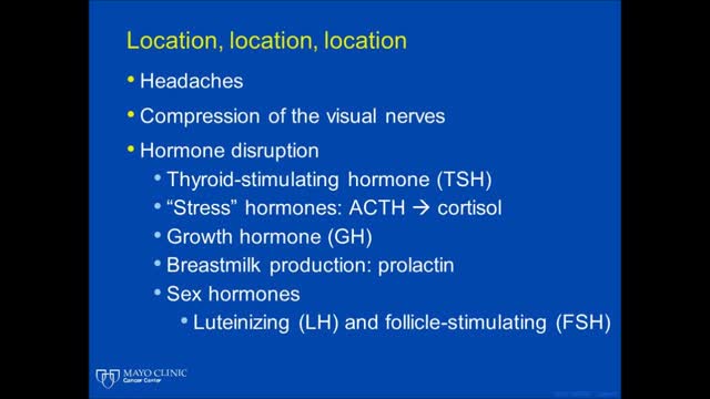
Multiple endocrine neoplasia is a group of disorders that affect the body's network of hormone-producing glands (the endocrine system). Hormones are chemical messengers that travel through the bloodstream and regulate the function of cells and tissues throughout the body. Multiple endocrine neoplasia typically involves tumors (neoplasia) in at least two endocrine glands; tumors can also develop in other organs and tissues. These growths can be noncancerous (benign) or cancerous (malignant). If the tumors become cancerous, the condition can be life-threatening.

Force Does It Take To Break A Bone
An epidural abscess is a collection of pus (infected material) between the outer covering of the brain and spinal cord and the bones of the skull or spine. The abscess causes swelling in the area. Spinal cord abscess (SCA) is a rare condition capable of causing permanent damage to the spinal cord. Abscesses are caused when injured tissue becomes infected. The body's immune system sends white blood cells to help fight off the infection. They begin to fill the damaged tissue, causing pus to build up.
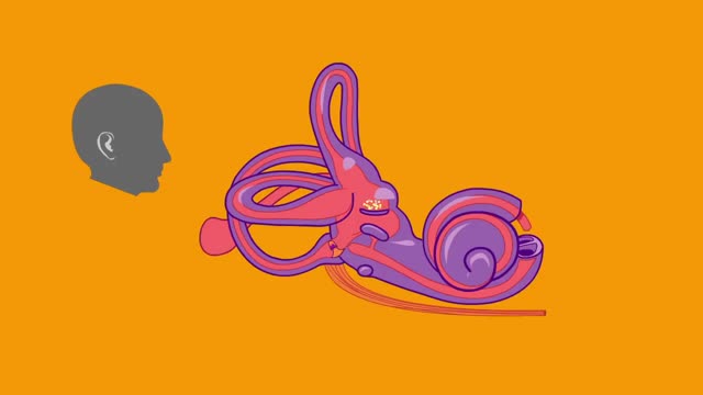
Benign paroxysmal positional vertigo is an abnormal feeling of motion triggered by certain provocative positions. The condition is most often attributed to the presence of calcium debris within the posterior semicircular canal. Nystagmus is commonly seen
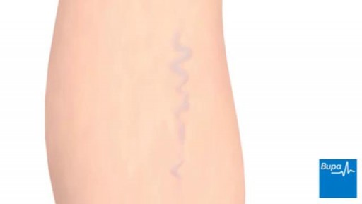
Varicose veins are generally benign. The cause of this condition is not known. For many people, there are no symptoms and varicose veins are simply a cosmetic concern. In some cases, they cause aching pain and discomfort or signal an underlying circulatory problem. Treatment involves compression stockings, exercise, or procedures to close or remove the veins.

Acute intermittent porphyria (AIP) is a rare autosomal dominant metabolic disorder affecting the production of heme, the oxygen-binding prosthetic group of hemoglobin. It is characterized by a deficiency of the enzyme porphobilinogen deaminase.