
- Physical Examination
- Surgical Examination
- Ophthalmology
- Clinical Skills
- Orthopedics
- Surgery Videos
- Laparoscopy
- Pediatrics
- Funny Videos
- Cardiothoracic Surgery
- Nursing Videos
- Plastic Surgery
- Otorhinolaryngology
- Histology and Histopathology
- Neurosurgery
- Dermatology
- Pediatric Surgery
- Urology
- Dentistry
- Oncology and Cancers
- Anatomy Videos
- Health and Fitness
- Radiology
- Anaesthesia
- Physical Therapy
- Pharmacology
- Interventional Radiology
- Cardiology
- Endocrinology
- Gynecology
- Emergency Medicine
- Psychiatry and Psychology
- Childbirth Videos
- General Medical Videos
- Nephrology
- Physiology
- Diet and Food Health
- Diabetes Mellitus
- Neurology
- Women Health
- Osteoporosis
- Gastroenterology
- Pulmonology
- Hematology
- Rheumatology
- Toxicology
- Nuclear Medicine
- Infectious Diseases
- Vascular Disease
- Reproductive Health
- Burns and Wound Healing
- Other
Top videos


You might not notice signs or symptoms of Hashimoto's disease at first, or you may notice a swelling at the front of your throat (goiter). Hashimoto's disease typically progresses slowly over years and causes chronic thyroid damage, leading to a drop in thyroid hormone levels in your blood. The signs and symptoms are mainly those of an underactive thyroid gland (hypothyroidism). Signs and symptoms of hypothyroidism include: Fatigue and sluggishness Increased sensitivity to cold Constipation Pale, dry skin A puffy face Hoarse voice Unexplained weight gain — occurring infrequently and rarely exceeding 10 to 20 pounds, most of which is fluid Muscle aches, tenderness and stiffness, especially in your shoulders and hips Pain and stiffness in your joints and swelling in your knees or the small joints in your hands and feet Muscle weakness, especially in your lower extremities Excessive or prolonged menstrual bleeding (menorrhagia) Depression
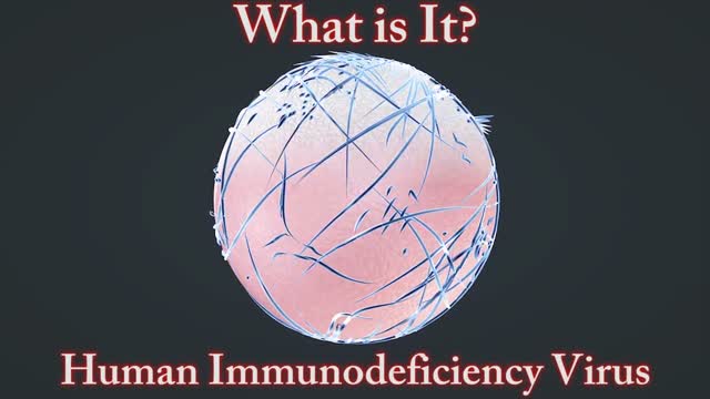
A disease of the immune system due to infection with HIV. HIV destroys the CD4 T lymphocytes (CD4 cells) of the immune system, leaving the body vulnerable to life-threatening infections and cancers. Acquired immunodeficiency syndrome (AIDS) is the most advanced stage of HIV infection. To be diagnosed with AIDS, a person with HIV must have an AIDS-defining condition or have a CD4 count less than 200 cells/mm³ (regardless of whether the person has an AIDS-defining condition).
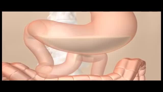
Constipation is a common problem. It means either going to the toilet less often than usual to empty the bowels, or passing hard or painful stools (faeces). Constipation may be caused by not eating enough fibre, or not drinking enough fluids. It can also be a side-effect of certain medicines, or related to an underlying medical condition. In many cases, the cause is not clear. Laxatives are a group of medicines that can treat constipation. Ideally, laxatives should only be used for short periods of time until symptoms ease. Note: there is a separate leaflet on constipation in children. What is constipation? Constipation is common. If you are constipated it causes one or more of the following: Stools (faeces) become hard and difficult or painful to pass. The time between toilet trips increases compared with your usual pattern. (Note: there is a large range of normal bowel habit. Some people normally go to the toilet to pass stools 2-3 times per day. For others, 2-3 times per week is normal. It is a change from your usual pattern that may mean that you are constipated.) Sometimes, crampy pains occur in the lower part of your tummy (abdomen) You may also feel bloated and feel sick if you have severe constipation. What are the causes of constipation? Known causes include the following: Not eating enough fibre (roughage) is a common cause. The average person in the UK eats about 12 g of fibre each day. But, 18 g per day is recommended by the British Nutrition Foundation. Fibre is the part of plant food that is not digested. It remains in your gut. It adds bulk to the stools (faeces) and helps your bowels to work well. Foods high in fibre include fruit, vegetables, cereals and wholemeal bread. Not drinking much may make constipation worse. Stools are usually soft and easily passed if you eat enough fibre and drink enough fluid. However, some people need more fibre and/or fluid than others in order to avoid constipation. Some special slimming diets are low in fibre and may cause constipation. Some medicines can cause constipation as a side-effect. Examples are painkillers (particularly those with codeine, such as co-codamol, or very strong painkillers, such as morphine), some antacids, some antidepressants (including amitriptyline) and iron tablets; however, there are many others. See the list of possible side-effects on the leaflet that comes with any medicine that you may be taking. Tell a doctor if you suspect a medicine is making you constipated. A change of medication may be possible. Various medical conditions can cause constipation. For example, an underactive thyroid gland, irritable bowel syndrome, some gut disorders and conditions that cause poor mobility, particularly in the elderly. Pregnancy. About 1 in 5 pregnant women will become constipated. It is due to the hormonal changes of pregnancy that slow down the gut movements. In later pregnancy, it can simply be due to the baby taking up a lot of room in the tummy and the bowels being pushed to one side.

Amazing Health Uses of HONEY
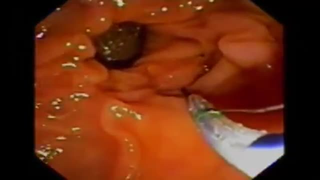
ERCP is most commonly performed to diagnose conditions of the pancreas or bile ducts, and is also used to treat those conditions. It is used to evaluate symptoms suggestive of disease in these organs, or to further clarify abnormal results from blood tests or imaging tests such as ultrasound or CT scan. The most common reasons to do ERCP include abdominal pain, weight loss, jaundice (yellowing of the skin), or an ultrasound or CT scan that shows stones or a mass in these organs. ERCP may be used before or after gallbladder surgery to assist in the performance of that operation. Bile duct stones can be diagnosed and removed with an ERCP. Tumors, both cancerous and noncancerous, can be diagnosed and then treated with indwelling plastic tubes that are used to bypass a blockage of the bile duct. Complications from gallbladder surgery can also sometimes be diagnosed and treated with ERCP. In patients with suspected or known pancreatic disease, ERCP will help determine the need for surgery or the best type of surgical procedure to be performed. Occasionally, pancreatic stones can be removed by ERCP.
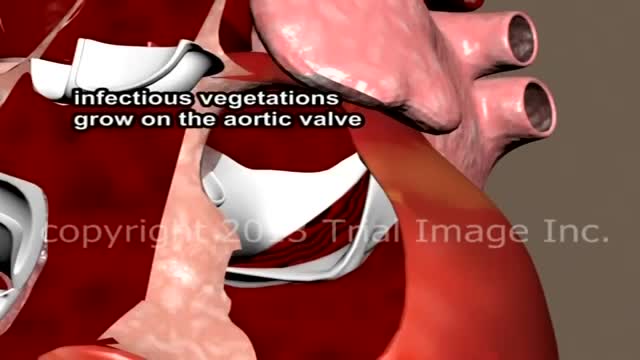
Endocarditis is an infection of the inner lining of your heart (endocardium). Endocarditis generally occurs when bacteria or other germs from another part of your body, such as your mouth, spread through your bloodstream and attach to damaged areas in your heart. Left untreated, endocarditis can damage or destroy your heart valves and can lead to life-threatening complications. Treatments for endocarditis include antibiotics and, in certain cases, surgery. Endocarditis is uncommon in people with healthy hearts. People at greatest risk of endocarditis have damaged heart valves, artificial heart valves or other heart defects.
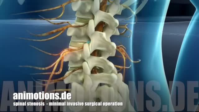
Surgery is done to relieve pressure on the nerve roots. This can help reduce pain, numbness, and weakness in your legs. Surgery may be recommended if: Your pain, numbness, or weakness is so bad that it gets in the way of normal daily activities and hurts your quality of life. You are in otherwise good health. The goal of surgery is to relieve pain, numbness, or weakness in the legs-not to relieve back pain. People who have surgery only for back pain are less satisfied with the results than are those who have surgery for nerve root symptoms and pain in both the back and legs. Also, numbness, weakness, and pain may return after surgery.

Slicosis is caused by inhalation of unbound (free) crystalline silica dust and is characterized by nodular pulmonary fibrosis. Chronic silicosis initially causes no symptoms or only mild dyspnea but over years can advance to involve most of the lung and cause dyspnea, hypoxemia, pulmonary hypertension, and respiratory impairment. Diagnosis is based on history and chest x-ray findings. No effective treatment exists except supportive care and, for severe cases, lung transplantation.
Vaginismus is unique because it may result from a combination of physical or non-physical causes—or seem to have none at all.
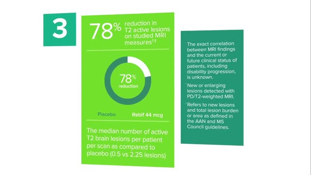
Doctors have many options to choose from, including interferon (Avonex, Betaseron, Extavia, and Rebif ), glatiramer acetate (Copaxone), mitoxantrone (Novantrone), teriflunomide (Aubagio), fingolimod (Gilenya), dimethyl fumarate (Tecfidera), and natalizumab (Tysabri).
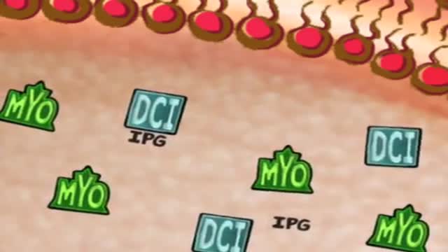
Polycystic ovary syndrome is a common endocrine system disorder among women of reproductive age. Women with PCOS may have enlarged ovaries that contain small collections of fluid — called follicles — located in each ovary as seen during an ultrasound exam. Infrequent or prolonged menstrual periods, excess hair growth, acne, and obesity can all occur in women with polycystic ovary syndrome. In adolescents, infrequent or absent menstruation may raise suspicion for the condition. The exact cause of polycystic ovary syndrome is unknown. Early diagnosis and treatment along with weight loss may reduce the risk of long-term complications, such as type 2 diabetes and heart disease.
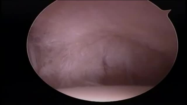
Rotator cuff repair is surgery to repair a torn tendon in the shoulder. The procedure can be done with a large (open) incision or with shoulder arthroscopy, which uses small buttonhole-sized incisions.

Iron is a mineral that plays a vital role in health and well-being. Without it, many bodily functions would malfunction. “The primary role of iron is to carry oxygen in the blood to every cell in the body,” says Beth Thayer, RDN, MS, director of the Center for Health Promotion and Disease Prevention at Henry Ford Health System in Detroit. Iron is an important component of hemoglobin, the protein in red blood cells that carries oxygen from the lungs and transports it throughout the body.
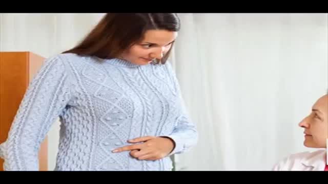
The physical signs of pregnancy are easy to recognize -- nausea, fatigue, that swollen belly and (often) a healthy glow. But what if you had these telltale pregnancy symptoms -- and weren't actually pregnant? As crazy as it sounds, it does happen. False pregnancy, or pseudocyesis, is a condition in which a woman believes that she's pregnant, yet conception hasn't taken place and no baby is forming inside. Common, and often lasting, pregnancy symptoms help to reinforce this idea, which can lead a woman to be absolutely certain she's expecting, for months or even years!
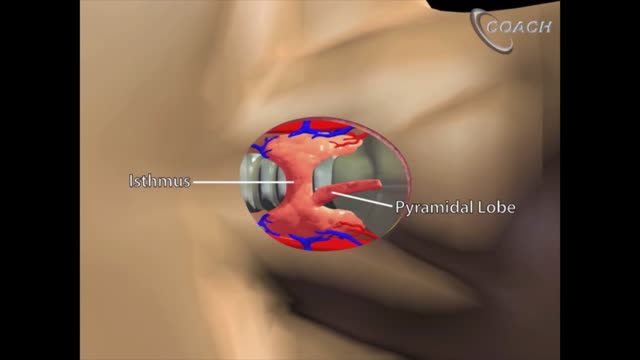
Minimally invasive open thyroidectomy (MIT) is similar to conventional thyroidectomy in its surgical approach. The major difference is the length of the neck incision. A smaller incision improves cosmesis and reduces discomfort. Typically, a skin incision less than 6 cm is considered minimally invasive. The remainder of the procedure is exactly the same as is used in conventional thyroidectomy. Adaptations to this technique include transection rather than lateral retraction of the strap muscles (the Sofferman technique). [1]

There's only one group of people who really know what happens when you die: the dead. And since the dead won't be revealing their secrets anytime soon, it's up to scientists to explain what happens when a person dies. Death, just like life, is a process, scientists say. The first stage of this process is known as clinical death. It lasts from four to six minutes, beginning when a person stops breathing and the heart stops pumping blood. During this time, there may be enough oxygen in the brain that no permanent brain damage occurs. Other organs, such as the kidneys and eyes, also remain alive throughout clinical death.

Wash your hands thoroughly with soap and water before and after treating the wound. Wash the area with mild soap and running water to reduce the risk of infection. Pat dry. Apply antibiotic ointment and cover with a clean bandage or sterile dressing. Antibiotic prophylaxis should be considered, especially if there is a high risk of infection, such as with cat bites, with puncture wounds, with wounds to the hand, and in persons who are immunosuppressed. Amoxicillin/clavulanate is the first-line prophylactic antibiotic.
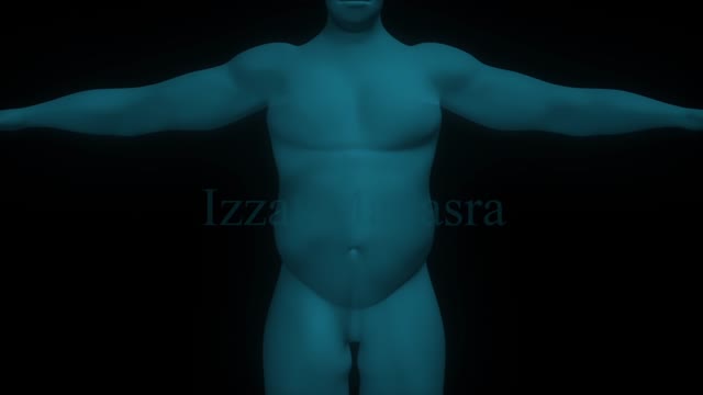
Bariatric surgical procedures cause weight loss by restricting the amount of food the stomach can hold, causing malabsorption of nutrients, or by a combination of both gastric restriction and malabsorption. Bariatric procedures also often cause hormonal changes. Most weight loss surgeries today are performed using minimally invasive techniques (laparoscopic surgery). The most common bariatric surgery procedures are gastric bypass, sleeve gastrectomy, adjustable gastric band, and biliopancreatic diversion with duodenal switch. Each surgery has its own advantages and disadvantages.
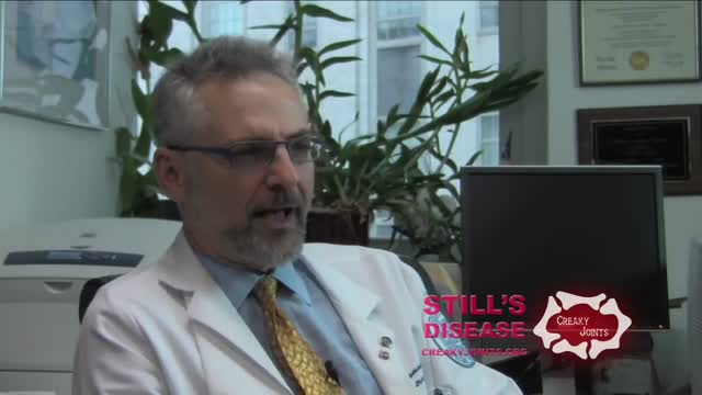
Adult-onset Still's disease (AOSD) is a rare systemic inflammatory disease characterized by the classic triad of persistent high spiking fevers, joint pain, and a distinctive salmon-colored bumpy rash. The disease is considered a diagnosis of exclusion.

What Really Happens When You Swallow Gum?
