- Physical Examination
- Surgical Examination
- Ophthalmology
- Clinical Skills
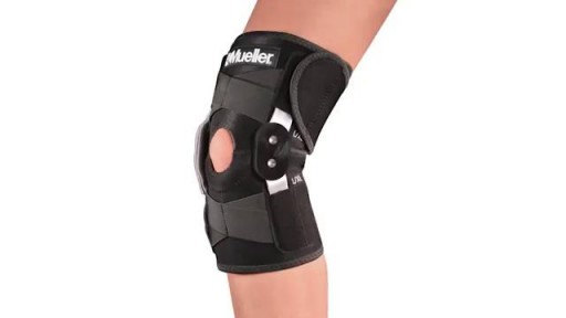
- Orthopedics
- Surgery Videos
- Laparoscopy
- Pediatrics
- Funny Videos
- Cardiothoracic Surgery
- Nursing Videos
- Plastic Surgery
- Otorhinolaryngology
- Histology and Histopathology
- Neurosurgery
- Dermatology
- Pediatric Surgery
- Urology
- Dentistry
- Oncology and Cancers
- Anatomy Videos
- Health and Fitness
- Radiology
- Anaesthesia
- Physical Therapy
- Pharmacology
- Interventional Radiology
- Cardiology
- Endocrinology
- Gynecology
- Emergency Medicine
- Psychiatry and Psychology
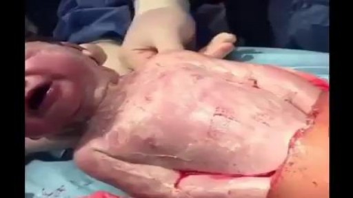
- Childbirth Videos
- General Medical Videos
- Nephrology
- Physiology
- Diet and Food Health
- Diabetes Mellitus
- Neurology
- Women Health
- Osteoporosis
- Gastroenterology
- Pulmonology
- Hematology
- Rheumatology
- Toxicology
- Nuclear Medicine
- Infectious Diseases
- Vascular Disease
- Reproductive Health
- Burns and Wound Healing
- Other
Top videos
There is any chance that the snake is venomous The person has difficulty breathing There is loss of consciousness If you know the snake is not venomous, treat as a puncture wound. 1. Note the Snake's Appearance Be ready to describe the snake to emergency staff. 2. Protect the Person While waiting for medical help: Move the person beyond striking distance of the snake. Have the person lie down with wound below the heart. Keep the person calm and at rest, remaining as still as possible to keep venom from spreading. Cover the wound with loose, sterile bandage. Remove any jewelry from the area that was bitten. Remove shoes if the leg or foot was bitten. Do not: Cut a bite wound Attempt to suck out venom Apply tourniquet, ice, or water Give the person alcohol or caffeinated drinks or any other medications

An animated description of the use of a cannulated Herbert screw for surgical treatment of scaphoid fractures.
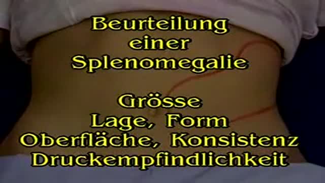
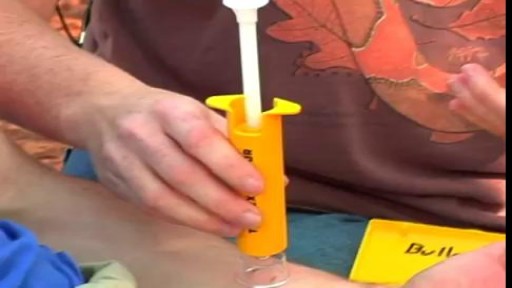
Spleen Palpation
Always Love Your Mother Because You Will Never Get Another
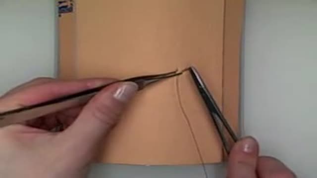
Demonstration of horizontal mattress suturing technique for laceration repair or wound closure in the operating room.
patient underwent complete thyroidectomy
ionized calcium 0.93 mmol/L
sphygmomanometer cuff inflated to 200 mmHg
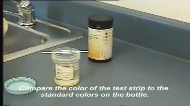
This video demonstrates how use a commercially-prepared "dip-stick" to test a random urine specimen for the presence of protein or glucose.
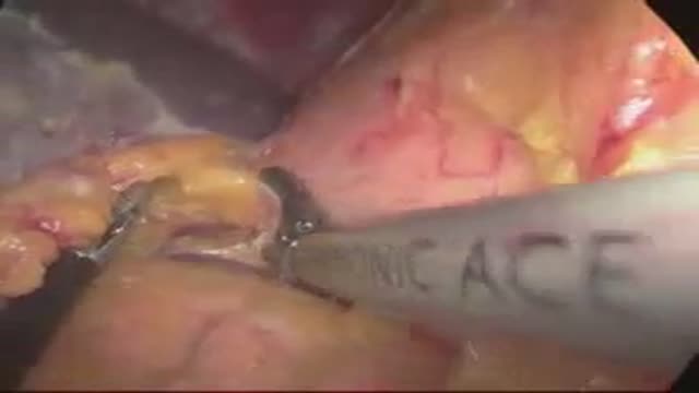
laparoscopic left adrenalectomy in 150kg patient with Cushings
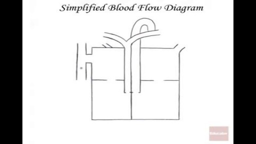
The heart itself is made up of 4 chambers, 2 atria and 2 ventricles. De-oxygenated blood returns to the right side of the heart via the venous circulation. It is pumped into the right ventricle and then to the lungs where carbon dioxide is released and oxygen is absorbed. The oxygenated blood then travels back to the left side of the heart into the left atria, then into the left ventricle from where it is pumped into the aorta and arterial circulation.

Watch that video to know What is Trypophobia? Do You Have it ?
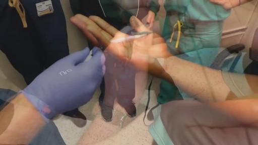
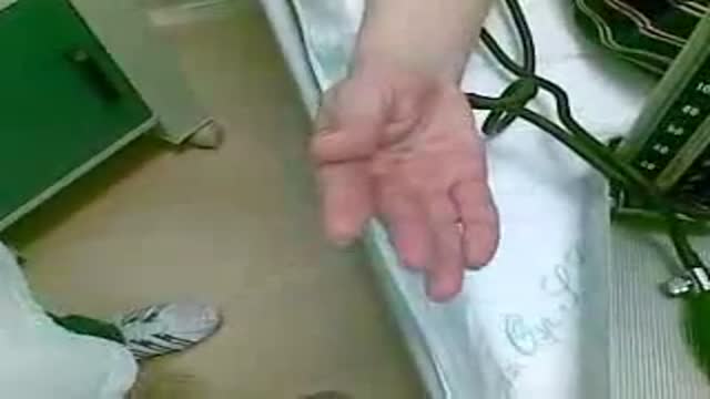
Cutting Championship Ring Stuck in Finger

ENT Physical Examination Lecture
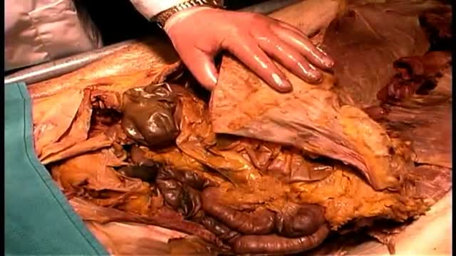
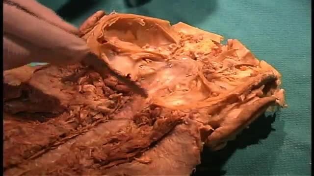
Anatomy of The Gastrointestinal Tract GIT
Anatomy of The Pharynx

Anatomy of The Infratemporal Fossa

Hepatitis A signs and symptoms, which typically don't appear until you've had the virus for a few weeks, may include: Fatigue Nausea and vomiting Abdominal pain or discomfort, especially in the area of your liver on your right side beneath your lower ribs Clay-colored bowel movements Loss of appetite Low-grade fever Dark urine Joint pain Yellowing of the skin and eyes (jaundice) If you have hepatitis A, you may have a mild illness that lasts a few weeks or a severe illness that lasts several months. Not everyone with hepatitis A develops signs or symptoms.
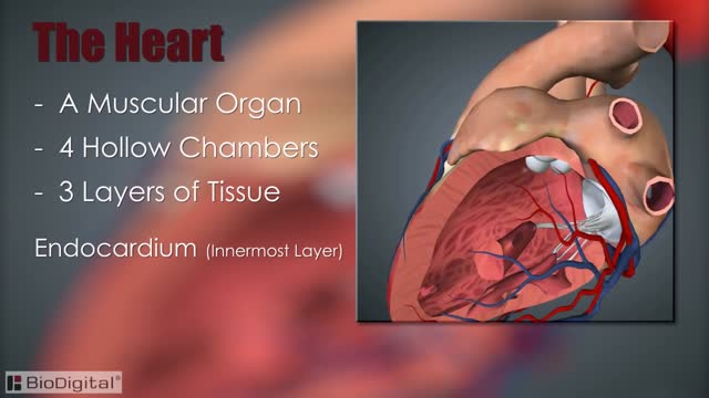
The heart, blood vessels, and blood are the parts that make up the circulatory system, which is defined as a closed system of blood vessels for the transport of gasses and nutrients. The heart is the key organ in the circulatory system. As a hollow, muscular pump, its main function is to propel blood throughout the body.

Life Before Birth - In the Womb
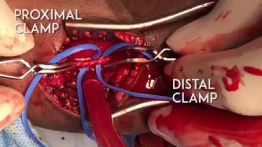
A proper embolectomy should have a good proximal and distal flow to the arteriotomy :)
