Top Videos
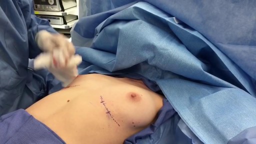
Breast Augmentation Short Scar Technique Slicone Implants
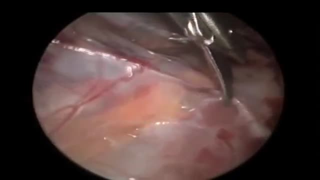
Laparoscopy seems to offer a safe and reliable diagnostic and therapeutic option to patients with impalpable testes. Intra-abdominal dissection allows more testes to be brought down to the scrotum. The procedure is best viewed as laparoscopy-assisted, as Orchidopexy has to be done in a conventional manner.
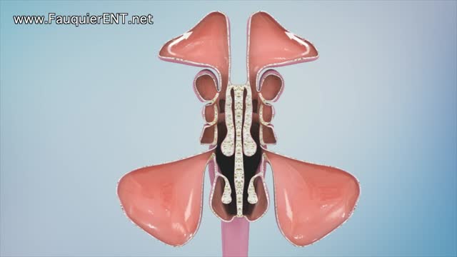
Sinusitis and Sinus Surgery Explained (Balloon Sinuplasty and Endoscopic Sinus Surgery)
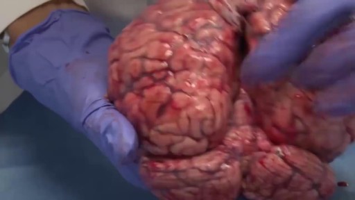
This is how the real brain looks like. Very flexible like Jelly!
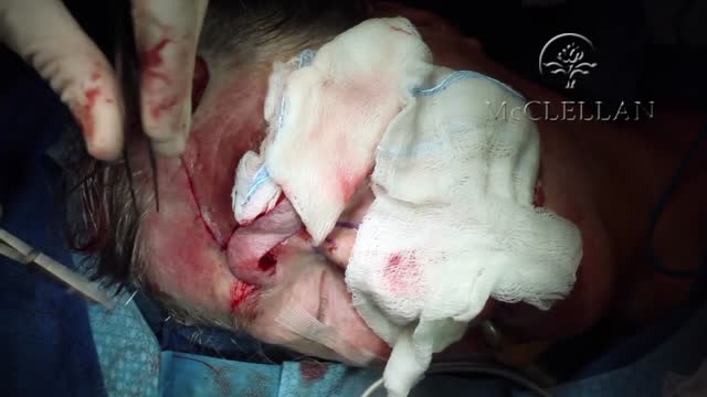
Forehead Flap Nasal Reconstruction
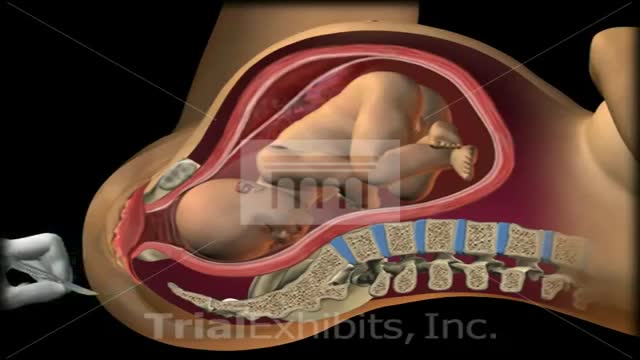
This medical 3D animation exhibit shows the left brachial plexus during birth and shoulder dystocia. Anatomy: symphysis pubis, uterus, sacrum, coccyx and fetus. "McRoberts Position". An episiotomy is cut. Brachial Plexus stretch injury. Retraction of head (turtle sign). Suprapubic pressure, gentle traction. To view our medical library of exhibits,
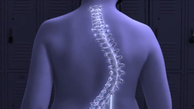
Pediatric orthopedic surgeons at Columbia are using a new device with magnetic technology that avoids the need for multiple spine-lengthening surgeries to correct early-onset scoliosis, a severe curvature of the spine in young children. In April 2014, Michael Vitale, MD, the Ana Lucia Professor of Pediatric Orthopedic Surgery at CUMC and 1995 graduate of P&S, performed the first procedure in the New York area, using the device to treat a 5-year-old boy. When braces and casts cannot control scoliosis in young children, surgeons turn to growing rods, which help correct the curve while allowing the spine to grow. When spinal maturity is near, the rods are removed and a spinal fusion can be performed. But during years of treatment with growing rods, patients must undergo surgery every six months to lengthen the rods to keep up with the patients’ growth. A patient may undergo eight to 10 procedures, which are costly and result in lost time for parents at work and children at school. The new device—MAGEC (MAGnetic Expansion Control) rods—contains a mechanism inside the growing rods that allows surgeons to lengthen the rods with a handheld external magnet, without surgery.
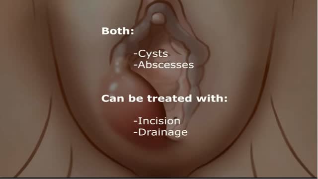
A fluid-filled swelling (cyst) in the Bartholin's glands, which lubricate the vagina.
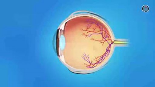
Most retinal tears need to be treated by sealing the retina to the back wall of the eye with laser surgery or cryotherapy (a freezing treatment). Both of these procedures create a scar that helps seal the retina to the back of the eye. This prevents fluid from traveling through the tear and under the retina, which usually prevents the retina from detaching. These treatments cause little or no discomfort and may be performed in your ophthalmologist’s office. With laser surgery, your ophthalmologist uses a laser to make small burns around the retinal tear. The scarring that results seals the retina to the underlying tissue, helping to prevent a retinal detachment.

ENT Physical Examination Lecture
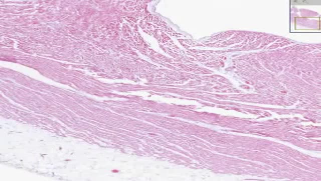
Histology of Heart Cardiac Muscle
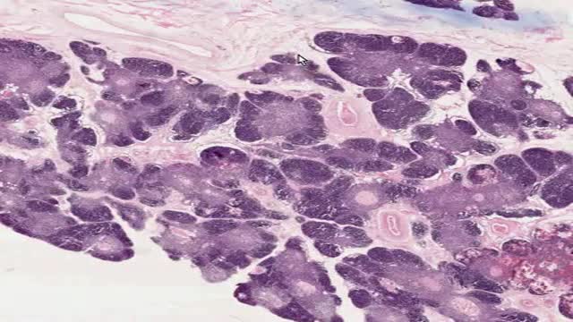
Histology of Thymus
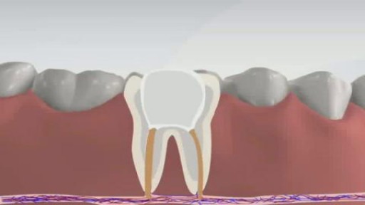
Root canal is a treatment to repair and save a badly damaged or infected tooth instead of removing it. The term "root canal" comes from cleaning of the canals inside a tooth's root. Decades ago, root canal treatments often were painful. With dental advances and local anesthetics, most people have little if any pain with a root canal. In fact, it's probably more painful living with a decayed tooth. Root canal alternatives include extracting the damaged tooth and replacing it with a dental implant, bridge or removable partial denture.
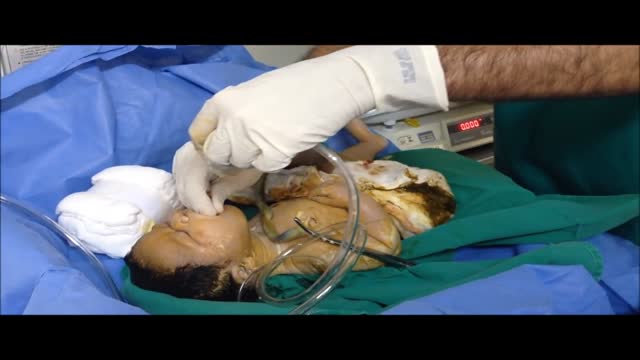
The following guidelines are an interpretation of the evidence presented in the 2010 International Consensus on Cardiopulmonary Resuscitation and Emergency Cardiovascular Care Science With Treatment Recommendations1). They apply primarily to newly born infants undergoing transition from intrauterine to extrauterine life, but the recommendations are also applicable to neonates who have completed perinatal transition and require resuscitation during the first few weeks to months following birth. Practitioners who resuscitate infants at birth or at any time during the initial hospital admission should consider following these guidelines. For the purposes of these guidelines, the terms newborn and neonate are intended to apply to any infant during the initial hospitalization. The term newly born is intended to apply specifically to an infant at the time of birth.
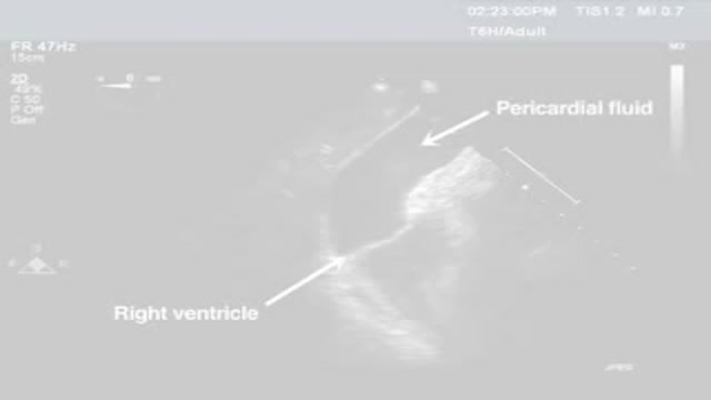
Pericardiocentesis is the aspiration of fluid from the pericardial space that surrounds the heart. This procedure can be life saving in patients with cardiac tamponade, even when it complicates acute type A aortic dissection and when cardiothoracic surgery is not available. [1] Cardiac tamponade is a time sensitive, life-threatening condition that requires prompt diagnosis and management. Historically, the diagnosis of cardiac tamponade has been based on clinical findings. Claude Beck, a cardiovascular surgeon, described 2 triads of clinical findings that he found associated with acute and chronic cardiac tamponade. The first of these triads consisted of hypotension, an increased venous pressure, and a quiet heart. It has come to be recognized as Beck's triad, a collection of findings most commonly produced by acute intrapericardial hemorrhage. Subsequent studies have shown that these classic findings are observed in only a minority of patients with cardiac tamponade. [2] The detection of pericardial fluid has been facilitated by the development and continued improvement of echocardiography. [3] Cardiac ultrasound is now accepted as the criterion standard imaging modality for the assessment of pericardial effusions and the dynamic findings consistent with cardiac tamponade. With echocardiography, the location of the effusion can be identified, the size can be estimated (small, medium, or large), and the hemodynamic effects can be examined by assessing for abnormal septal motion, right atrial or right ventricular inversion, and decreased respiratory variation of the diameter of the inferior vena cava.
ESCLEROTERAPIA
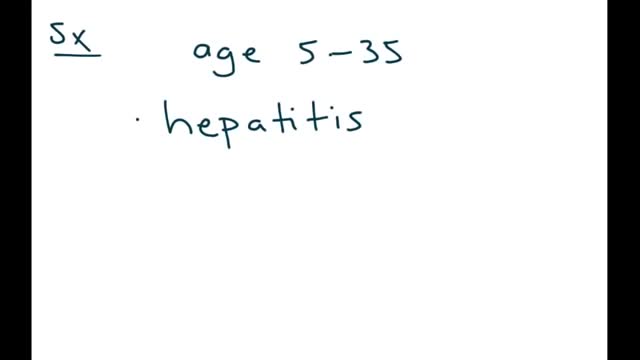
Wilson's disease is a rare inherited disorder that causes too much copper to accumulate in your liver, brain and other vital organs. Symptoms typically begin between the ages of 12 and 23. Copper plays a key role in the development of healthy nerves, bones, collagen and the skin pigment melanin. Normally, copper is absorbed from your food, and any excess is excreted through bile — a substance produced in your liver.
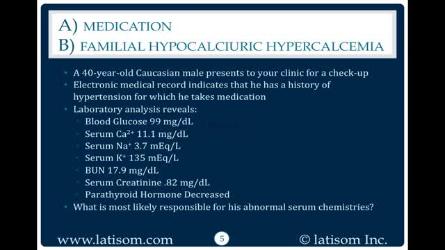
There are 3 genetic types of FHH based on chromosome location. FHH type 1 accounts for 65% of cases and is due to inactivating mutations in the CASR gene, localized to 3q21.1. This gene encodes the calcium-sensing receptor (CaSR). Loss of CaSR function results in a reduction in the sensitivity of parathyroid and renal cells to calcium levels so hypercalcemia is perceived as normal. The other 35% have either a mutation GNA11 (19p13.3) seen in FHH type 2 or AP2S1 (19q13.2-q13.3) seen in FHH type 3 (see these terms) or in genes not yet discovered. FHH is rarely caused by auto-antibodies against CaSR in those without a mutation.
