- Physical Examination
- Surgical Examination
- Ophthalmology
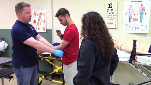
- Clinical Skills
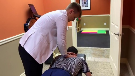
- Orthopedics
- Surgery Videos
- Laparoscopy
- Pediatrics
- Funny Videos
- Cardiothoracic Surgery
- Nursing Videos
- Plastic Surgery
- Otorhinolaryngology
- Histology and Histopathology
- Neurosurgery
- Dermatology
- Pediatric Surgery
- Urology
- Dentistry
- Oncology and Cancers
- Anatomy Videos
- Health and Fitness
- Radiology
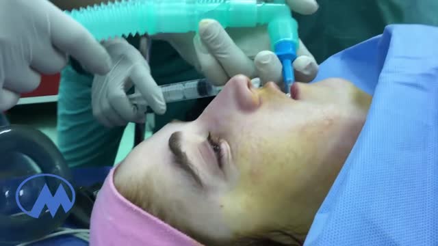
- Anaesthesia
- Physical Therapy
- Pharmacology
- Interventional Radiology
- Cardiology
- Endocrinology
- Gynecology
- Emergency Medicine
- Psychiatry and Psychology
- Childbirth Videos
- General Medical Videos
- Nephrology
- Physiology
- Diet and Food Health
- Diabetes Mellitus
- Neurology
- Women Health
- Osteoporosis
- Gastroenterology
- Pulmonology
- Hematology
- Rheumatology
- Toxicology
- Nuclear Medicine
- Infectious Diseases
- Vascular Disease
- Reproductive Health
- Burns and Wound Healing
- Other
Top videos
https://www.youtube.com/watch?v=0N7Yy1UYEWk
Every day, specialists deliver high-quality care in 68 disciplines in health centres across Canada. Yet many Canadians know very little about what many specialists actually do, and the important role these disciplines play in Canada’s health care system.
A pheochromocytoma (fee-o-kroe-moe-sy-TOE-muh) is a rare, usually noncancerous (benign) tumor that develops in cells in the center of an adrenal gland. You have two adrenal glands, one above each kidney. Your adrenal glands produce hormones that give instructions to virtually every organ and tissue in your body. If you have a pheochromocytoma, an adrenal gland releases hormones that cause persistent or episodic high blood pressure. If left untreated, a pheochromocytoma can result in severe or life-threatening damage to other body systems, especially the cardiovascular system. Most people with a pheochromocytoma are between the ages of 20 and 50, but the tumor can develop at any age. Surgical treatment to remove a pheochromocytoma usually returns blood pressure to normal.
If a patient comes to you with a painful, throbbing, swollen, red face (a ''fat face'), perhaps with fever, trismus and lymphadenitis, he is probably suffering from an acute dental or oral infection, most probably an alveolar abscess. He may have: (1) An alveolar abscess begins as an infection in the bone around a non-vital infected tooth. He has severe pain, which becomes less as pus is released into more superficial tissues and his face starts to swell. After 36 hours of cellulitis he usually has a fluctuant abscess which needs draining. If drainage is delayed, the pus in his abscess discharges spontaneously through a sinus (26-8) in his gum or face, which may become chronic. First, control infection with antibiotics, and then drain the abscess, either by incising it where it is pointing, or by removing the infected tooth, which acts as a cork to prevent the pus escaping, or by doing both these things. If you remove a tooth before you have controlled the infection with antibiotics, and while his face is still severely swollen, you may spread the infection; your task will also be more difficult. (2) A periodontal abscess at the side of a tooth, caused by spread from an infected gum. (3) A pericoronal abscess caused by infection of the gum over the crown of an unerupted and impacted tooth, usually a lower third molar (''an infected wisdom tooth'). Often, an abscess does not form, and the gum round the tooth is merely inflamed.
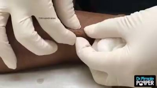
Epidermoid cysts, also called sebaceous, keratin, or epithelial cysts, are small, hard lumps that develop under the skin. These cysts are common. They grow slowly. They do not cause other symptoms and are nearly never cancerous. Epidermoid cysts are often found on the face, head, neck, back, or genitals
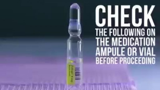
Learn how to give an intramuscular injection
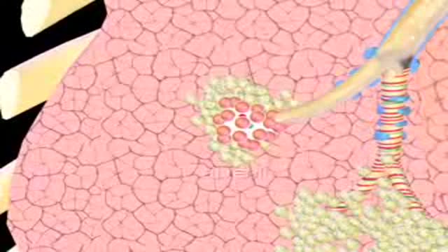
Emphysema gradually damages the air sacs (alveoli) in your lungs, making you progressively more short of breath. Emphysema is one of several diseases known collectively as chronic obstructive pulmonary disease (COPD). Smoking is the leading cause of emphysema. Your lungs' alveoli are clustered like bunches of grapes. In emphysema, the inner walls of the air sacs weaken and eventually rupture — creating one larger air space instead of many small ones. This reduces the surface area of the lungs and, in turn, the amount of oxygen that reaches your bloodstream. When you exhale, the damaged alveoli don't work properly and old air becomes trapped, leaving no room for fresh, oxygen-rich air to enter. Treatment may slow the progression of emphysema, but it can't reverse the damage.
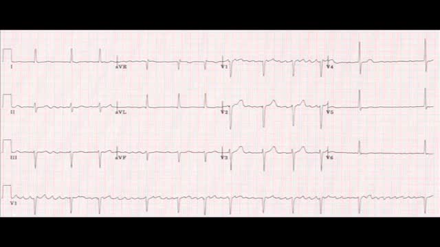
AFib is caused by abnormal electrical impulses in the atria, which are the upper chambers of the heart. The result is a rapid and irregular pumping of blood through the atria. These chambers fibrillate, or quiver, rapidly.

The preferred route of access for temporary transvenous pacing is the internal jugular vein followed by subclavian and femoral veins. However, all the major venous access sites (internal and external jugular, subclavian, brachial, femoral) have been used and each is associated with particular problems.
What is a mole? Many people refer to a mole as any dark spot or irregularity in the skin. Doctors use different terms. But the following types of skin marks such as these are not treated the same way moles are and are not discussed here: Birthmarks Abnormal formations of blood vessels (hemangiomas) Keratoses (benign or precancerous spots, which appear after about age 30 years)
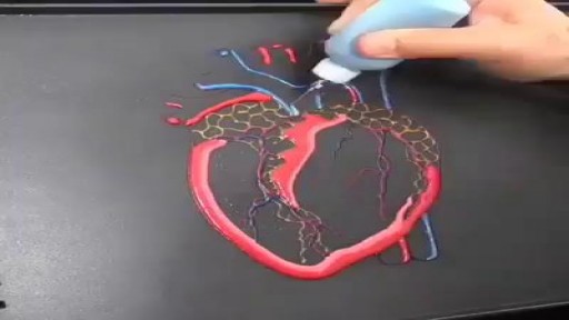
Pancake by a Cardiologist

Hereditary hemochromatosis (he-moe-kroe-muh-TOE-sis) causes your body to absorb too much iron from the food you eat. Excess iron is stored in your organs, especially your liver, heart and pancreas. Too much iron can lead to life-threatening conditions, such as liver disease, heart problems and diabetes.

Sliced Fingertip Makeup Tutorial
Woman suffers allergic reaction to nut protein in boyfriend's sperm.A British woman has become the first recorded case of someone suffering an allergic...
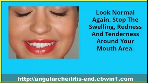
Cracked Corners Of Mouth, Cheilitis, Angular Cheilitis Remedy, Angular Cheilitis Medicine, Cheilitis--- http://angularcheilitis-end.cbwin1.com --- Foods Which Can Limit the Occurrence of Angular Cheilitis. People suffering from Angular Cheilitis know that this is one of the most troubling and annoying skin condition one can experience. It prevents you from eating, drinking and speaking normally. Many people even refuse to go out of the house when suffering from this condition, thus becoming isolated from the rest of the world. This is why it is better to prevent it then having to treat it. If you have had it long time ago and are afraid that will come back, if you have it and want to treat it faster or if you do not want to have this terrible experience ever, you should start by eating the foods listed below. They will provide your body with all the vitamins and nutrients necessary to effectively fight this disease and prevent it from appearing ever again. Most of the times, Angular Cheilitis appears as a result of a weak immune system. Thus, you will need to have a balanced diet, filled with fruits and vegetables that will supply you with all the things you need to remain healthy and have a strong immune system. The first thing that you will need to have in your body to fight Angular Cheilitis is iron. If you no longer want to have those anesthetic and painful cracks around your mouth, if you want to eat, drink and speak normally without experiencing any pain when opening your mouth, then check out this new and revolutionary treatment! It will get you rid of Angular Cheilitis in just a few days and you will be able to enjoy life to its fullest again, without worrying about those otiose cracks! Click Here. http://angularcheilitis-end.cbwin1.com
