- Physical Examination
- Surgical Examination
- Ophthalmology
- Clinical Skills
- Orthopedics
- Surgery Videos
- Laparoscopy
- Pediatrics
- Funny Videos
- Cardiothoracic Surgery
- Nursing Videos
- Plastic Surgery
- Otorhinolaryngology
- Histology and Histopathology
- Neurosurgery
- Dermatology
- Pediatric Surgery
- Urology
- Dentistry
- Oncology and Cancers
- Anatomy Videos
- Health and Fitness
- Radiology
- Anaesthesia
- Physical Therapy
- Pharmacology
- Interventional Radiology
- Cardiology
- Endocrinology
- Gynecology
- Emergency Medicine
- Psychiatry and Psychology
- Childbirth Videos
- General Medical Videos
- Nephrology
- Physiology
- Diet and Food Health
- Diabetes Mellitus
- Neurology
- Women Health
- Osteoporosis
- Gastroenterology
- Pulmonology
- Hematology
- Rheumatology
- Toxicology
- Nuclear Medicine
- Infectious Diseases
- Vascular Disease
- Reproductive Health
- Burns and Wound Healing
- Other
Top videos

What Your Handwriting Says About You

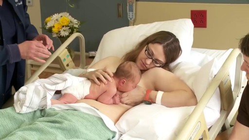
Millions of sperms are deposited into the vagina during sexual intercourse. The sperms make their way through the cervix into the uterus and then on to the fallopian tubes. As they swim along this way their numbers decline. Only a few hundred sperm will get close to the egg. During the trip, sperm prepare themselves to meet the egg by subtle alterations of their heads and movement patterns. Once inside the fallopian tube, the sperm attracts the egg by releasing a chemical. The egg is surrounded by a protective covering called the zona pellucida, which allows only one sperm to penetrate it. Once inside the egg, the head of the fertilizing sperm releases its genetic contents, which fuses with the nucleus of the egg. Fertilisation is now complete. Sperm are able to survive for 2-3 days within the female's reproductive tract. The length of the time that a woman's egg can be fertilized by a man's sperm ranges from 12-24 hours.

Scientists reveal how LSD alters your mind.

This video narrates the story of a girl who travels to Iran for doing a nose surgery.
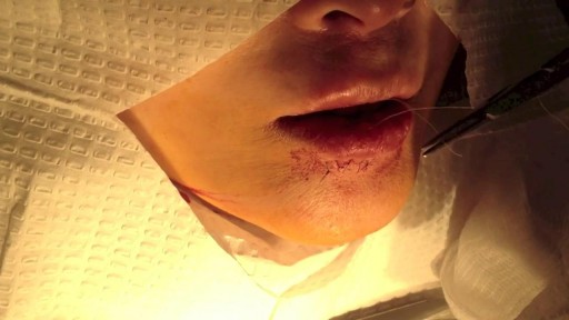
This video details the layered closure of a through-and-through facial laceration
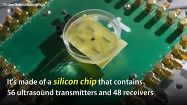
The term "miniaturization" is widely accepted in our vernacular as a positive step in product development. Reducing components to create less space, product footprint and more affordable medical devices are ongoing objectives for manufacturers today. Jabil strives to integrate new innovative technologies into product design and manufacturing as continual miniaturization of medical devices is a focus of the healthcare thought process. Miniaturization is a constantly moving target. Once a novel, new technology sets a higher bar for miniaturization standards, the next ambitious goal is to achieve an even thinner and smaller device. Industry trends, including minimally invasive surgical devices and home health care delivery, demand more sophisticated medical portable devices and easy-to-use electronics which may not be a core competency of medical device manufacturers.
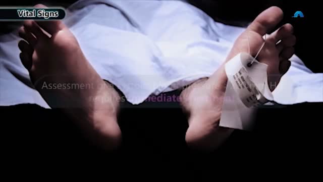
Hypertensive emergencies encompass a spectrum of clinical presentations in which uncontrolled blood pressures lead to progressive or impending end-organ dysfunction. In these conditions, the BP should be lowered aggressively over minutes to hours. Neurologic end-organ damage due to uncontrolled BP may include hypertensive encephalopathy, cerebral vascular accident/cerebral infarction, subarachnoid hemorrhage, and/or intracranial hemorrhage.[1] Cardiovascular end-organ damage may include myocardial ischemia/infarction, acute left ventricular dysfunction, acute pulmonary edema, and/or aortic dissection. Other organ systems may also be affected by uncontrolled hypertension, which may lead to acute renal failure/insufficiency, retinopathy, eclampsia, or microangiopathic hemolytic anemia.[1] With the advent of antihypertensives, the incidence of hypertensive emergencies has declined from 7% to approximately 1% of patients with hypertension.[2] In addition, the 1-year survival rate associated with this condition has increased from only 20% (prior to 1950) to a survival rate of more than 90% with appropriate medical treatment
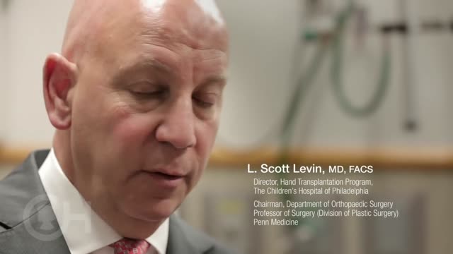
Surgeons at The Children’s Hospital of Philadelphia were the first to perform a bilateral hand transplant on a child. Our research and work in this groundbreaking field of medicine led us to establish the Hand Transplantation Program. Combining the expertise of the Penn Transplant Institute and the Hospital’s Division of Plastic and Reconstructive Surgery and Division of Orthopedics, the program aims to improve quality of life for children who may benefit from this procedure.

Acute leukaemias develop quickly and need to be treated urgently. Chronic leukaemias develop more slowly and may not need to be treated for some time after they are diagnosed. Some forms may not require any treatment. Myeloid leukaemias arise from myeloid stem cells and are characterised by the accumulation of cancerous myeloid cells. Lymphoid leukaemias arise from lymphoid stem cells and are characterised by the accumulation of cancerous lymphoid cells such as B-cells and T-cells. The most common forms of leukaemia in adults are CLL and AML, and the common cancer in children is ALL. Leukaemia is more common in adults.
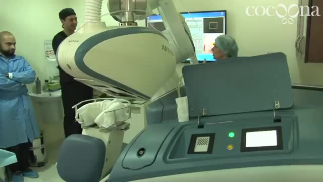
Introducing ARTAS in Cocoona Delhi & Dubai by Dr Sanjay Parashar - Hair Transplant Robot

Mommy Makeover Plastic Surgery Boca Raton FL

Chemokuren geven neuropathische pijnen. Behandeling met het supplement palmitoylethanolamide en topicale analgetische creme geven het volgende resultaat

When you’re depressed, it can feel like you’ll never get out from under a dark shadow. However, even the most severe depression is treatable. So, if your depression is keeping you from living the life you want to, don’t hesitate to seek help. Learning about your depression treatment options will help you decide what approach is right for you. From therapy to medication to healthy lifestyle changes, there are many effective treatments that can help you overcome depression and reclaim your life.
complications from using a urinary catheter include: allergic reaction to the material used in the catheter, such as latex. bladder stones. blood in the urine. injury to the urethra. kidney damage (with long-term indwelling catheters) septicemia, or infection of the urinary tract, kidneys, or blood.

The eyes A close up of a young person's eyes. The eyes are responsible for four-fifths of all the information our brain receives. Here you can find out a bit more about how they work, common problems that affect vision and the work Sightsavers does to treat and prevent avoidable blindness. You can also find out more about the people whose lives have been changed thanks to donations from people like you. How do eyes work? (click image to see enlarged version or click here for text alternative) Graphic of an eye with information about its different parts The images we see are made up of light reflected from the objects we look at. This light enters the eye through the cornea. Because this part of the eye is curved, it bends the light, creating an upside down image on the retina (this is eventually put the right way up by the brain). The retina is a complex part of the eye, but only the very back of it is light sensitive. This part of the retina has roughly the area of a 10p coin, and is packed with photosensitive cells called rods and cones. Cones are the cells responsible for daylight vision. There are three kinds – each responding to a different wavelength of light: red, green and blue. The cones allow us to see images in colour and detail. Rods are responsible for night vision. They are sensitive to light but not to colour. In darkness, the cones do not function at all. How do we see an image? The lens focuses the image. It can do this because it is adjustable – using muscles to change shape and help us focus on objects at different distances. The automatic focusing of the lens is a reflex response and is not controlled by the brain. Once the image is clearly focused on the sensitive part of the retina, energy in the light that makes up that image creates an electrical signal. Nerve impulses can then carry information about that image to the brain through the optic nerve.
What Causes Chest Pain ?
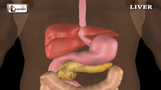
The liver regulates most chemical levels in the blood and excretes a product called bile. ... Production of bile, which helps carry away waste and break down fats in the small intestine during digestion. Production of certain proteins for blood plasma.
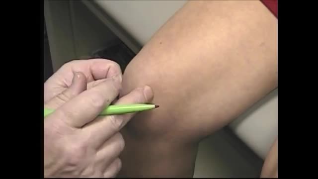
When oral medications do not relieve knee pain, but you're not to the point of pursuing knee surgery, one of the following injections or procedures may help. Hyaluronic acid supplements – Although not technically medications, these substances are injected into knee joints to supplement naturally occurring hyaluronic acid. In healthy joints hyaluronic acid acts as a shock absorber and lubricant, allowing joints to move smoothly over each other. However, the acid appears to break down in people with osteoarthritis. Injecting it into a joint may lessen pain and inflammation. The injections are given weekly for three or five weeks, depending on the product (examples are Synvisc and Hyalgan). A small amount of joint fluid is removed first to make room for the hyaluronic acid. Corticosteroid Injections – Doctors sometimes inject corticosteroids directly into the knee joint for quick relief of pain and inflammation. Their benefits may last anywhere from a few days to more than six months. While the injections bring targeted relief to the joint and lack many of the side effects of oral corticosteroid medications, they are not without risks. Repeated knee injections may actually contribute to cartilage breakdown. For that reason your doctor will likely put a limit on the number of injections you can receive. Read a report from the British Medical Journal on corticosteroid injections for knee osteoarthritis. Arthrocentesis – Also called joint fluid aspiration, arthrocentesis is removal of joint fluid through a hollow needle inserted into the joint space of the knee. Although the purpose of removing joint fluid from the knee is usually so that it can be tested in the lab, removing excess fluid can also quickly ease pain and swelling. Often after withdrawing fluid, doctors use the same puncture site where the fluid was removed to inject a corticosteroid preparation and/or anesthetic into the knee joint to further relieve pain and inflammation.
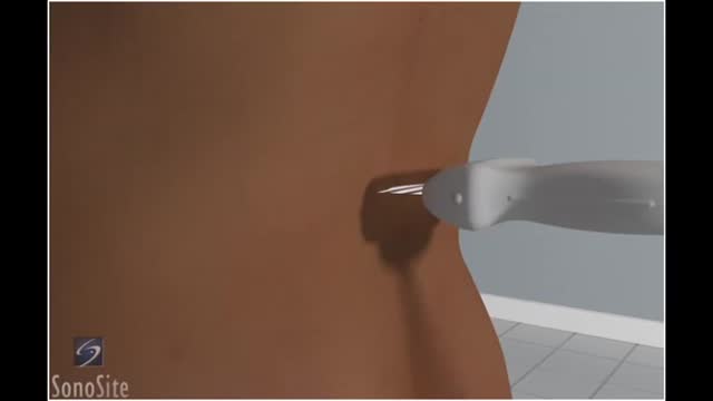
Lumbar puncture is a common emergency department procedure used to obtain information about the cerebrospinal fluid (CSF) for diagnostic and, less commonly, therapeutic reasons. Please refer to the full article on Lumbar Puncture for more details on the lumbar puncture procedure. Lumbar puncture is typically performed via “blind” surface landmark guidance. The surface landmark technique is reported to be successful in a high percentage of attempted lumbar punctures; however, surface landmark identification of underlying structures has been shown to be accurate only 30% of the time. [1] Unsuccessful identification of proper landmarks often leads to increased difficulty in obtaining CSF, if the procedure is performed, and a higher rate of complications. Few alternatives are available in these cases. If available, fluoroscopic-guided lumbar puncture may be performed. If not, treatment is sometimes initiated empirically without obtaining CSF. Disadvantages of using fluoroscopy include limited availability or necessary transport of the patient outside of the emergency department, inability to directly visualize the spinal canal, and inherent radiation exposure
