أهم مقاطع الفيديو

There are 3 major parts of the respiratory system: the airway, the lungs, and the muscles of respiration. The airway, which includes the nose, mouth, pharynx, larynx, trachea, bronchi, and bronchioles, carries air between the lungs and the body's exterior.

This Unorthodox Procedure Makes Short People A Foot Taller
blood transfusion performance
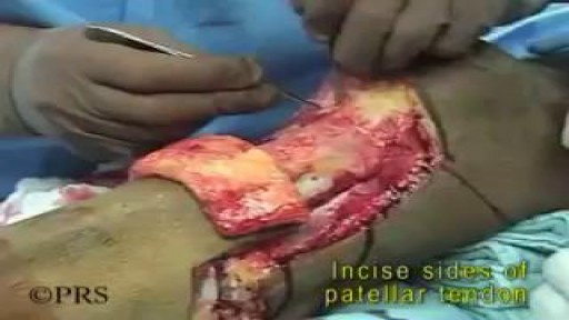
Pathology: Previous spinal cord injury, diabetes, renal failure, dynamic knee contracture, open left ankle disarticulation for sepsis and severe foot infection

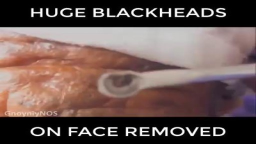
Blackheads are small bumps that appear on your skin due to clogged hair follicles. These bumps are called “blackheads” because the surface looks dark or black. Blackheads are a mild type of acne that usually form on the face, but they can also appear on the back, chest, neck, arms, and shoulders

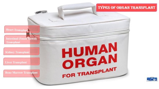
This process involves surgical removing of an #organ or tissue from one person (organ donor) & placing into another person (recipient) body. https://goo.gl/JfoN8y
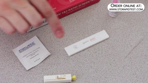
How to use a HIV rapid test kit for self-diagnosis of HIV (fingerstick blood). Convenient, Easy to Use, and over 99% Accurate. USAID approved. Test yourself at home with Complete Privacy. Buy online today at: http://www.stdrapidtest.com
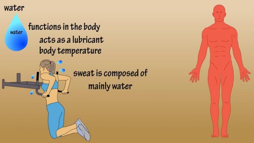
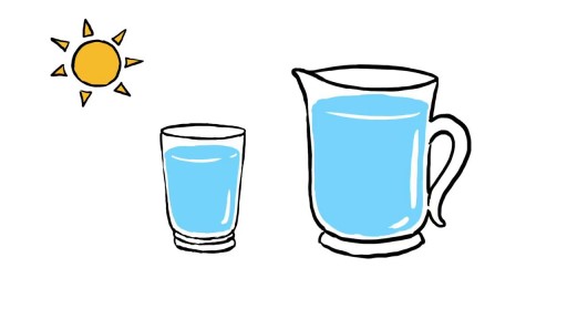
Water is an essential nutrient for the body, as the body loses water through perspiration, breathing, bowel movements, and in urine. Water must be consumed regularly to maintain a sufficient level. Water has many vital functions in the body, including… Serving as a lubricant. Water is a main component of saliva, which helps moisten food making it easier to swallow. Water also helps lubricate joints, reducing friction and inflammation. Water is important in body temperature regulation. When body heat rises, such as during strenuous activities, the body starts to sweat to cool itself. And sweat is made up almost entirely of water.

http://vencer-la-diabetes-rapido.info-pro.co/ Como Controlar La Diabetes Tipo 2 Naturalmente Sin Medicamentos, Pre Diabetes Y Diabetes Tipo 1. https://youtu.be/BOSkQ5MnjT0 Que es la Insulina? Una definición practica sin adentrarnos en terminos estrictamente medicos es que la insulina es una hormona formada por 51 aminoácidos. Dentro del páncreas, las células beta producen la hormona llamada insulina. Con cada comida, las células beta liberan insulina para ayudar al cuerpo a utilizar o almacenar en la sangre la glucosa que se obtiene de los alimentos. Su déficit provoca la diabetes mellitus y su exceso provoca hiperinsulinismo con hipoglucemia. En las personas con diabetes tipo 1, el páncreas no produce insulina. Las células beta han sido destruidas y se necesitan inyecciones de insulina para utilizar la glucosa de las comidas. Las personas con diabetes tipo 2 producen insulina, pero sus cuerpos no responden bien a la misma. Algunas personas con diabetes tipo 2 necesitan medicamentos para la diabetes o inyecciones de insulina para ayudar a su cuerpo a utilizar la glucosa para obtener energía. * La insulina no se puede tomar como una píldora, ya que se descompone durante la digestión al igual que la proteína en los alimentos. Se debe inyectar en la grasa debajo de la piel para que llegue a la sangre. Existen diferentes tipos de insulina en función de la rapidez con que trabajan, y en funcion de su duración. La insulina viene en diferentes concentraciones, la más común es U-100. Tipos de insulina: * De Acción Rápida: Comienza a trabajar unos 15 minutos después de la inyección, con picos en aproximadamente 1 hora, y continúa trabajando por un tiempo de 2 a 4 horas. Tipos: Insulina glulisina (Apidra), la insulina lispro (Humalog) y la insulina aspart (NovoLog). * Regular o de Acción Corta: Generalmente llega al torrente sanguíneo a los 30 minutos después de la inyección, picos de entre 2 a 3 horas después de la inyección, y es efectiva durante aproximadamente 3 a 6 horas. Tipos: Humulin R, Novolin R * De Acción Intermedia: Generalmente llega al torrente sanguíneo de aproximadamente 2 a 4 horas después de la inyección, picos de 4 a 12 horas y eseficaz durante aproximadamente 12 a 18 horas. Tipos: NPH (Humulin N, Novolin N) * De Acción Prolongada: Alcanza el torrente sanguíneo varias horas después de la inyección y tiende a disminuir los niveles de glucosa de manera bastante uniforme durante un período de 24 horas. Tipos: La insulina detemir (Levemir) y la insulina glargina (Lantus) Nota: Esta información debes consultarla siempre con tu medico especialista. La insulina Tiene 3 Características: El inicio: Es el tiempo antes de que la insulina alcance el torrente sanguíneo y se inicie la reducción de la glucosa en sangre. Pico: Es el tiempo durante el cual la insulina está surtiendo el máximo efecto en términos de reducción de la glucosa en sangre. La duración: Es cuánto tiempo la insulina continúa reduciendo la glucosa sanguínea.

http://plantar-fasciitis-solution.info-pro.co Plantar Fasciitis Symptoms, Foot Pain Running, Foot Pain Ball Of Foot, Taping For Plantar Fasciitis Home Treatments. Knowing what the cause of the pain is and why the pain is occurring enables a person more effectively tackle home treatments and remedies for plantar fasciitis. Dedicated exercise rehabilitation is one home treatment technique that has been proven to address the deficiencies identified in the plantar fascia tissue. Determine the severity of the pain being experienced and this may provide an idea regarding the level of exercise the affected foot can accommodate at a time. It can be possible to use anti-inflammatory medications or natural nutritional substances that contain anti-inflammatory properties to relieve pain symptoms associated with plantar fasciitis. While gently exercising the affected foot or feet, it is important to avoid activity that can exacerbate the condition. This is why a person with plantar fasciitis can notice pain when resuming activity with the feet after being in a resting position for a period of time. Also, an important home treatment for plantar fasciitis is rest! The affected foot needs rest and this can help the healing process. If you would like more information Click HERE To Learn More About Plantar Fasciitis. http://plantar-fasciitis-solution.info-pro.co

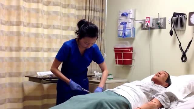
Live in Caregiver Toronto - https://medwayhealthcare.com/ Foot Care Nurse - https://medwayhealthcare.com/foot-care/ Respite Care - https://medwayhealthcare.com/respite-care/

Watch that video of The Most Amazing Plastic Surgeries
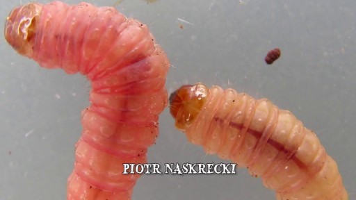
Watch that video to know When Botflies Attack

baby wrapping
Watch that video of a Black Salve Left an Inch Hole In Man's Hole
Watch that Female Breasts Augmentation Plastic Surgery
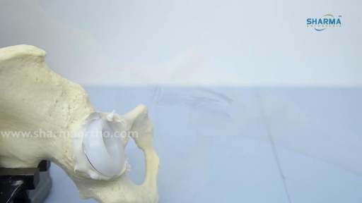
Hip replacement is a surgical procedure in which the hip joint is replaced by a prosthetic implant, that is, a hip prosthesis. Hip replacement surgery can be performed as a total replacement or a hemi replacement
