- Physical Examination
- Surgical Examination
- Ophthalmology
- Clinical Skills
- Orthopedics
- Surgery Videos
- Laparoscopy
- Pediatrics
- Funny Videos
- Cardiothoracic Surgery
- Nursing Videos
- Plastic Surgery
- Otorhinolaryngology
- Histology and Histopathology
- Neurosurgery
- Dermatology
- Pediatric Surgery
- Urology
- Dentistry
- Oncology and Cancers
- Anatomy Videos
- Health and Fitness
- Radiology
- Anaesthesia
- Physical Therapy
- Pharmacology
- Interventional Radiology
- Cardiology
- Endocrinology
- Gynecology
- Emergency Medicine
- Psychiatry and Psychology
- Childbirth Videos
- General Medical Videos
- Nephrology
- Physiology
- Diet and Food Health
- Diabetes Mellitus
- Neurology
- Women Health
- Osteoporosis
- Gastroenterology
- Pulmonology
- Hematology
- Rheumatology
- Toxicology
- Nuclear Medicine
- Infectious Diseases
- Vascular Disease
- Reproductive Health
- Burns and Wound Healing
- Other
Top videos
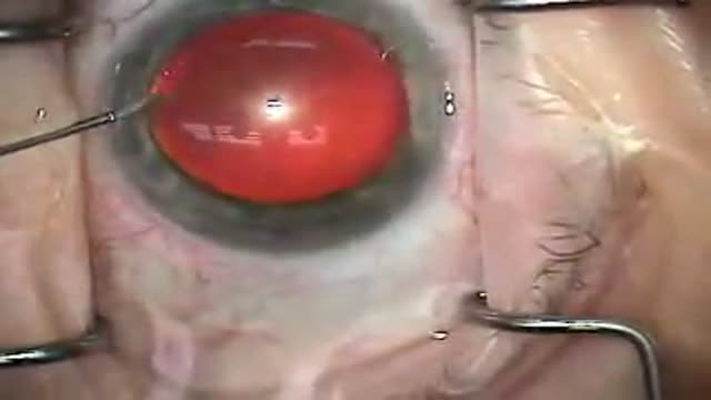
real time video of modern cataract surgery employing a temporal, clear-corneal approach with topical anesthesia and ultrasound phacoemulsification; an aspheric silicone lens implant is inserted
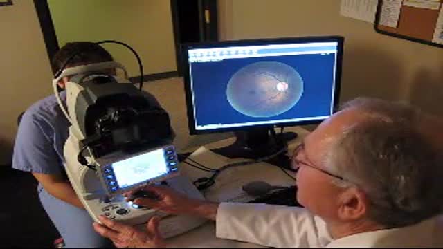
new fundus camera for examining the retina without dilating the pupil

Endoscopic Brain Surgery, third Ventriculostomy

Laparoscopic Gastric Bypass Surgery

This is an Abdominal Liposuction surgery performed by Dr. Art Foley. Liposuction is a procedure that can help sculpt the body by removing unwanted fat from specific areas including the abdomen, hips, buttocks, thighs, knees, upper arms and neck. Although no type of liposuction is a substitute for dieting and exercise, liposuction can remove stubborn areas of fat that don't respond to traditional weight loss methods.

This video features a testimonial of Okino Mosses from Nigeria recovers from nerve decompression after his Lumber spine decompression surgery at Mumbai in India who recovered from nerve decompression after his lumber spine surgery at Mumbai in India. Okino was suffering from nervous spine decompression and was in need of a good doctor plus medical solution and then he came to know of international quality spine treatment available in India at a reduced cost. Availing the assistance of medical tourism in India Okino was able to get an international quality and cost effective lumber spine decompression surgery at Mumbai in India. Lumber spine decompression surgery is a surgical procedure that is performed to alleviate pain caused by pinched nerves (neural impingement). This surgery provides assured medical recovery to medical patients who suffer from nervous decompression disorder. In the procedure of lumber spine decompression surgery a small portion of the bone over the nerve root and/or disc material from under the nerve root is removed to give the nerve root more space and provide a better healing environment. Several conditions may cause neural impingement, including spinal stenosis, a disc herniation, isthmic spondylolisthesis, degenerative spondylolisthesis, or (rarely) a spinal tumor. And lumber spine decompression surgery provides medical recovery from these spine disorders. Indian spine surgery hospitals of Delhi, Mumbai and Chennai have got good medical state of art facilities for abroad patients who want to get lumber spine surgery in India at a reduced price budget. The price of spine surgery procedure in India is affordable and the best doctors operate them to give patients a positive medical feed back after the surgery. 24/7 hours patient care provided by well trained Indian medical staff makes India a reliable medical destination. Medical tourism in India provides good care and assistance to patients who far in abroad to plan a cost effective medical trip to India. You may get more details about lumber spine surgery in India at http://www.dheerajbojwani.com or mail your queries at contact@dheerajbojwani.com

Laparoscopy by Dr. Emadi in Qatar

Details about the nature and procedure for this "something" will be in the next video ..soon.

The Small Intestine

Your body is a brilliant machine with many important parts. Watch movies to learn more

Blood type (or blood group) is determined, in part, by the ABO blood group antigens present on red blood cells. A blood type (also called a blood group) is a classification of blood based on the presence or absence of inherited antigenic substances on the surface of red blood cells (RBCs).

A ventricular septal defect (VSD) is an opening or hole in the wall that separates the two lower chambers of the heart. This wall is called the ventricular septum. The hole causes oxygen-rich blood to leak from the left side of the heart to the right side. This causes extra work for the right side of the heart, since more blood than necessary is flowing through the right ventricle to the lungs. The hole is usually closed with surgery. However, in certain situations, your child's cardiologist and surgeon may think it is best to close the hole with a special device. This procedure is done in the heart catheterization lab.

The pain of ovulation can range from a mild twinge to severe discomfort and usually lasts from minutes to hours. It is generally felt on one side of the abdomen and may vary each month, depending on which ovary is releasing the egg during that cycle.

Cancer, also called malignancy, is an abnormal growth of cells. There are more than 100 types of cancer, including breast cancer, skin cancer, lung cancer, colon cancer, prostate cancer, and lymphoma. Symptoms vary depending on the type. Cancer treatment may include chemotherapy, radiation, and/or surgery.

Such foods include carrots, eggplant, cauliflower, green beans, broccoli, peppers, onions, lettuce, zucchini, tomatoes, peanuts and walnuts. These foods are generally safe for you to eat at each meal without spiking your blood sugar.

The eyes A close up of a young person's eyes. The eyes are responsible for four-fifths of all the information our brain receives. Here you can find out a bit more about how they work, common problems that affect vision and the work Sightsavers does to treat and prevent avoidable blindness. You can also find out more about the people whose lives have been changed thanks to donations from people like you. How do eyes work? (click image to see enlarged version or click here for text alternative) Graphic of an eye with information about its different parts The images we see are made up of light reflected from the objects we look at. This light enters the eye through the cornea. Because this part of the eye is curved, it bends the light, creating an upside down image on the retina (this is eventually put the right way up by the brain). The retina is a complex part of the eye, but only the very back of it is light sensitive. This part of the retina has roughly the area of a 10p coin, and is packed with photosensitive cells called rods and cones. Cones are the cells responsible for daylight vision. There are three kinds – each responding to a different wavelength of light: red, green and blue. The cones allow us to see images in colour and detail. Rods are responsible for night vision. They are sensitive to light but not to colour. In darkness, the cones do not function at all. How do we see an image? The lens focuses the image. It can do this because it is adjustable – using muscles to change shape and help us focus on objects at different distances. The automatic focusing of the lens is a reflex response and is not controlled by the brain. Once the image is clearly focused on the sensitive part of the retina, energy in the light that makes up that image creates an electrical signal. Nerve impulses can then carry information about that image to the brain through the optic nerve.

Hemoglobin is the protein molecule in red blood cells that carries oxygen from the lungs to the body's tissues and returns carbon dioxide from the tissues back to the lungs. Hemoglobin is made up of four protein molecules (globulin chains) that are connected together.
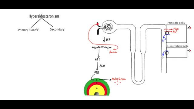
Primary aldosteronism, also known as primary hyperaldosteronism or Conn's syndrome, is excess production of the hormone aldosterone by the adrenal glands resulting in low renin levels. Often it produces few symptoms. Most people have high blood pressure which may cause poor vision or headaches.

Cardiovascular disease (CVD) is a general term that describes a disease of the heart or blood vessels. Blood flow to the heart, brain or body can be reduced as the result of a blood clot (thrombosis), or by a build-up of fatty deposits inside an artery that cause the artery to harden and narrow (atherosclerosis).

What Happens When You're In a Coma?
