- Physical Examination
- Surgical Examination
- Ophthalmology
- Clinical Skills
- Orthopedics
- Surgery Videos
- Laparoscopy
- Pediatrics
- Funny Videos
- Cardiothoracic Surgery
- Nursing Videos
- Plastic Surgery
- Otorhinolaryngology
- Histology and Histopathology
- Neurosurgery
- Dermatology
- Pediatric Surgery
- Urology
- Dentistry
- Oncology and Cancers
- Anatomy Videos
- Health and Fitness
- Radiology
- Anaesthesia
- Physical Therapy
- Pharmacology
- Interventional Radiology
- Cardiology
- Endocrinology
- Gynecology
- Emergency Medicine
- Psychiatry and Psychology
- Childbirth Videos
- General Medical Videos
- Nephrology
- Physiology
- Diet and Food Health
- Diabetes Mellitus
- Neurology
- Women Health
- Osteoporosis
- Gastroenterology
- Pulmonology
- Hematology
- Rheumatology
- Toxicology
- Nuclear Medicine
- Infectious Diseases
- Vascular Disease
- Reproductive Health
- Burns and Wound Healing
- Other
Top videos
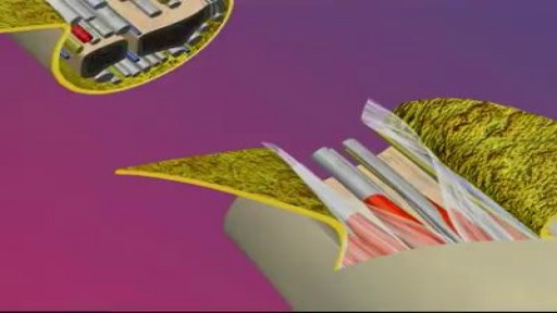
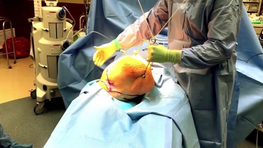
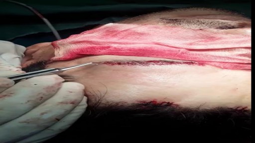
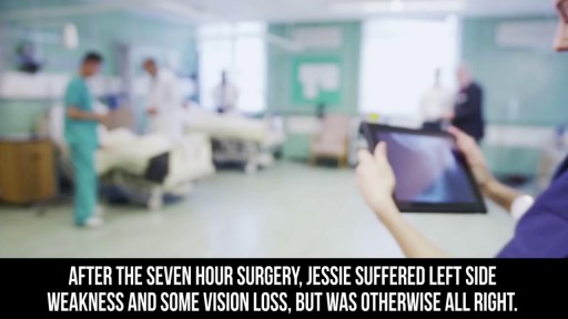
A reconstructive transplant, also called a composite tissue transplant or composite tissue allograft, is an operation that involves transplantation of bone, tissue, muscle and blood vessels. A reconstructive hand transplant is an operation tailored to each patient’s individual needs, type of injury and anatomy. This transplants an upper extremity, usually at the level of the forearm and wrist, but sometimes above the elbow, to help restore function after the loss of a hand or arm.
This video was taken 2 weeks after this lovely patient had a Endoscopic Brow Lift, Face and Neck lift, and Fat Grafting. Know more about Handal Plastic Surgery Call us at (561) 912-9888 for more info!
The treatment of cardiac arrest
Each kidney contains around 1 million individual nephrons, the kidneys' microscopic functional units that filter blood to produce urine. The nephron is made of 2 main parts: the renal corpuscle and the renal tubule.

http://plantar-fasciitis-solution.info-pro.co Pain In Arch Of Foot, Severe Heel Pain, Best Running Shoes For Plantar Fasciitis, Foot Pain Heel Know the Symptoms of Plantar Fasciitis An injury to the plantar fascia can manifest in different ways. Initially, it may be a gradual pain that can progressively become worse, especially if the injured foot remains in active use. Sometimes, the pain from plantar fasciitis can be quite severe and seem like the stab of a knife of a sharp and sudden cut. The pain of plantar fasciitis may also occur more frequently after injured feet have been at rest for a while. For instance, after a person wakes up and tries to use his or her feet, pain may be experienced. It can be dangerous to ignore pain that is associated with the feet or any pain felt in the body. Sometimes, symptoms of plantar fasciitis include more subtle pain that may appear as a throbbing sensation which may be radial in nature or isolated to a particular part of the foot. If the pain from plantar fasciitis starts off mildly and is ignored, continued use of the affected foot or feet will cause further damage. The pain from plantar fasciitis is debilitating and it is essential that treatment is sought immediately. get instant plantar fasciitis pain relief in just 5 minutes! click here. http://plantar-fasciitis-solution.info-pro.co
Huge Pimple Draining
Watch that Massive Big Skin Wart Removal
Watch that Huge Skin Tag Removal Procedure
Watch that Massive Skin Jiggers Removals
Come Rimanere Incinta Velocemente, Per Restare Incinta, Rimanere Incinta A 45 Anni, In Gravidanza--- http://come-rimanere-incinta.info-pro.co -- Farti rimanere incinta rapidamente e allo stesso tempo invertire l'infertilità. E' un dato di fatto. Il 92% delle donne che usano trattamenti convenzionali per aumentare le loro probabilità di concepire non riescono a rimanere incinta e, a volte, la loro situazione peggiora anzichè migliorare. Ora tu puoi decidere di far parte del 8% delle donne che sono guarite dall'infertilità per sempre, imparando a lavorare sinergicamente con il tuo corpo. Contrariamente agli approcci convenzionali, lavorando con il tuo corpo, eliminando la causa principale e specifica della tua infertilità (come: cisti ovariche, fibromi uterini, endometriosi, livelli di follitropina alti, sindrome dell'ovaio policistico, ecc), migliorando contemporaneamente la tua mentalità, il tuo stato emotivo e biologico-riproduttivo, rimarrai velocemente incinta e darai alla luce un bimbo sano e forte, indipendentemente dalla tua età, dal numero di tentativi andati male o dalla gravità della tua situazione. Farti rimanere incinta olisticamente. E' un dato di fatto, non potrai mai rimanere incinta naturalmente e curare la tua infertilità affrontando solo uno dei tanti fattori responsabili dell'infertilità. Ad esempio, se hai già provato trattamenti come le pillole ormonali, posizioni sessuali o diete differenti, e non hai ottenuto nessun risultato probabilmente è perchè ti sei concentrata solo su un aspetto della tua condizione. Il mio sistema non ti insegnerà solo l'unico modo per rimanere incinta naturalmente, ma imparerai anche l'unico modo per invertire la tua infertilità per sempre, in modo olistico. Questo Rivoluzionario Sistema E' Talmente Unico ed Efficace che Ha il Potere di... Clicca sul link http://come-rimanere-incinta.info-pro.co

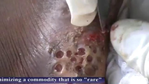
Watch that video of an Ingrown Hair Causes Huge Tumor in a Man's Stomach
Como Aumentar La Libido, Aumentar Niveles De Testosterona, Como Aumentar El Deseo Masculino ---- http://aumentar-testosterona.good-info.co/ --- ¿Se puede tener una erección con bajos niveles de testosterona? Mi libido está quedando atrás y estoy teniendo dificultades para conseguir una erección, así que estoy tratando de averiguar qué está pasando aquí. La disfunción eréctil rara vez es causada sólo por la deficiencia de testosterona. Por lo general es un grupo de cosas que funcionan en concierto juntos, que se alimentan entre sí, que conducen a la incapacidad del hombre para lograr una erección. La aterosclerosis (estrechamiento y endurecimiento de las arterias) es uno de los mayores impulsores de la disfunción eréctil, pero estas arterias dañadas no aparecen de la nada. Otras cosas tienen que estar sucediendo en el cuerpo para que ésta aterosclerosis pase, y como estamos a punto de ver, estas otras cosas contribuyen al problema también. Así que vamos a repasar esta lista… Nivel de azúcar alto – baja testosterona y disfunción eréctil La azúcar elevada en la sangre es un arma de doble filo, porque los hombres que sufren de esta condición son mucho más propensos a ser afectados por la disfunción eréctil y la testosterona baja. Una Investigación de John Hopkins encontró que las ratas diabéticas presentaron una respuesta eréctil 30% inferior, sus erecciones fueron como máximo 40% más pequeñas y las erecciones tomaron 70% más tiempo para lograrse en comparación con los controles que no eran diabéticos. Otros estudios han confirmado que los hombres con diabetes tipo 2 son dos veces más propensos a sufrir de disfunción eréctil, y la condición les golpeará una década antes, en comparación con los hombres sin tipo 2. Este vínculo es tan fuerte porque el azúcar en la sangre hace un daño directo a las arterias cuando se tiene demasiado de él, y las arterias en el pene suelen ser afectados en primer lugar, porque son muy pequeñas y estrechas. Por lo tanto, tiene todo el sentido que éstas pueden dañarse primero. El ejercicio que baja la testosterona haga click aqui http://aumentar-testosterona.good-info.co/
Eye Brow Transplant Procedure
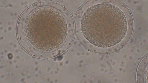
Sperm Meets Egg: Weeks 1 to 3 of Pregnancy. Something magical is about to happen! Watch as the ovulation process occurs, and then millions of sperm swim upstream on a quest to fertilize an egg. Your due date is calculated from the first day of your last menstrual period
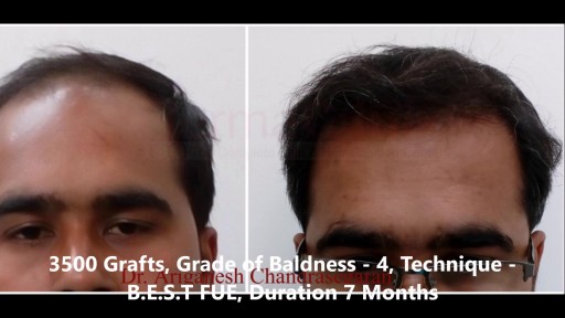
Hair Transplant Results Before and After Photos who undergone Hair Transplant. View our patient's successful results with the FUE, Bio - FUE and B.E.S.T FUE hair transplant technique. Comparable before & after photos! For More Visit Here:- https://www.hairtransplantchennai.org/hair-transplant-results-chennai.php or call us:- +91-8939636222

Case of ITP with persistent very low platelet count despite best medical management
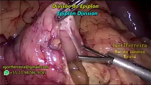
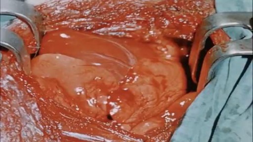
a sleeve gastrectomy with very few edditing. During the start 3 smal spleen perforations caused by Veres Needle were identified, caused by a giant spleen undentified on pre operatory ultrasound. They were controled with gauze compression and at the end of the surgery surgicel was placed and no complications were observed. Patient discharged 3 days after the surgery.
top 10 most incredible surgeries ever done
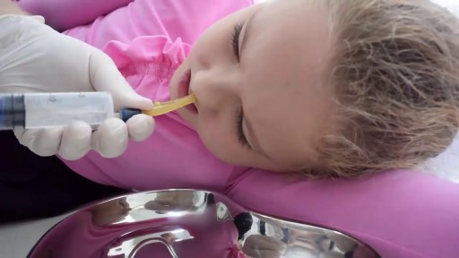
Antibiotic therapy is the mainstay of medical treatment for pediatric rhinosinusitis.] Because of increasing prevalence of beta-lactam–resistant bacteria in the community, administer antibiotics only for suspected infection as based on a careful history and physical examination. Direct the therapeutic regimen against the prevalent pathogens in the community and carefully consider suspicion for highly resistant bacteria. Typically, uncomplicated cases of acute sinusitis are responsive to amoxicillin. Most patients respond to this initial regimen. For children allergic to penicillin, a second- or third-generation cephalosporin can be used (only if the allergic reaction is not a type 1 hypersensitivity reaction). In cases of serious allergic reaction, a macrolide or clindamycin can be used.