- Physical Examination
- Surgical Examination
- Ophthalmology
- Clinical Skills
- Orthopedics
- Surgery Videos
- Laparoscopy
- Pediatrics
- Funny Videos
- Cardiothoracic Surgery
- Nursing Videos
- Plastic Surgery
- Otorhinolaryngology
- Histology and Histopathology
- Neurosurgery
- Dermatology
- Pediatric Surgery
- Urology
- Dentistry
- Oncology and Cancers
- Anatomy Videos
- Health and Fitness
- Radiology
- Anaesthesia
- Physical Therapy
- Pharmacology
- Interventional Radiology
- Cardiology
- Endocrinology
- Gynecology
- Emergency Medicine
- Psychiatry and Psychology
- Childbirth Videos
- General Medical Videos
- Nephrology
- Physiology
- Diet and Food Health
- Diabetes Mellitus
- Neurology
- Women Health
- Osteoporosis
- Gastroenterology
- Pulmonology
- Hematology
- Rheumatology
- Toxicology
- Nuclear Medicine
- Infectious Diseases
- Vascular Disease
- Reproductive Health
- Burns and Wound Healing
- Other
Top videos

How to Improve Sexual Health or Stamina Part 4 All Solution of Male Disorder Male Infertility Diagnostic and Treatment Re-Slim Care Latest Technology in Pakistan Dr. Aslam Naveed is a well known sexologist in Pakistan. He has treated more than 1 Lac patients since last 30 years of clinical Practice in sexology, he knows how to help the people facing sexual disorders. Contact: 02134965050, 03432821919 https://www.facebook.com/menssexcareclinic/ ADDRESS: Men’s Care Modern Hospital, Opposite, Safari Park, University Road, Karachi, Pakistan.
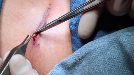
Removal of Infected Hernia Mesh

At URBN Dental, we offer the best dental services and highest quality care for your gum tissue health. Proper flossing techniques prevent your gum tissue from swelling, which often occurs from food and debris catching between your teeth. A routine dental cleaning every 6 months is recommended to maintain gum tissue health. Skipping bi annual checkups and improper flossing techniques often lead to periodontal disease which usually require a dental deep cleaning to undo tissue damage.
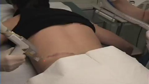
aser treatment for scars reduces the appearance of scars. It uses focused light therapy to either remove the outer layer of the skin’s surface or stimulate the production of new skin cells to cover damaged skin cells. Laser treatment for scars can reduce the appearance of warts, skin wrinkles, age spots, scars, and keloids. It doesn’t completely remove a scar.

What is hemodialysis, and why would someone need it? How does hemodialysis work? Can people perform hemodialysis at home? John Kevin Tucker, M.D., Nephrologist at Brigham and Women's Hospital and Vice President for Education at Mass General Brigham, discusses hemodialysis and how it helps people who have lost their kidney function to maintain normal lives.
Subscribe Link: https://www.youtube.com/channe....l/UCYrLjATd88gPwIKnt
0:00 - Intro
0:26 - The Condition
2:06 - Hemodialysis: How It Works
4:37 - In-Center Hemodialysis Care Team
About Mass General Brigham:
Mass General Brigham combines the strength of two world-class academic medical centers, five nationally ranked specialty hospitals, 11 community hospitals, and dozens of health centers. Our doctors and researchers accelerate medical breakthroughs and drive innovations in patient care. They are leaders in medical education, serving as Harvard Medical School faculty and training the next generation of physicians. Mass General Brigham’s mission is to deliver the best, affordable health care to patients everywhere. Together, we transform the health of our communities and beyond.
#MassGeneralBrigham #MGB #Hemodialysis
Visit Mass General Brigham: https://www.massgeneralbrigham.org/
Find us on social:
Twitter: https://twitter.com/MassGenBrigham
Instagram: https://www.instagram.com/massgeneralbrigham/
Facebook: https://www.facebook.com/MassGeneralBrigham/
LinkedIn: https://www.linkedin.com/compa....ny/mass-general-brig
Mass General Brigham:
https://www.youtube.com/massgeneralbrigham
Kidney Failure: Signs, Dialysis Options, and Hemodialysis Explained | Mass General Brigham
https://youtu.be/azy7yc19QYQ
![What does a fistula for dialysis look like? [CHT CERTIFICATION REVIEW] 2022](https://i.ytimg.com/vi/gjB5eKo4eh8/maxresdefault.jpg)
If this is the first time visiting us, make sure to subscribe to our channel here: https://bit.ly/2yXNBYp
What does a fistula for dialysis look like?
A fistula for dialysis is a surgical connection between a vein and an artery.
In this video, I will show you a real fistula and how we should evaluate it before a dialysis connection.
Additional videos:
💉How to properly cannulate a fistula: https://youtu.be/IqoHnzFyhJQ
💉 What is a fistula for dialysis treatment: https://youtu.be/B5EEf-MklFk
💉 The 10-second assessment for fistulas: https://youtu.be/Uqo0LhjZSI8
💉 If you would like to be trained as a dialysis professional focused on offering quality of care to renal patients, visit our program details here: https://utopiahcc.com/hemodialysis-technician/
For nursing and technician schools😷 🩺 🎓, we can offer a special renal failure class to your students. For inquiries please contact us: info@utopiahcc.com
Where to find us:
Facebook: https://www.facebook.com/utopiahealth
Email: info@utopiahcc.com
Website: utopiahcc.com
🤔 Looking for renal and dialysis continuing education for your certification renewal? Check out our CE package where you will get a little over 40 contact hours for a small price and receive your certificates immediately.
Here's the link: https://bit.ly/3dbPvDZ
Want to watch *Free Dialysis Training Videos*?
https://utopiahcc.com/free-dia....lysis-video-training
__________________________________________________________
Additional resources:
What Does a Healthy AV Fistula Look Like? | Azura Vascular ...
www.azuravascularcare.com infodialysisaccess healt...
Jul 17, 2018 — An AV fistula is a surgically-created permanent access located under the skin, making a direct connection between a vein and an artery. An AV fistula is typically created in the non-dominant arm. If the veins in your arm are not large or healthy enough to support a fistula, it may be created in your leg.
Preparing for Dialysis (AV Fistula) Fact Sheets Yale ...
www.yalemedicine.org › conditions › preparing-dialysi...
To undergo dialysis, patients need a surgical procedure to create an access point for the dialysis machine. An AV fistula is the most common access point.
Vascular Access for Hemodialysis - Life Options
lifeoptions.org living-with-kidney-failure vascular-a...
Jump to How a Catheter Looks and Feels — This makes a pattern that looks a bit like a rope ladder. The next best way—for fistulas ONLY—is the “Buttonhole ...
Fistula or Graft Surgery · Needle Fear · How a Fistula or Graft Looks...
Taking Care of Your Fistula - DaVita
www.davita.com dialysis preparing-for-dialysis › ta...
An arteriovenous (AV) fistula is a type of access used for hemodialysis. ... access because it utilizes the patient's own vessels and does not require permanent placement of foreign materials such ... Look for redness or swelling around the fistula area. ... This sound may change from a whooshing noise to a whistle-like sound.
Vascular Access for Hemodialysis - Department of Surgery
surgery.ucsf.edu conditions--procedures vascular-ac...
The patient does not need anesthesia for this procedure. ... A vascular surgeon performs AV graft surgery, much like AV fistula surgery, in an outpatient center or ...
Frequently Asked Questions about Dialysis Access Surgery ...
www.bidmc.org transplant-institute frequently-aske...
Dialysis access surgery creates the vascular opening so a needle can be inserted for ... fluid and to correct electrolytes like potassium, sodium, phosphate and calcium, to name a few. ... Where are AV fistulas located and how long do they last?
Fistula and Graft Placement (Eric K. Peden, MD) - YouTube
www.youtube.com watch
Mar 28, 2016 — ... Bootcamp 2015 August 14 - 16, 2015 "Dialysis Access" Fistula and Graft Placement (Eric K. Peden, MD) DICET@Houstonmethodist.org.

Dialysis lecture 1. Dialysis Study: EXPERT NOTES for DHA, Bonent, CHT, B.Sc in Dialysis, Diploma in Dialysis https://amzn.eu/d/35Ui1kT
2. Dialysis Study : Q & A: MCQs, Fill in the blanks, True or False https://amzn.eu/d/gGn8u73
1. Dialysis Study :EXPERT NOTES for DHA, Bonent, CHT, B.Sc in Dialysis, Diploma in Dialysis, Naseha Helal.
https://play.google.com/store/....books/details?id=D_7
2. Dialysis Study: Q & A MCQ https://play.google.com/store/....books/details?id=T_3
Whatsapp
https://chat.whatsapp.com/DKCHbgsNwXS1wd7xI31tpr
Telegram
https://t.me/dialysislife PRINCIPLE OF dialysis
https://youtu.be/cfOm0aFmbe8
Dialysis machine alarms
https://youtu.be/-1A1INyDEOg
DDS dialysis disequilibrium syndrome
https://youtu.be/8AqVFiBOkIc
Peritoneal Dialysis
https://youtu.be/iHPPadGmsv0
Itching
https://youtu.be/T83Wm3HHU4M
What is CRRT
https://youtu.be/jPgFnoSEBMU
LVH
https://youtu.be/ZhFL3Z6LHeA
Sorbent dialysis
https://youtu.be/-rie5dC_FkY
RO Water
https://youtu.be/3jlEsK4Lg_I
Carbon filter RO water
https://youtu.be/mJrgtjNafQw
Hemoperfusion
https://youtu.be/UkbBm8rm9Ww
AV fistula or Dialysis fistula
https://youtu.be/uDbyfqCkCbo
Dialysis MCQ
https://youtu.be/zmOj0BL6jVY
AVF cannulation
https://youtu.be/PyqMcHA07zY
Complications of AV fistula
https://youtu.be/a_CXIvuOO_s
Blood clotting during Dialysis
https://youtu.be/9hYNepiO2o8
Muscle crapms
https://youtu.be/09s07Eiqr2k
Hepatitis C
https://youtu.be/qdNj_GhmnSE
Dialysis procedure
https://youtu.be/u1mGqXO5pzQ
Hypotension
https://youtu.be/4EVPmWTSyN8
Heparin free dialysis
https://youtu.be/rFqAn7HcWwM
Plasmapheresis
https://youtu.be/kbgsjjs9krg
Isolated ultrafiltration
https://youtu.be/xp5I5--uWb0
High flux dialyzer
https://youtu.be/gCNsErn1HHM
Urea and Creatinine
https://youtu.be/Id9AIySMQ6c
Practical RO water demo
https://youtu.be/2pXKGMDNS84
Sodium profiling
https://youtu.be/bE_DcBXNB5g
Peritoneal Dialysis
https://youtu.be/vtK6VZsi8AY
Air embolism
https://youtu.be/WJE-xqnQfd8
Dialysate
https://youtu.be/z_nb43bcWsM
How to stop Bleed from fistula
https://youtu.be/N_inLKPhPUc
Dialysis short form
https://youtu.be/3BqB-gODb5o
Dialyzer reprocessing
https://youtu.be/XelfkKsndlc
Dialysis catheter
https://youtu.be/V7y90m4xlv8
How to set KT/V
https://youtu.be/hWXjU8VTQdk
Mircera injection
https://youtu.be/STtd3I3EijA
Dialysis procedure
https://youtu.be/MIdhIgcKRZ8
Dialysis in snake bite poison
https://youtu.be/niA9RI38jyY
Uf profiling
https://youtu.be/wyjpFjD5Hi0
Heparin dose
https://youtu.be/kB56MkzHIQ0
Hyperkalemia
https://youtu.be/1rWWNlcAuio
Change bandages of leaking fistula
https://youtu.be/_0cebWWdjM8
AvF needle
https://youtu.be/GvUxbXxftTk
Polycystic kidney disease
https://youtu.be/IhsMbHFXZG8
Nephrotic syndrome
https://youtu.be/FEEOsIrXxV8
Diabetic nephropathy
https://youtu.be/v-FBIQ7MA4k
Hemodialysis permanent access
https://youtu.be/_YrwxwiR0f8
Sex and dialysis
https://youtu.be/vvl8UT8lK4k
Albumin and dialysis
https://youtu.be/yzG7yD45Nwg
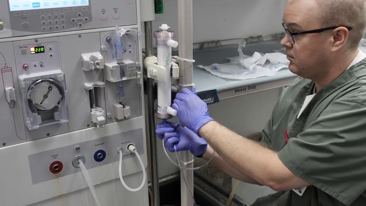
In this instructional video, Director of Critical Care Nephrology, Sevag Demirjian, MD goes over the steps for in-hospital production of ultra-pure continuous hemodialysis fluid.
By using the information in this video and/or any other materials made available by Cleveland Clinic related to the dialysate solution, you agree to comply with and be bound by the terms of the Permissive Use Agreement, a copy of which is available at https://bit.ly/3f9lN4j
![Knee Injury Rehabilitation [Early Stage] - (1st Two Weeks After Injury)](https://i.ytimg.com/vi/35zoRRUDVYo/maxresdefault.jpg)
I have shared with you in this video couple of exercises that you can follow immediately after your Knee injury.
As I promised here are 2 protocols to follow in this routine. I have also added my blog on how to strengthen your glutes and why that can help you with your knee pain.
1- Avoid Harm ( https://dublinsportsinjuryclin....ic.com/acute-injury-
2- POLICE PROTOCOL (https://dublinsportsinjuryclin....ic.com/acute-injury-
3- Read my blog and check how to strengthen your glutes (https://dublinsportsinjuryclin....ic.com/knee-injury-r
Please make sure to watch the video until the end since I'm sharing with you a couple of tips at the end of this video.
References:
1- https://www.ncbi.nlm.nih.gov/pmc/arti...
2- https://www.sciencedirect.com/science...
3- https://www.sciencedirect.com/science...
_______________________________
Music: See You
Musician: @iksonofficial
---------------------------------------------------
**MEDICAL DISCLAIMER**
All information, content, and material of this video or website are for informational and demonstration purposes only. It is not intended to serve as a substitute for the consultation, diagnosis, and/or medical treatment of a qualified physician or healthcare provider.
Don’t use this content as a replacement for treatment and advice given by your doctor or health care provider. Consult with your physical therapist or healthcare professional before doing anything contained in this content.
By watching this video, you agree to indemnify and hold harmless Dublin Sports Injury Clinic(and its representatives) for any and all losses, injuries, or damages resulting from any and all claims that arise from your use or misuse of this content. Dublin Sports Injury Clinic makes no representations about the accuracy or suitability of this content.
USE OF THIS VIDEO'S CONTENT IS AT YOUR OWN RISK.
𝐂𝐨𝐧𝐧𝐞𝐜𝐭 𝐰𝐢𝐭𝐡 𝐁𝐨𝐛 𝐚𝐭 𝐃𝐮𝐛𝐥𝐢𝐧 𝐒𝐩𝐨𝐫𝐭𝐬 𝐈𝐧𝐣𝐮𝐫𝐲 𝐂𝐥𝐢𝐧𝐢𝐜
𝐖𝐄𝐁𝐒𝐈𝐓𝐄→ https://www.dublinsportsinjuryclinic.com
𝐄𝐌𝐀𝐈𝐋 → Rehab@dublinsportsinjuryclinic.com
𝐓𝐄𝐋 → 0879276712
𝐅𝐀𝐂𝐄𝐁𝐎𝐎𝐊 → https://www.facebook.com/dublinphysicaltherapy/
𝐈𝐍𝐒𝐓𝐀𝐆𝐑𝐀𝐌 → https://www.instagram.com/dubl....in_sports_injury_cli
Tags:
Knee Injury Rehabilitation [Early Stage] - (1st Two Weeks After Injury)
Knee injury exercises, knee exercises, knee rehabilitation, Sore knee rehabilitation, Twisted knee exercises, sore kneecap exercises, runners knee injury, #kneeinjury #soreknee #runnersknee #Kneerehabilitation #kneeexercices #dublinsportsinjuryclinic
#anteriorkneepain #kneepain #kneephysio #injureknee #exerciseforknee #kneerehab #swollenknee
#runnersknee #kneeminiscus #acl #Mcl #kneeligaments#dublinsportsinjuryclinic #dublinsportsphysio #bobfiro #dulin2phyiso #bobyourphysio #bobonlinecare #Sportsinjurydublinclinic#dublinsportsinjuryclinic #dublinsportsphysio #bobfiro #dulin2phyiso #bobyourphysio #bobonlinecare #Sportsinjurydublinclinic

Thought a snake in your boot was bad? That old 19th-century idiom is nothing compared to one in your ear.
Shocking footage captured the alleged moment that a “surgeon” tried to remove a live snake that infiltrated a woman’s ear. Video of the herpetological surgery has racked up more than 125,000 views as viewers speculate whether or not the squirm-inducing footage is authentic.
“The snake has gone in the ear,” reads the caption to the bizarre Facebook clip, which was posted Sept. 1 by an India-based social media star named Chandan Singh to his 20,126 followers. However, it’s unclear where, when or how this unfortunate event transpired, local outlet the Economic Times reported.
In the nearly four-minute clip, an alleged medical practitioner can be seen using tweezers in a desperate attempt to extract a black and yellow serpent that’s peeking its head out from a female patient’s ear.

https://bit.ly/3HIStRc #shorts
Coloscopy | Colon Polyp Resection | Polypectomy
Colonoscopies are essential for detecting colorectal abnormalities, including colon polyps. Polypectomy, the surgical removal of these growths, can prevent them from becoming cancerous. This article offers a brief overview of colonoscopies, colon polyps, and polypectomy procedures.
A colonoscopy is an endoscopic examination allowing healthcare providers to visualize the colon and rectum using a colonoscope. The colonoscope, a flexible tube with a camera and light source, helps detect abnormalities, including polyps or tumors.
Colon polyps are abnormal growths arising from the colon's inner lining. While most polyps are benign, some can become malignant. Adenomatous polyps have a higher potential to become cancerous, whereas hyperplastic and inflammatory polyps pose a lower risk.
Polypectomy involves removing colon polyps during a colonoscopy. Two primary techniques include snare polypectomy, using a wire loop to cut the polyp, and cold forceps polypectomy, which employs forceps to grasp and remove smaller polyps.
Following a polypectomy, patients may experience mild discomfort or bleeding. Regular surveillance is crucial to minimize colorectal cancer risk. The frequency of surveillance colonoscopies depends on the number, size, and type of polyps found, as well as the patient's overall risk factors.
Colonoscopies and polypectomies play vital roles in detecting and removing colon polyps, reducing the risk of colorectal cancer, and maintaining optimal colon health.
Do you want to learn more about colon polyps and colonoscopy? check our:
Article @ https://bit.ly/41w5Ooq
For more free resources, find us on Pinterest & Facebook pages:
https://www.pinterest.ca/medicalartsofficial/
https://www.facebook.com/Medicalartsofficial
https://www.youtube.com/@medic....alarts?sub_confirmat
https://www.instagram.com/medicalartsofficial/
https://www.tiktok.com/@medicalarts
#endoscopicsurgery #digestivesystem
coloscopy
polyp
colon polyp
polypectomy
colonoscopy
colon polyp animation
gi endoscopy
@MedicalArts , 2023.
Click here to get 2 free filet mignons and $15 off your first ButcherBox: https://butcherbox.com/doctormike
Includes FREE Shipping. Be sure to enter your email to access the deal. Thanks to ButcherBox for sponsoring this video.
I’ll teach you how to become to media’s go-to expert in your field. Enroll in The Professional’s Media Academy now: https://www.professionalsmediaacademy.com
Listen to my podcast, @DoctorMikeCheckup here:
YouTube: https://go.doctormikemedia.com..../youtube/channel/The
Spotify: https://go.doctormikemedia.com..../spotify/CheckUpSpot
Apple Podcasts: https://go.doctormikemedia.com..../applepodcast/AppleP
Survivor is coming up on its 43rd season this fall (whaaat??), and with all that reality TV goodness in the can already I knew there would be some medical moments to react to. Turns out, I was right, in that there have been a bunch of ailments on the show over the years! These injuries span the entire length of the whole series, so if you're a long time Survivor and Jeff Probst fan, this one is for you!
I LOVE reading your comments and take your suggestions seriously. If there’s a subject you want me to discuss or something you’d like for me to react to, leave a comment down below. Many of my videos have been born out of suggestions directly from you, so don’t hold back!
-Doctor Mike Varshavski
Help us continue the fight against medical misinformation and change the world through charity by becoming a Doctor Mike Resident on Patreon where every month I donate 100% of the proceeds to the charity, organization, or cause of your choice! Residents get access to bonus content, an exclusive discord community, and many other perks for just $10 a month. Become a Resident today:
https://www.patreon.com/doctormike
Please SUBSCRIBE for new videos every Wednesday afternoon and Sunday morning! https://goo.gl/87kYq6
Let’s connect:
IG https://goo.gl/41ZS7w - Doctor Mike
Reddit https://www.reddit.com/r/DoctorMike/
Twitter https://goo.gl/kzmGs5 - Real Doctor Mike
Facebook https://goo.gl/QH4nJS - Real Doctor Mike
Contact Email: DoctorMikeMedia@Gmail.com
Executive Producer: Doctor Mike
Production Director and Editor: Dan Owens
Managing Editor and Producer: Sam Bowers
Editor and Designer: Caroline Weigum
* Select photos/videos provided by Getty Images *
** The information in this video is not intended nor implied to be a substitute for professional medical advice, diagnosis or treatment. All content, including text, graphics, images, and information, contained in this video is for general information purposes only and does not replace a consultation with your own doctor/health professional **

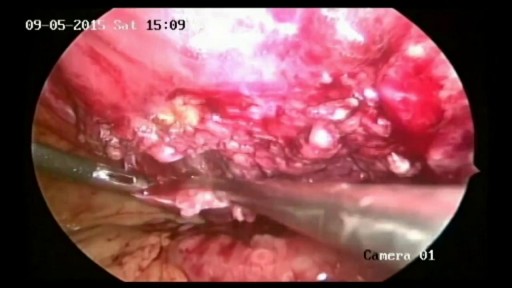
Intestinal obstruction.....
This video is only educational purposes and this is not for entertainment....this is surgery time

This video provides a guide peforming a respiratory examination in an OSCE station, including real-time auscultation sounds of common pathology such as coarse crackles, fine crackles, wheeze and stridor.
You can access our step-by-step OSCE guide to accompany this video here: https://geekymedics.com/respiratory-examination-2/
Check out our other awesome clinical skills resources including:
• 🔥 Geeky Medics Bundles (discounted products): https://app.geekymedics.com/purchase/bundles/
• ✨ 1000+ OSCE Stations: https://app.geekymedics.com/pu....rchase/osce-stations
• 🏥 Geeky Medics OSCE Revision Book: https://app.geekymedics.com/purchase/book/
• 📝 150+ PDF OSCE Checklists: https://geekymedics.com/pdf-osce-checklists/
• 🗂️ 3000+ OSCE Flashcards: https://app.geekymedics.com/pu....rchase/flashcard-col
• 📱 Geeky Medics OSCE App: https://geekymedics.com/geeky-medics-app/
• 🩺 Medical Finals SBA Question Pack: https://app.geekymedics.com/pu....rchase/medical-stude
• 💊 PSA Question Pack: https://app.geekymedics.com/pu....rchase/prescribing-s
Chapters:
- Introduction 00:00
- General inspection 00:40
- Inspection of the hands 00:50
- Schamroth's window test 01:09
- Heart rate and respiratory rate 01:50
- Jugular venous pressure 02:02
- Face, eyes and mouth 02:13
- Anterior chest inspection 02:36
- Trachea and cricosternal distance 03:01
- Palpation of apex beat 03:16
- Chest expansion 03:28
- Lung percussion 03:50
- Auscultation of lungs 04:21
- Vocal resonance 05:03
- Lymph node palpation 05:32
- Inspection of posterior chest 06:04
- Posterior chest expansion 06:10
- Percussion of posterior chest 06:32
- Auscultation of posterior chest 06:55
- Sacral and pedal oedema 08:04
- Summary of findings 08:39
Subscribe to our newsletter to be the first to know about our latest content: https://geekymedics.com/newsletter/ ✉️
Join the Geeky Medics community: 👩👩👧👧
Twitter: http://www.twitter.com/geekymedics
Instagram: https://instagram.com/geekymedics
Facebook: http://www.facebook.com/geekymedics
Always adhere to your medical school/local hospital guidelines when performing examinations or clinical procedures. DO NOT perform any examination or procedure on patients based purely upon the content of these videos. Geeky Medics accepts no liability for loss of any kind incurred as a result of reliance upon the information provided in this video.
Some people have found this video useful for ASMR purposes.
Special thanks to www.easyauscultation.com and Andy Howes for providing some of the respiratory sounds.

Watch this clinical examination video to learn how to diagnose cervical spine pathology.
This video clip is part of the FIFA Diploma in Football Medicine and the FIFA Medical Network. To enrol or to find our more click on the following link http://www.fifamedicalnetwork.com
The Diploma is a free online course designed to help clinicians learn how to diagnose and manage common football-related injuries and illnesses. There are a total of 42 modules created by football medicine experts. Visit a single page, complete individual modules or finish the entire course.
The network provides the opportunity for clinicians around the world to meet and share ideas relating to football medicine. Ask about an interesting case, debate current practice and discuss treatment strategies. Create a profile and log on to interact with other health professionals from around the globe.
This is not medical advice. The content is intended as educational content for health care professionals and students. If you are a patient, seek care of a health care professional.

Elbow Exam - Orthopaedic OSCE - Clinical Skills - Dr Gill
The elbow examination is a core skill - in this video, we demonstrate how to perform an elbow EXAM for an Orthopaedic Clinical Skills OSCE, which should be one of the more accessible examination stations for medical students.
For a passing grade in your Clinical Skills OSCE, an elbow assessment should follow the LOOK, FEEL, MOVE approach
Initially looking for erythema, scars, swelling and position
Palpating the elbow - specifically the olecranon, medial and lateral epicondyles, and radial head for heat, oedema and crepitus
Finally assess range of movement with flexion and extension at the elbow, before determining for tennis and golfers' elbows
Watch further orthopaedic examinations for your OSCE revision:
The Elbow - Deep Dive
https://youtu.be/SX5buhtCVDw
The Spine Examination:
https://youtu.be/pJxMHa6SCgU
The Knee examination
https://youtu.be/oyKH4EYfJDM
The Hip examination
https://youtu.be/JC9GKq5nSdQ
The GALS examination
https://youtu.be/5qJaf7gW-B0 - Gait, Arms, Legs, Spine - GALS screen
------------
Please note that there is no ABSOLUTE way to perform a clinical examination. Different institutions and even clinicians will have differing degrees of variations - the aim is the effectively identify medically relevant signs.
However during OSCE assessments. Different medical schools, nursing colleges and other health professional courses will have their own preferred approach to a clinical assessment - you should concentrate on THEIR marks schemes for your assessments.
The examination demonstrated here is derived from Macleods Clinical Examination - a recognised standard textbook for clinical skills.
Some people viewing this medical examination video may experience an ASMR effect
#clinicalskills #Elbow #DrGill

Ear Examination ENT is often a challenging examination, crossing over with the cranial nerve examination of the vestibular cochlear exam as well at other neurological assessments of balance
Here we will review the ear examination, looking both at the use of the otoscope, but also the Dix-Hallpike Manoeuvre, along with HINTS assessment. the Webers and Rinne's test is also included to determine types of hearing loss.
Often these ear examination techniques are performed separately, depending on the patients presenting complaint
#EARExamination #DrGill #ClinicalSkills
