
- Physical Examination
- Surgical Examination
- Ophthalmology
- Clinical Skills
- Orthopedics
- Surgery Videos
- Laparoscopy
- Pediatrics
- Funny Videos
- Cardiothoracic Surgery
- Nursing Videos
- Plastic Surgery
- Otorhinolaryngology
- Histology and Histopathology
- Neurosurgery
- Dermatology
- Pediatric Surgery
- Urology
- Dentistry
- Oncology and Cancers
- Anatomy Videos
- Health and Fitness
- Radiology
- Anaesthesia
- Physical Therapy
- Pharmacology
- Interventional Radiology
- Cardiology
- Endocrinology
- Gynecology
- Emergency Medicine
- Psychiatry and Psychology
- Childbirth Videos
- General Medical Videos
- Nephrology
- Physiology
- Diet and Food Health
- Diabetes Mellitus
- Neurology
- Women Health
- Osteoporosis
- Gastroenterology
- Pulmonology
- Hematology
- Rheumatology
- Toxicology
- Nuclear Medicine
- Infectious Diseases
- Vascular Disease
- Reproductive Health
- Burns and Wound Healing
- Other
Top videos
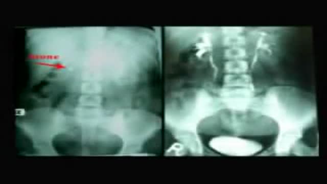

Totally US Guided PCNL in Flank Position

Guest: Dr. Vicente Santos Jr., Ophthalmologist/President - Fatima University Medical Center
Date Aired: August 14, 2015
Visit http://www.untvweb.com/programs/good-morning-kuya
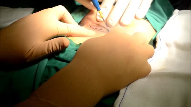
Excision of Pilonidal Cyst. Open method.

Mini-Laparoscopic Cholecystectomy with Intraoperative Cholangiogram for Symptomatic Cholelithiasis (Gallstones) - Extended
Authors: Brunt LM1, Singh R1, Yee A2
Published: September 26, 2017
AUTHOR INFORMATION
1 Department of Surgery, Washington University, St. Louis, Missouri
2 Division of Plastic and Reconstructive Surgery, Washington University, St. Louis, Missouri
DISCLOSURE
No authors have a financial interest in any of the products, devices, or drugs mentioned in this production or publication.
ABSTRACT
Minimal invasive laparoscopic cholecystectomy is the typical surgical treatment for cholelithiasis (gallstones), where patients present with a history of upper abdominal pain and episodes of biliary colic. The classic technique for minimal invasive laparoscopic cholecystectomy involves four ports: one umbilicus port, two subcostal ports, and a single epigastric port. The Society of American Gastrointestinal and Endoscopic Surgeons (SAGES) has instituted a six-step strategy to foster a universal culture of safety for cholecystectomy and minimize risk of bile duct injury. The technical steps are documented within the context of the surgical video for (1) achieving a critical view of safety for identification of the cystic duct and artery, (2) intraoperative time-out prior to management of the ductal structures, (3) recognizing the zone of significant risk of injury, and (4) routine intraoperative cholangiography for imaging of the biliary tree. In this case, the patient presented with symptomatic biliary colic due to a gallstone seen on the ultrasound in the gallbladder. The patient was managed a mini-laparoscopic cholecystectomy using 3mm ports for the epigastric and subcostal port sites with intraoperative fluoroscopic cholangiogram. Specifically, the senior author encountered a tight cystic duct preventing the insertion of the cholangiocatheter and the surgical video describes how the author managed the cystic duct for achieving a cholangiogram, in addition to the entire technical details of laparoscopic cholecystectomy.
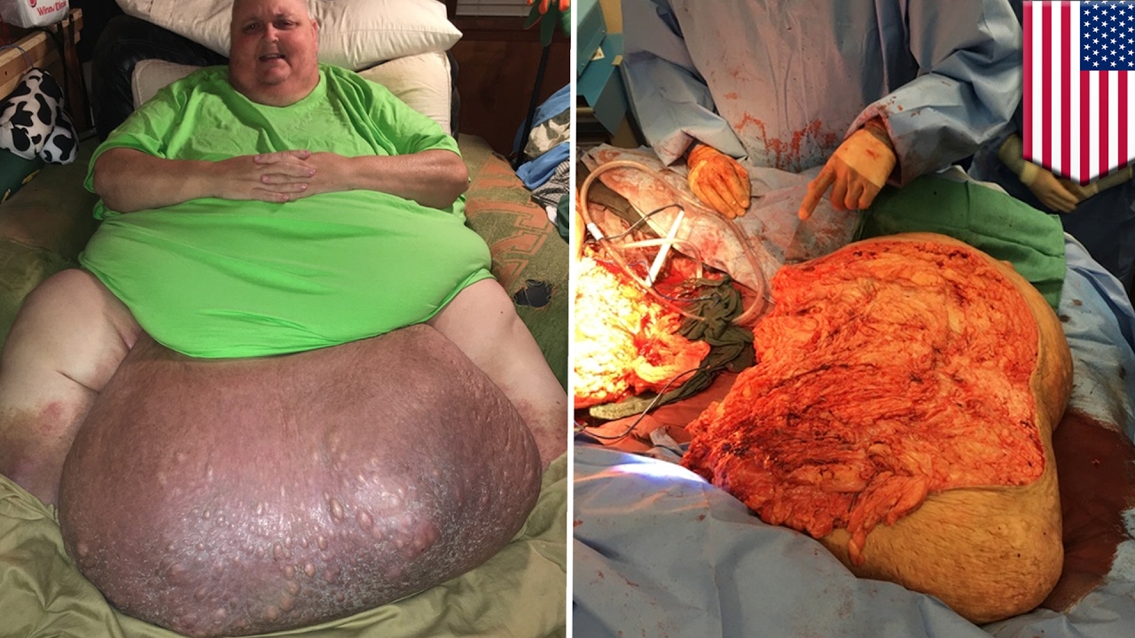
Watch that video of an Ingrown hair turned into 140-pound tumor in man’s stomach
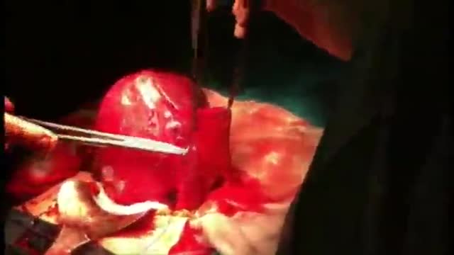
Pomeroy Tubal Ligation during a C-Sections

The MINI tummy-tuck is a lesser variant of the classic tummy tuck. The MINI tummy-tuck always involved skin excision (often a scar revision and skin excision of the flabby skin over a C-section scar or hysterectomy or laparotomy scar) but may also involve liposuction, umbilical floating, etc. Commonly it will not include any muscle repair otherwise it it now a classic tummy tuck (aka abdominoplasty). Cost varies depending on the components involved. Here, Toronto Aesthetic Plastic Surgeon Dr Marc DuPéré describes a MINI tummy-tuck done on a patient who had a Brazilian Butt Lift before (and skin harvesting from abdomen) and a recent 20 lbs weight loss, a patient who wants more liposuction to abdomen and flanks and whose skin has now lost elasticity, hence the requirement for this small skin excision. Dr DuPéré also explains what UMBILICAL floating means. Dr DuPéré performs more than 5 different techniques of tummy-tucks in Toronto and the technique chosen reflects the patient’s expectations and anatomy. Call us if interested in learning about YOUR options for a flatter tummy! 📱 416-929-9800
Patient consent obtained. Thank you to my patient.
Visage Clinic Toronto
https://www.visageclinic.com/
(416) 929-9800
101-133 Hazelton Avenue, Toronto, ON M5R 0A6
https://www.facebook.com/VisageClinic/
https://www.instagram.com/VisageClinicDrDuPere/
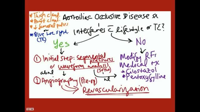
Aortoiliac occlusive disease (AIOD) occurs commonly in patients with PAD. Significant lesions in the aortoiliac arterial segment are exposed easily by palpation of the femoral pulses. Any diminution of the palpable femoral pulse indicates that a more proximal obstruction exists. Obstructive lesions may be present in the infrarenal aorta, common iliac, internal iliac (hypogastric), external iliac, or combinations of any or all of these vessels. Occasionally, degenerated nonstenotic atheromatous disease exists in these vessels and may manifest by atheroembolism to the foot, the "blue toe" or "trash foot" syndrome. Generally, patients with aortoiliac PAD have a poorer general prognosis than those with more distal PAD.

The video demonstrates complete excision of endometrosis in a variety of challenging situations.
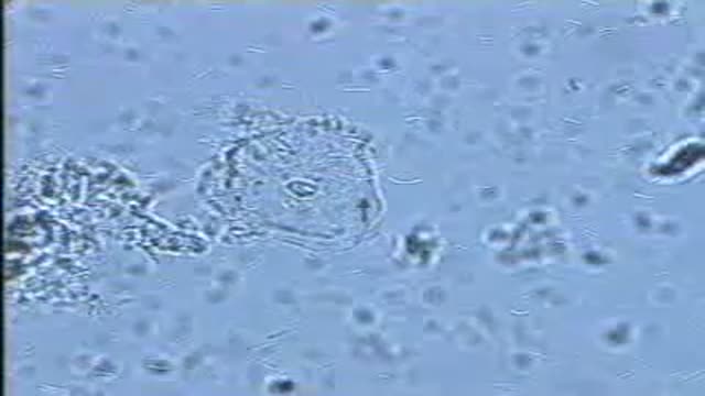
A normal vaginal epithelial cell is clear, with recognizable contents, and sharp, distinct cell borders.
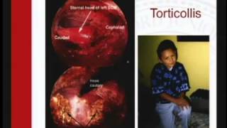
Sanjeev Dutta, MD, FACS discusses the fascinating new world of surgical technology. The pediatric general surgeon shares how medicine and technology have combined to achieve less invasive procedures and healthier outcomes for surgical patients.
Dr. Dutta is a pediatric general surgeon at Lucile Packard Children's Hospital. He is also an Associate Professor of Surgery at Stanford School of Medicine and Surgical Director of the Multidisciplinary Initiative for Surgical Technology Research.
Learn more about Stanford Children's Health. http://www.stanfordchildrens.org.
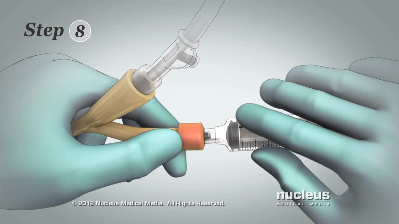
This 3D medical animation contains the discharge instructions for removal of a Foley catheter from a man. The step-by-step procedures for emptying the Foley bag and removing the Foley catheter are shown. Symptoms requiring a follow-up call to the surgeon are listed.
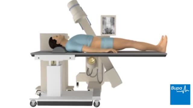
Extracorporeal shock wave lithotripsy (ESWL) uses shock waves to break a kidney stone into small pieces that can more easily travel through the urinary tract camera.gif and pass from the body. See a picture of ESWL camera.gif. You lie on a water-filled cushion, and the surgeon uses X-rays or ultrasound tests to precisely locate the stone. High-energy sound waves pass through your body without injuring it and break the stone into small pieces. These small pieces move through the urinary tract and out of the body more easily than a large stone. The process takes about an hour. You may receive sedatives or local anesthesia. Your surgeon may use a stent if you have a large stone. A stent is a small, short tube of flexible plastic mesh that holds the ureter open. This helps the small stone pieces to pass without blocking the ureter.
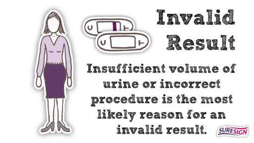
How to Perform Pregnancy Test at Home Video
Symptoms Burning stomach pain Feeling of fullness, bloating or belching Fatty food intolerance Heartburn Nausea The most common peptic ulcer symptom is burning stomach pain. Stomach acid makes the pain worse, as does having an empty stomach. The pain can often be relieved by eating certain foods that buffer stomach acid or by taking an acid-reducing medication, but then it may come back. The pain may be worse between meals and at night. Nearly three-quarters of people with peptic ulcers don't have symptoms. Less often, ulcers may cause severe signs or symptoms such as: Vomiting or vomiting blood — which may appear red or black Dark blood in stools, or stools that are black or tarry Trouble breathing Feeling faint Nausea or vomiting Unexplained weight loss Appetite changes
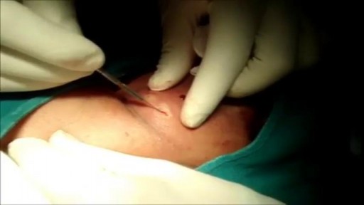
This is a 60 year man having large swelling of size 7cm x 5 cm behind neck for one year. Patient complained pain and tenderness over local area for 7 days and came to us.On examination punctum found in the centre of swelling and fluctuation positive.Infected sebaceous cyst diagnosis made. Incision and drainage surgery done under local anesthesia.all infected pultaceous material evacuated.Pus culture sent and antibiotics given as per sensitivity report. Patient improved with daily dressing.
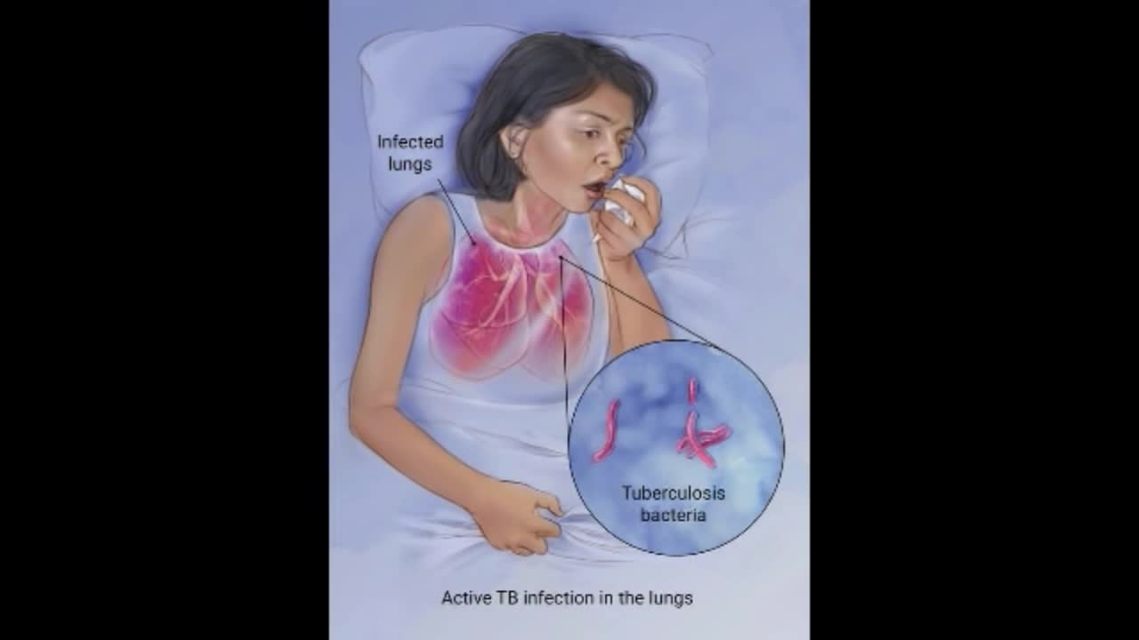
Although your body may harbor the bacteria that cause tuberculosis, your immune system usually can prevent you from becoming sick. For this reason, doctors make a distinction between: Latent TB. In this condition, you have a TB infection, but the bacteria remain in your body in an inactive state and cause no symptoms. Latent TB, also called inactive TB or TB infection, isn't contagious. It can turn into active TB, so treatment is important for the person with latent TB and to help control the spread of TB. An estimated 2 billion people have latent TB. Active TB. This condition makes you sick and can spread to others. It can occur in the first few weeks after infection with the TB bacteria, or it might occur years later. Signs and symptoms of active TB include: Coughing that lasts three or more weeks Coughing up blood Chest pain, or pain with breathing or coughing Unintentional weight loss Fatigue Fever Night sweats

You can now test your knowledge with a free lesson quiz on NURSING.com!
Click here for your free quiz: https://bit.ly/3HwJr8t
Stoma Care- Changing a Colostomy Bag (Nursing Skills)
FREE Nursing School Cheat Sheets at: http://www.NURSING.com
Get the full lesson on Stoma Care here:
05.01 Stoma Care (Colostomy bag) | NURSING.com
Check out our new Nurse Care Plan Lessons here:
https://bit.ly/3BPRfPL
Watch the Nursing Skills Course Introduction here:
https://nursing.com/lesson/ski....lls-00-01-course-int
Get Access to Thousands of Lessons here:
https://nursing.com/courses/
Welcome to the NURSING Family, we call it the most supportive nursing cohort on the planet.
At NURSING.com, we want to help you remove the stress and overwhelm of nursing school so that you can focus on becoming an amazing nurse.
Check out our freebies and learn more at: (http://www.nursing.com)
Stoma Care- Changing a Colostomy Bag (Nursing Skills)
In this video, we’re going to talk about stoma care. Now, the wafer and bag for an ostomy only NEEDS to be changed every 3 days, or if it’s leaking. But, you still need to be able to assess the stoma itself. In this case we’re going to show you how to replace the bag and clean and assess the stoma. Start by putting a towel under the patient on the side of the stoma. We love you guys! Go out and be your best selves today! And, as always, happy nursing!
Bookmarks:
0.05 Introduction to Stoma Care
0:20 Assessing the stoma
0:47 Cleaning the stoma
1:12 Inspecting the stoma
1:25 Measuring and cutting the stoma
2:00 Applying and sealing the bag
2:35 Documentation
2:41 Outro
Visit us at https://nursing.com/medical-disclaimer/ for disclaimer information.
NCLEX®, NCLEX-RN® are registered trademarks of the National Council of State Boards of Nursing, INC. and hold no affiliation with NURSING.com.

Scientists don't know what causes canker sores. Most believe that there is a problem with the body's immune system. Emotional stress, menstruation or injury to the mouth are common triggers for simple canker sores. Certain foods such as citrus or acidic foods may trigger a canker sore or make one more uncomfortable.
