トップ動画
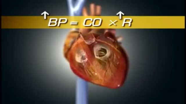
High blood pressure is a common condition in which the long-term force of the blood against your artery walls is high enough that it may eventually cause health problems, such as heart disease. Blood pressure is determined both by the amount of blood your heart pumps and the amount of resistance to blood flow in your arteries. The more blood your heart pumps and the narrower your arteries, the higher your blood pressure. You can have high blood pressure (hypertension) for years without any symptoms. Even without symptoms, damage to blood vessels and your heart continues and can be detected. Uncontrolled high blood pressure increases your risk of serious health problems, including heart attack and stroke. High blood pressure generally develops over many years, and it affects nearly everyone eventually. Fortunately, high blood pressure can be easily detected. And once you know you have high blood pressure, you can work with your doctor to control it.
ost of us come across this particular sign quite often. Of course, you can just jump to the numerous investigations and one after another, rule out the possible causes, finally getting to the diagnosis. For me, that’s no fun at all. Although I still don’t know whether I am going to become a surgeon or not (embarassing for me, since I’m going to be done with med-school this year), its pretty fascinating. If I were to work in a country whether investigations aren’t that expensive, I would definitely just perform a small examination and take a short history, sending off my patient to get a myriad of investigations, reporting to me after a while, with the diagnosis in his reports.
A cesarean delivery is a surgical procedure in which a fetus is delivered through an incision in the mother's abdomen and uterus. ... According to the CDC, in 2010, almost 33% of births were by cesarean delivery.

Subcutaneous Injection

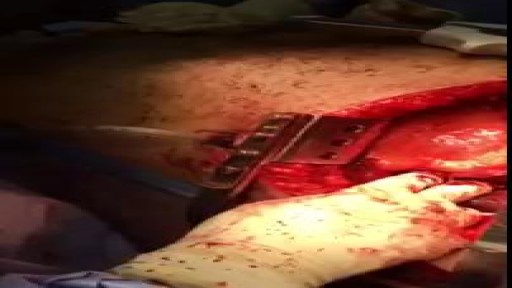
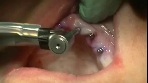
bilateral tubal ligation as modified Pomeroy technique during a C-Section


The operation of vasectomy
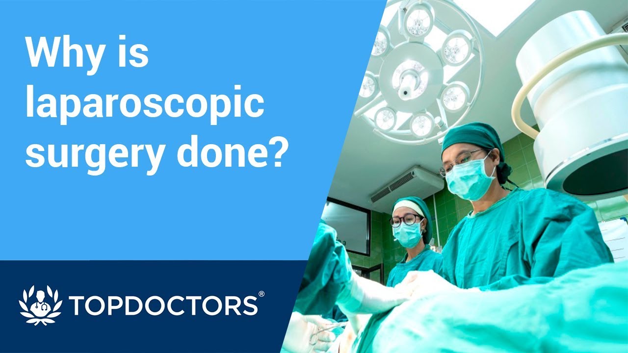
Laparoscopic surgery is minimally-invasive (keyhole) surgery and it is performed through very small incisions, using a camera to guide the surgeon during the procedure. Miss Sarah Mills, a top colorectal surgeon, explains why laparoscopic surgery is performed over alternative methods.
Make an appointment with Miss Sarah Mills here: https://www.topdoctors.co.uk/doctor/sarah-mills
The Combitube is a twin lumen device designed for use in emergency situations and difficult airways. It can be inserted without the need for visualization into the oropharynx, and usually enters the esophagus. It has a low volume inflatable distal cuff and a much larger proximal cuff designed to occlude the oro- and nasopharynx.
If the tube has entered the trachea, ventilation is achieved through the distal lumen as with a standard ETT. More commonly the device enters the esophagus and ventilation is achieved through multiple proximal apertures situated above the distal cuff. In the latter case the proximal and distal cuffs have to be inflated to prevent air from escaping through the esophagus or back out of the oro- and nasopharynx.

McRoberts Maneuver for Shoulder Dystocia Birth
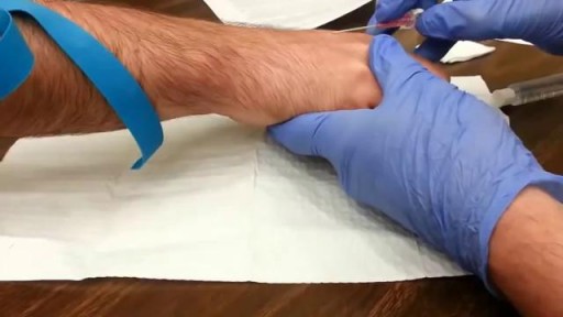

How to inject IM: How to draw substance
Cesarean VS Vaginal Birth Recovery
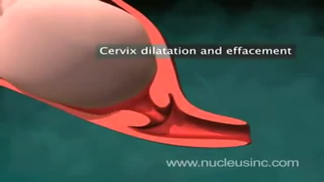
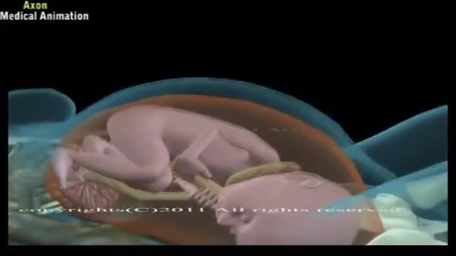
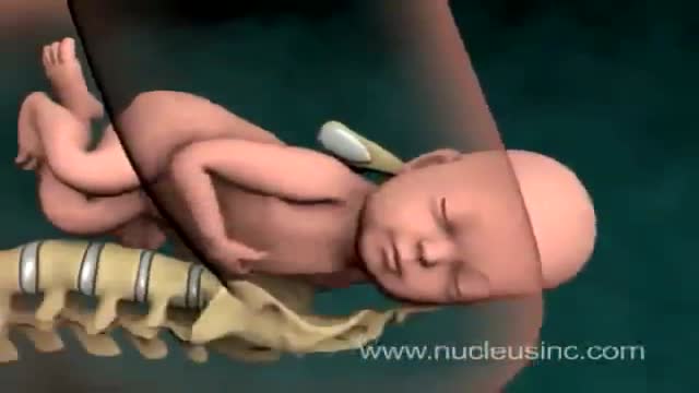
An animation showing vaginal childbirth (delivery)
A Beautiful Smile at Lake Pointe is Sugar Land premier dentistry practice. Dr. Lance Jue has been serving patients' preventive, restorative and cosmetic dental needs here in Sugar Land for over 19 years. Book an appointment online now with Dr. Lance Jue
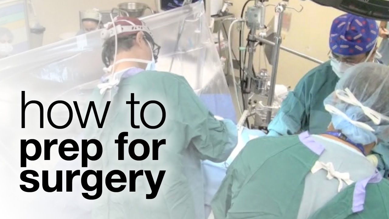
Thousands of Canadians undergo surgery every year, so how can you best prepare? The first step is having a dialogue, says Sunnybrook anesthesiologist Dr. Colin McCartney. Read the blog for more: http://sunnyview.sunnybrook.ca
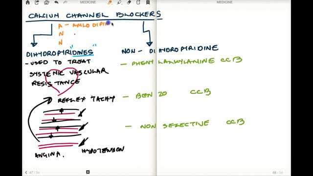
Calcium channel blockers prevent calcium from entering cells of the heart and blood vessel walls, resulting in lower blood pressure. Calcium channel blockers, also called calcium antagonists, relax and widen blood vessels by affecting the muscle cells in the arterial walls. Some calcium channel blockers have the added benefit of slowing your heart rate, which can further reduce blood pressure, relieve chest pain (angina) and control an irregular heartbeat. Examples of calcium channel blockers Some calcium channel blockers are available in short-acting and long-acting forms. Short-acting medications work quickly, but their effects last only a few hours. Long-acting medications are slowly released to provide a longer lasting effect. Several calcium channel blockers are available. Which one is best for you depends on your health and the condition being treated. Examples of calcium channel blockers include: Amlodipine (Norvasc) Diltiazem (Cardizem, Tiazac, others) Felodipine Isradipine Nicardipine Nifedipine (Adalat CC, Afeditab CR, Procardia) Nisoldipine (Sular) Verapamil (Calan, Verelan) In some cases, your doctor might prescribe a calcium channel blocker with other high blood pressure medications or with cholesterol-lowering drugs such as statins.
What Is an Appendectomy? An appendectomy is the surgical removal of the appendix. It’s a common emergency surgery that’s performed to treat appendicitis, an inflammatory condition of the appendix. The appendix is a small, tube-shaped pouch attached to your large intestine. It’s located in the lower right side of your abdomen. The exact purpose of the appendix isn’t known. However, it’s believed that it may help us recover from diarrhea, inflammation, and infections of the small and large intestines. These may sound like important functions, but the body can still function properly without an appendix. When the appendix becomes inflamed and swollen, bacteria can quickly multiply inside the organ and lead to the formation of pus. This buildup of bacteria and pus can cause pain around the belly button that spreads to the lower right section of the abdomen. Walking or coughing can make the pain worse. You may also experience nausea, vomiting, and diarrhea. It’s important to seek treatment right away if you’re having symptoms of appendicitis. When the condition goes untreated, the appendix can burst (perforated appendix) and release bacteria and other harmful substances into the abdominal cavity. This can be life-threatening, and will lead to a longer hospital stay. Appendectomy is the standard treatment for appendicitis. It’s crucial to remove the appendix right away, before the appendix can rupture. Once an appendectomy is performed, most people recover quickly and without complications. Why Is an Appendectomy Performed? An appendectomy is often done to remove the appendix when an infection has made it inflamed and swollen. This condition is known as appendicitis. The infection may occur when the opening of the appendix becomes clogged with bacteria and stool. This causes your appendix to become swollen and inflamed. The easiest and quickest way to treat appendicitis is to remove the appendix. Your appendix could burst if appendicitis isn’t treated immediately and effectively. If the appendix ruptures, the bacteria and fecal particles within the organ can spread into your abdomen. This may lead to a serious infection called peritonitis. You can also develop an abscess if your appendix ruptures. Both are life-threatening situations that require immediate surgery. Symptoms of appendicitis include: stomach pain that starts suddenly near the belly button and spreads to the lower right side of the abdomen abdominal swelling rigid abdominal muscles constipation or diarrhea nausea vomiting loss of appetite low-grade fever Although pain from appendicitis typically occurs in the lower right side of the abdomen, pregnant women may have pain in the upper right side of the abdomen. This is because the appendix is higher during pregnancy. Go to the emergency room immediately if you believe you have appendicitis. An appendectomy needs to be performed right away to prevent complications. What Are the Risks of an Appendectomy? An appendectomy is a fairly simple and common procedure. However, there are some risks associated with the surgery, including: bleeding infection injury to nearby organs blocked bowels It’s important to note that the risks of an appendectomy are much less severe than the risks associated with untreated appendicitis. An appendectomy needs to be done immediately to prevent abscesses and peritonitis from developing. How Do I Prepare for an Appendectomy? You’ll need to avoid eating and drinking for at least eight hours before the appendectomy. It’s also important to tell your doctor about any prescription or over-the-counter medications you’re taking. Your doctor will tell you how they should be used before and after the procedure. You should also tell your doctor if you: are pregnant or believe you may be pregnant are allergic or sensitive to latex or certain medications, such as anesthesia have a history of bleeding disorders You should also arrange for a family member or friend to drive you home after the procedure. An appendectomy is often performed using general anesthesia, which can make you drowsy and unable to drive for several hours after surgery. Once you’re at the hospital, your doctor will ask you about your medical history and perform a physical examination. During the exam, your doctor will gently push against your abdomen to pinpoint the source of your abdominal pain. Your doctor may order blood tests and imaging tests if appendicitis is caught early. However, these tests may not be performed if your doctor believes an emergency appendectomy is necessary. Before the appendectomy, you’ll be hooked up to an IV so you can receive fluids and medication. You’ll likely be put under general anesthesia, which means you’ll be asleep during surgery. In some cases, you’ll be given local anesthesia instead. A local anesthetic numbs the area, so even though you’ll be awake during the surgery, you won’t feel any pain. How Is an Appendectomy Performed? There are two types of appendectomy: open and laparoscopic. The type of surgery your doctor chooses depends on several factors, including the severity of your appendicitis and your medical history. Open Appendectomy During an open appendectomy, a surgeon makes one incision in the lower right side of your abdomen. Your appendix is removed and the wound is closed with stiches. This procedure allows your doctor to clean the abdominal cavity if your appendix has burst. Your doctor may choose an open appendectomy if your appendix has ruptured and the infection has spread to other organs. It’s also the preferred option for people who have had abdominal surgery in the past. Laparoscopic Appendectomy During a laparoscopic appendectomy, a surgeon accesses the appendix through a few small incisions in your abdomen. A small, narrow tube called a cannula will then be inserted. The cannula is used to inflate your abdomen with carbon dioxide gas. This gas allows the surgeon to see your appendix more clearly. Once the abdomen is inflated, an instrument called a laparoscope will be inserted through the incision. The laparoscope is a long, thin tube with a high-intensity light and a high-resolution camera at the front. The camera will display the images on a screen, allowing the surgeon to see inside your abdomen and guide the instruments. When the appendix is found, it will be tied off with stiches and removed. The small incisions are then cleaned, closed, and dressed. Laparoscopic surgery is usually the best option for older adults and people who are overweight. It has fewer risks than an open appendectomy procedure, and generally has a shorter recovery time. What Happens After an Appendectomy? When the appendectomy is over, you’ll be observed for several hours before you’re released from the hospital. Your vital signs, such your breathing and heart rate, will be monitored closely. Hospital staff will also check for any adverse reactions to the anesthesia or the procedure. The timing of your release will depend on: your overall physical condition the type of appendectomy performed your body’s reaction to the surgery In some cases, you may have to remain in the hospital overnight. You may be able to go home the same day as the surgery if your appendicitis wasn’t severe. A family member or friend will need to drive you home if you received general anesthesia. The effects of general anesthesia usually take several hours to wear off, so it can be unsafe to drive after the procedure. In the days following the appendectomy, you may feel moderate pain in the areas where incisions were made. Any pain or discomfort should improve within a few days. Your doctor may prescribe medication to relieve the pain. They might also prescribe antibiotics to prevent an infection after surgery. You can further reduce your risk for infection by keeping the incisions clean. You should also watch for signs of infection, which include: redness and swelling around the incision fever above 101°F chills vomiting loss of appetite stomach cramps diarrhea or constipation that lasts for more than two days Although there’s a small risk of infection, most people recover from appendicitis and an appendectomy with little difficulty. Full recovery from an appendectomy takes about four to six weeks. During this time, your doctor will probably recommend that you limit physical activity so your body can heal. You’ll need to attend a follow-up appointment with your doctor within two to three weeks after the appendectomy.
