- Physical Examination
- Surgical Examination
- Ophthalmology
- Clinical Skills
- Orthopedics
- Surgery Videos
- Laparoscopy
- Pediatrics
- Funny Videos
- Cardiothoracic Surgery
- Nursing Videos
- Plastic Surgery
- Otorhinolaryngology
- Histology and Histopathology
- Neurosurgery
- Dermatology
- Pediatric Surgery
- Urology
- Dentistry
- Oncology and Cancers
- Anatomy Videos
- Health and Fitness
- Radiology
- Anaesthesia
- Physical Therapy
- Pharmacology
- Interventional Radiology
- Cardiology
- Endocrinology
- Gynecology
- Emergency Medicine
- Psychiatry and Psychology
- Childbirth Videos
- General Medical Videos
- Nephrology
- Physiology
- Diet and Food Health
- Diabetes Mellitus
- Neurology
- Women Health
- Osteoporosis
- Gastroenterology
- Pulmonology
- Hematology
- Rheumatology
- Toxicology
- Nuclear Medicine
- Infectious Diseases
- Vascular Disease
- Reproductive Health
- Burns and Wound Healing
- Other
Top videos
A video showing the process of childbirth via vaginal delivery.
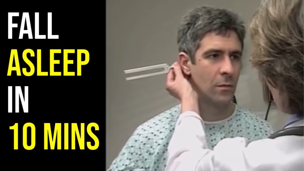
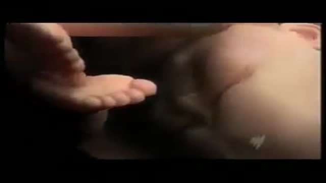
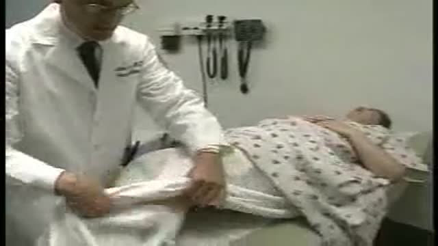
The MORE subscribers we have the MORE unintentional ASMR videos we can post, so please SUBSCRIBE http://bit.ly/UASMR, hit that 🔔 and LIKE this video for more medical exam ASMR like this! So... here is the BEST medical exam ASMR on YouTube edited specifically for unintentional ASMR to become the best video to help fall asleep at night. Our medical exam ASMR playlist: http://bit.ly/MedicalASMR
🔴 Support the channel and get access to extra perks by becoming a member here: https://www.youtube.com/channe....l/UCWCk81c_nQOsm5M-x
Try out the MOST comfortable sleep mask I've ever worn here ➜ https://bit.ly/MantaDiscount.
Prefer SOOTHING music for sleep? Here's what I use ➜ https://bit.ly/3lOPqe2
This hour long medical exam has a quiet doctor performing a routine medical exam with minimal talking. The video is 100% accidental ASMR and features a real doctor doing a head to toe assessment. This is honestly one of the best medical exam ASMR videos and on my all time favourite unintentional ASMR videos. This audio for this video has been significantly edited for maximum ASMR.
EXCLUSIVE: You can get 10% off any SleepPhones products with the code SLEEPNOW here: https://bit.ly/2YrrKQC
For the best unintentional ASMR and sleep aid videos we can't post here see http://ineedtosleepnow.com/
Medical ASMR: http://bit.ly/MedicalASMR
Female ASMR: http://bit.ly/FemaleASMR
Best of the best: http://bit.ly/BestUnintentionalASMR
Celeb ASMR: http://bit.ly/CelebASMR
Show and tell ASMR: http://bit.ly/ShowTellASMR
Relaxing interviews: http://bit.ly/InterviewsASMR
Original un-edited videos:
Part 1: https://youtu.be/S5tK3Ikb7ug
Part 2: https://youtu.be/m_-ALfUmmiA
WHAT IS UNINTENTIONAL ASMR?
https://ineedtosleepnow.com/what-is-u...
💤 For MORE of the Best (Intentional) ASMR videos on YouTube click here: https://goo.gl/mTsEBN
🙂 Finding, editing and uploading these videos does take some time and I try to upload a new video each night. If you find these videos useful, please consider supporting us by doing your Amazon shopping through our affiliate link: https://www.amazon.ca/shop/pur....eunintentionalasmrby
Best Microphone for recording ASMR: https://amzn.to/34WJhn2
▬▬▬▬▬▬▬▬▬▬▬▬▬▬▬▬▬▬▬▬▬▬▬▬▬
WANT TO MAKE A RECOMMENDATION? Contact us via:
Facebook ► https://www.facebook.com/uASMR/
Twitter ► https://twitter.com/AccidentalASMR
Instagram ► https://www.instagram.com/pureunintentionalasmr/
See what's coming next by checking out our FB group: https://www.facebook.com/groups/1649835798657192/
🔴 In case this channel is removed, please subscribe to our newsletter for updates on new videos locations: http://eepurl.com/ds-orr. We will never email you about anything other than a new channel.
▬▬▬▬▬▬▬▬▬▬▬▬▬▬▬▬▬▬▬▬▬▬▬▬▬
More of the Pure ASMR channels:
Pure ASMR: http://bit.ly/PureASMR and hit that 🔔.
Pure Unintentional ASMR 2: https://bit.ly/PureUASMR2
Pure Natural ASMR: http://bit.ly/NatureASMR
Pure Audiobook ASMR: http://bit.ly/AudiobookChannel
Purr ASMR: http://bit.ly/CatASMR
▬▬▬▬▬▬▬▬▬▬▬▬▬▬▬▬▬▬▬▬▬▬▬▬▬
NOTE: This video is being shared for #unintentionalASMR reasons only. The video has been edited from its original format for this purpose.
WHAT IS ASMR? http://ineedtosleepnow.com/what-is-asmr
ASMR definition: http://ineedtosleepnow.com/asmr-definition-what-is-the-definition-for-asmr
http://iNeedToSleepNow.com is a YouTube channel and website that curates calm softly spoken voices that serve as great unintentional #ASMR. This channel focuses primarily on soft speaking, calm softly spoken voices, unintentional ASMR interviews, relaxing voices, ASMR tingles and anything that helps you sleep.
Disclaimer: Some of these links are affiliate links where this channel could earn a small commission if you make a purchase. Shopping through these links is a great way to support the channel so we can keep making more videos for you. Thanks!

Patient Greg Grindley communicates with host Bryant Gumbel and his wife for the first time while undergoing deep brain stimulation surgery at University Hospital's Case Medical Center in Cleveland, Ohio.
➡ Subscribe: http://bit.ly/NatGeoSubscribe
About National Geographic:
National Geographic is the world's premium destination for science, exploration, and adventure. Through their world-class scientists, photographers, journalists, and filmmakers, Nat Geo gets you closer to the stories that matter and past the edge of what's possible.
Get More National Geographic:
Official Site: http://bit.ly/NatGeoOfficialSite
Facebook: http://bit.ly/FBNatGeo
Twitter: http://bit.ly/NatGeoTwitter
Instagram: http://bit.ly/NatGeoInsta
Greg's First In-Surgery Conversation | Brain Surgery Live
https://youtu.be/zvqV_2zncNU
National Geographic
https://www.youtube.com/natgeo

Physical Assessment of a Child
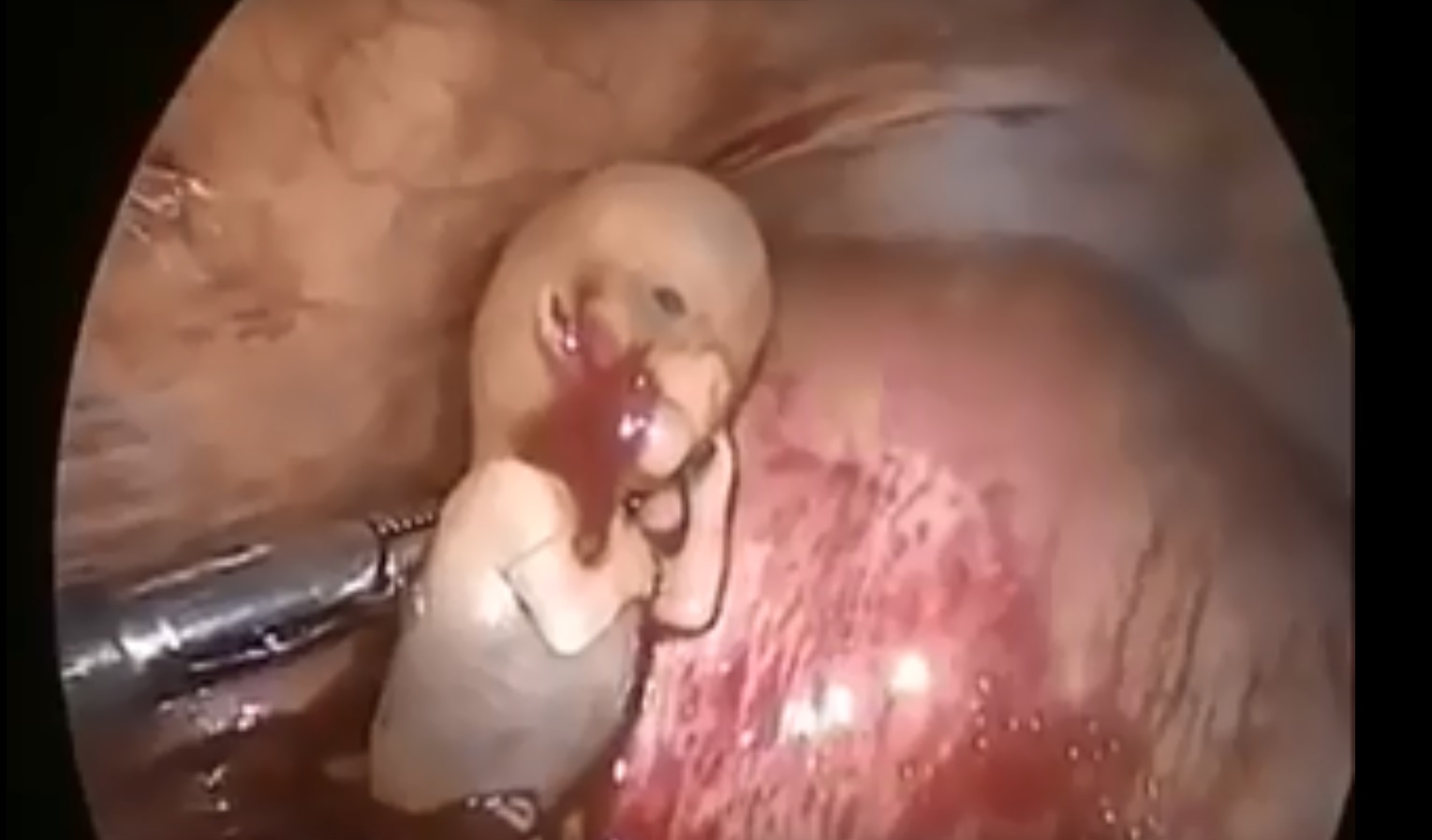
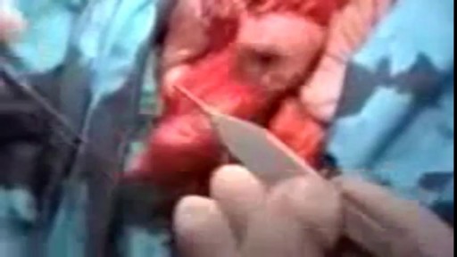
Watch that Ectopic Pregnancy Abortion Surgery
This is a video of a Gender Reassignment Surgery, watch as surgeons change a male to a female its an extremely interesting procedure

Acclaimed sexologist Hanny Lightfoot-Klein, author of several highly illuminating books on genital mutilation, discusses compromises in orgasm after male circumcision. Also commenting is cultural anthropologist James De Meo.From the groundbreaking documentary film, "Whose Body, Whose Rights?"
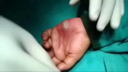
Draining a Hand Abscess
Part 2. Full Obstetric examination and normal delivery by Egyptian doctor Hussein Sulayman and the video is in English showing: Obstetric Examination Episiotomy Obstetric Forceps Obstetric Instruments
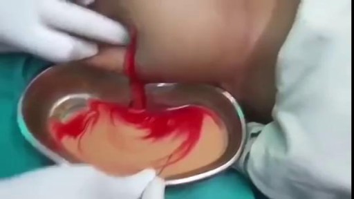
Draining HUGE back abscess
"The act of cutting off the prepuce or foreskin of males, or the internal labia of females." Webster's Revised Unabridged Dictionary (1913)
Full examination of the female from head to toe by Loyola Medical School, Chicago. Part 4

Female breast exam video

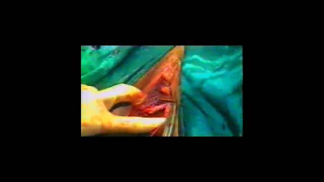
Surgical cutting and removal of a deep skin cyst Medical Videos
childbirth twin baby
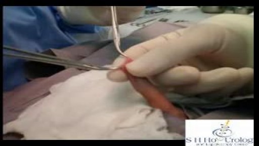
Popping Cyst in the Ear Lobe

This video provides a demonstration of how to assess for transillumination when assessing scrotal swelling.
Read our step-by-step guide here: https://geekymedics.com/testic....ular-examination-osc
Check out our other awesome clinical skills resources, including:
• 🔥 Geeky Medics Bundles (discounted products): https://app.geekymedics.com/purchase/bundles/
• ✨ 1000+ OSCE Stations: https://app.geekymedics.com/pu....rchase/osce-stations
• 🏥 Geeky Medics OSCE Revision Book: https://app.geekymedics.com/purchase/book/
• 📝 150+ PDF OSCE Checklists: https://geekymedics.com/pdf-osce-checklists/
• 🗂️ 3000+ OSCE Flashcards: https://app.geekymedics.com/pu....rchase/flashcard-col
• 📱 Geeky Medics OSCE App: https://geekymedics.com/geeky-medics-app/
• 🩺 Medical Finals SBA Question Pack: https://app.geekymedics.com/pu....rchase/medical-stude
• 💊 PSA Question Pack: https://app.geekymedics.com/pu....rchase/prescribing-s
Subscribe to our newsletter to be the first to know about our latest content: https://geekymedics.com/newsletter/ ✉️
Join the Geeky Medics community: 👩👩👧👧
Twitter: http://www.twitter.com/geekymedics
Instagram: https://instagram.com/geekymedics
Facebook: http://www.facebook.com/geekymedics
Always adhere to your medical school/local hospital guidelines when performing examinations or clinical procedures. DO NOT perform any examination or procedure on patients based purely on the content of these videos. Geeky Medics accepts no liability for loss of any kind incurred as a result of reliance upon the information provided in this video.

Alexandra J. Golby, MD, Director, Image-guided Neurosurgery at Brigham and Women’s Hospital, discusses technological advancements to improve the precision of surgery to remove brain tumors.
It’s estimated that each year nearly 80,000 people are diagnosed with primary brain tumors and 100,000 with metastatic brain tumors. Nearly everybody is at risk for developing a brain tumor. Brain tumors can affect people from childhood to the last years of their lives. Men are slightly more affected than women and the causes of most brain tumors are not known.
There are a number of unique challenges in treating brain tumors. One challenge is that primary tumors can have indistinct margins that are difficult to see. Another challenge is that the tissue around a brain tumor is uniquely important and may impact things like language, visual and motor function.
The AMIGO Suite, opened in 2011 at Brigham and Women’s Hospital, is the Advanced Multimodality Image Guided Operating Suite. It's an NIH-funded national center which was developed with the goal of translating technological advances into improvements in surgical and interventional care for patients. In the AMIGO Suite, there is an intraoperative MRI scanner which can be brought in and out of the operating room during surgery to help surgeons visualize a patient’s tumor better.
Image-guided surgery uses the information obtained from advanced imaging and translates that into the planning and execution of surgery by acquiring high resolution and specialty structural images of the brain and also functional images of the brain. These images can be registered to one another and then to the patient's head during surgery. This allows surgeons to pinpoint the location of the tumor as well as the areas that we would like to preserve, areas that serve critical brain functions are located.
One of the big challenges, even with image-guided surgery, is that as we perform the surgery, the configuration of the brain is changing, and we call that brain shift. And it's due to changes in the brain itself and also as we remove tissue, things are constantly shifting and moving. When we're talking about doing brain tumor surgery, a few millimeters of movement can be a big difference. How to measure and track brain shift is an important area of research and a number of technologies are being studied to understand how to measure brain shift during surgery.
The development of various intraoperative imaging technologies allows surgeons to provide the most accurate surgical treatment for each individual patient.
Learn more about precision brain surgery at Brigham and Women’s Hospital:
https://www.brighamandwomens.o....rg/neurosurgery/brai
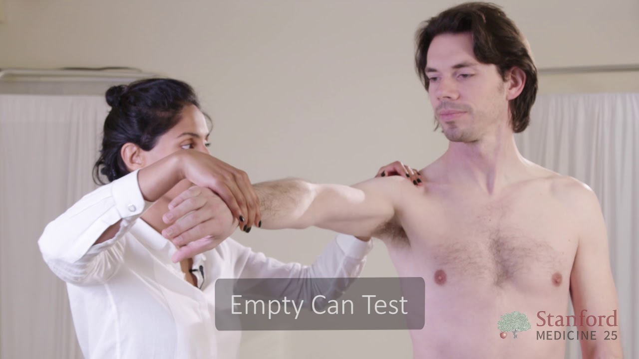
This video is brought to you by the Stanford Medicine 25 to teach you the common causes of shoulder pain and how to diagnose them by the physical exam.
The Stanford Medicine 25 program for bedside medicine at the Stanford School of Medicine aims to promote the culture of bedside medicine to make current and future clinicians and other healthcare provides better at the art of physical diagnosis and more confident at the bedside of their patients.
Visit us:
Website: http://stanfordmedicine25.stanford.edu/
Blog: http://stanfordmedicine25.stanford.edu/blog.html
Facebook: https://www.facebook.com/StanfordMedicine25
Twitter: https://twitter.com/StanfordMed25
Diagnoses covered in this video:
Rotator Cuff Pathology
Impingement Syndrome
Biceps Tendinopathy
Adhesive Capsulitis (Frozen Shoulder)
Acromioclavicular (AC) Joint Disease
Shoulder Instability
Labral Tears (SLAP Lesions)
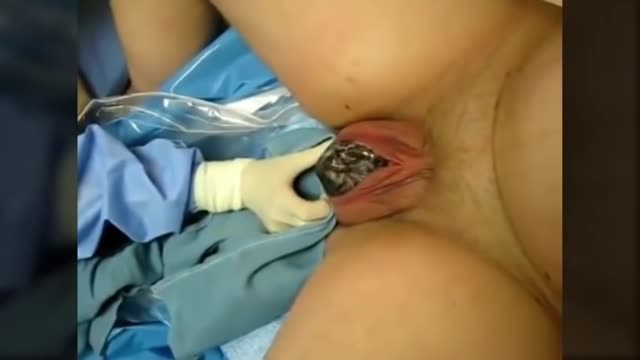
Gynecological Examination
