- Physical Examination
- Surgical Examination
- Ophthalmology
- Clinical Skills
- Orthopedics
- Surgery Videos
- Laparoscopy
- Pediatrics
- Funny Videos
- Cardiothoracic Surgery
- Nursing Videos
- Plastic Surgery
- Otorhinolaryngology
- Histology and Histopathology
- Neurosurgery
- Dermatology
- Pediatric Surgery
- Urology
- Dentistry
- Oncology and Cancers
- Anatomy Videos
- Health and Fitness
- Radiology
- Anaesthesia
- Physical Therapy
- Pharmacology
- Interventional Radiology
- Cardiology
- Endocrinology
- Gynecology
- Emergency Medicine
- Psychiatry and Psychology
- Childbirth Videos
- General Medical Videos
- Nephrology
- Physiology
- Diet and Food Health
- Diabetes Mellitus
- Neurology
- Women Health
- Osteoporosis
- Gastroenterology
- Pulmonology
- Hematology
- Rheumatology
- Toxicology
- Nuclear Medicine
- Infectious Diseases
- Vascular Disease
- Reproductive Health
- Burns and Wound Healing
- Other
Top videos
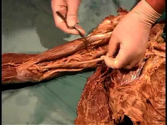
Anatomy of The Shoulder and Arm Muscles
Since the first replant more than 50 years ago, thousands of severed body parts have been reattached, preserving the quality of life for thousands of patients through improved function and appearance that the void remaining after amputation cannot provide. Ronald Malt performed the first replantation on May 23, 1962 at Massachusetts General Hospital on a 12-year-old boy who had his right arm amputated in a train accident. [1, 2] This amputation occurred at the level of the humeral neck.
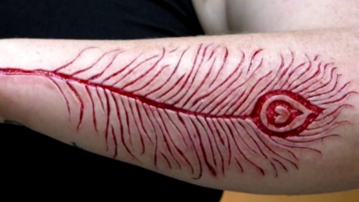
Bizarre Body Modifications
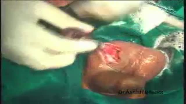
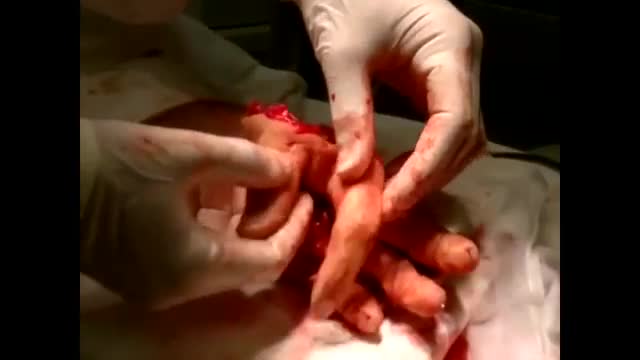
Orchidectomy and Orchidopexy in testis Torsion
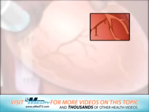
Coronary artery bypass grafting (CABG) is a type of surgery that improves blood flow to the heart. Surgeons use CABG to treat people who have severe coronary heart disease (CHD). CHD is a disease in which a waxy substance called plaque (plak) builds up inside the coronary arteries.

The menstrual cycle is the regular natural change that occurs in the female reproductive system that makes pregnancy possible. The cycle is required for the production of oocytes, and for the preparation of the uterus for pregnancy.
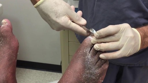
Presence of several characteristic diabetic foot pathologies such as infection, diabetic foot ulcer and neuropathic osteoarthropathy is called diabetic foot syndrome. Due to the peripheral nerve dysfunction associated with diabetes (diabetic neuropathy), patients have a reduced ability to feel pain.
Watch that video of an Ingrown hair turns into 140-pound tumor in man’s stomach
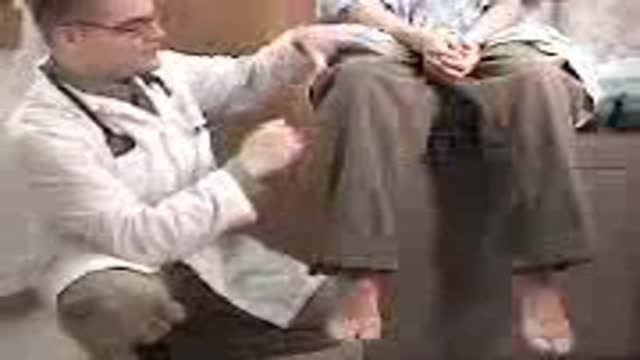
Motor examination of Lower Limb from the USMLE collection

peptic ulcer
Female ejaculation is characterized as an expulsion of fluid from or near the vagina during or before an orgasm
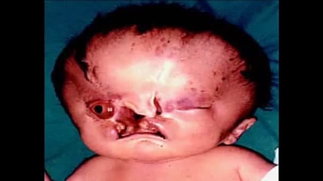
Holoprosencephaly (HPE, once known as arhinencephaly) is a cephalic disorder in which the prosencephalon (the forebrain of the embryo) fails to develop into two hemispheres. Normally, the forebrain is formed and the face begins to develop in the fifth and sixth weeks of human pregnancy. The condition also occurs in other species, as with Cy, the Cyclops kitten.

Nissen Fundoplication
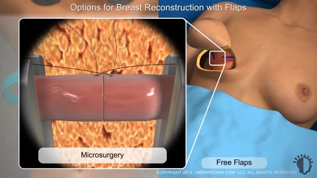
Typically, breast reconstruction takes place during or soon after mastectomy, and in some cases, lumpectomy. Breast reconstruction also can be done many months or even years after mastectomy or lumpectomy. During reconstruction, a plastic surgeon creates a breast shape using an artificial implant (implant reconstruction), a flap of tissue from another place on your body (autologous reconstruction), or both.
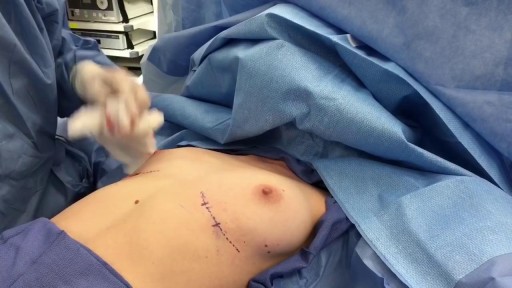
Breast Augmentation Short Scar Technique Slicone Implants

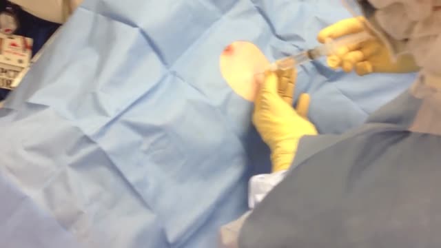
Amniocentesis,before the actual procedure, a local anesthetic is sometimes given to relieve the pain when inserting the needle used to withdraw the fluid. A needle is usually inserted through the mother's abdominal wall or at the end of the vagina, and through the wall of the uterus into the amniotic sac. With assistance from ultrasound, a physician aims towards an area of the sac that is away from the fetus and extracts a small amount of amniotic fluid for testing. The puncture heals, and the amniotic sac replenishes the liquid over a day or so. After the amniotic fluid is extracted, the fetal cells are separated from it using a centrifuge, and the fetal chromosomes are examined for abnormalities. Various genetic testing may be performed, but the three most common abnormalities tested for are Down's syndrome, Trisomy 18 and spina bifida. Amniocentesis can be performed as soon as sufficient amniotic fluid surrounds the fetus to allow a sample to be recovered relatively safely, usually no earlier than the 14th week of pregnancy. Often, genetic counseling is offered in conjunction with amniocentesis.
Defecography showing Anterior Rectal Wall Prolapse
A pneumothorax is usually caused by an injury to the chest, such as a broken rib or puncture wound. It may also occur suddenly without an injury. A pneumothorax can result from damage to the lungs caused by conditions such as chronic obstructive pulmonary disease (COPD), asthma, cystic fibrosis, and pneumonia.
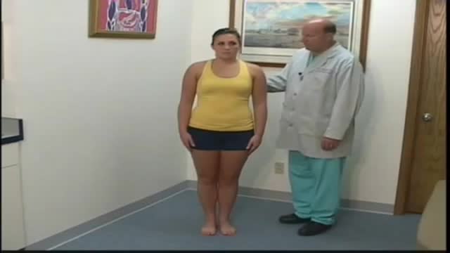
A patient who has a problem with proprioception can still maintain balance by using vestibular function and vision. In the Romberg test, the standing patient is asked to close his or her eyes. A loss of balance is interpreted as a positive Romberg's test.

This video shows the process of development and growth of the fetus intrauterine.
