Najbolji videi
Watch that video of Super Model's Butt and Leg Implants Exploded
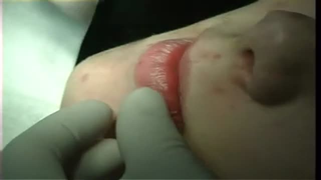
Surgical removal of mucocele from lower lip
Polycystic ovary syndrome is a common endocrine system disorder among women of reproductive age. Women with PCOS may have enlarged ovaries that contain small collections of fluid — called follicles — located in each ovary as seen during an ultrasound exam. Infrequent or prolonged menstrual periods, excess hair growth, acne, and obesity can all occur in women with polycystic ovary syndrome. In adolescents, infrequent or absent menstruation may raise suspicion for the condition. The exact cause of polycystic ovary syndrome is unknown. Early diagnosis and treatment along with weight loss may reduce the risk of long-term complications, such as type 2 diabetes and heart disease.
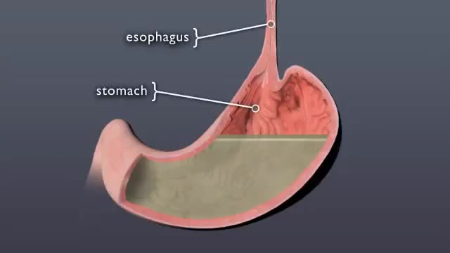
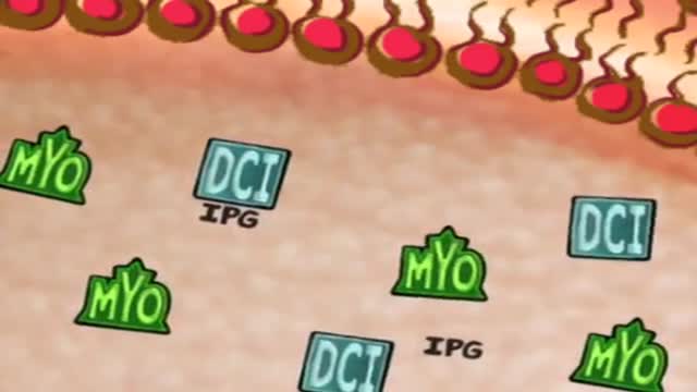
Discover what happens to pill when it swallowed
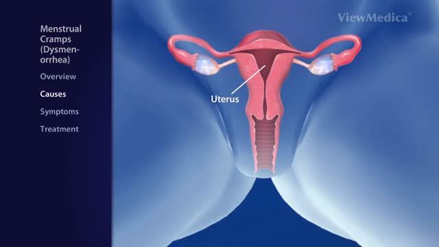
Menstrual cramps (dysmenorrhea) are throbbing or cramping pains in the lower abdomen. ... Menstrual cramps may be caused by identifiable problems, such as endometriosis or uterine fibroids. Treating any underlying cause is key to reducing the pain
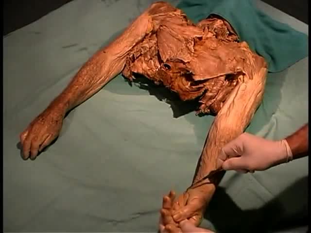
Anatomy of The Superficial Dissection of The Upper and Lower Limbs
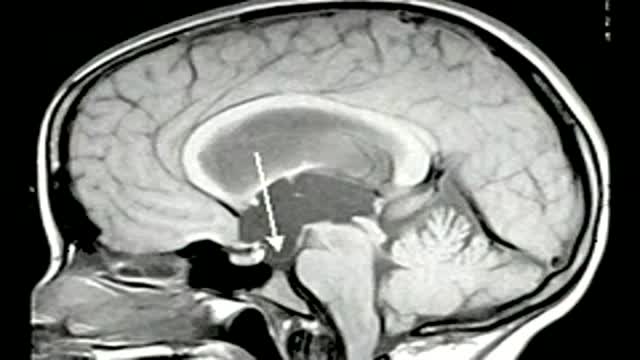
Endoscopic third ventriculostomy in a patient with obstructive hydrocephalus
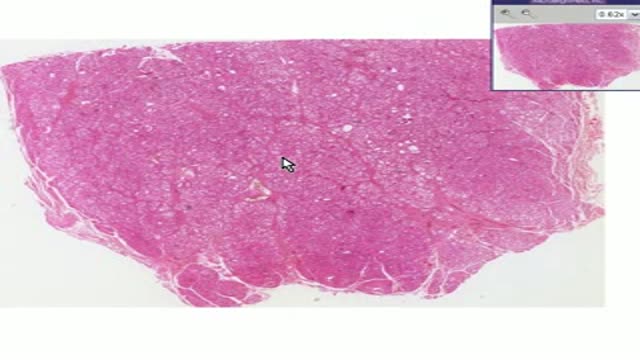
Histopathology of Graves Disease
Laparoscopic Assisted Right Hemicolectomy
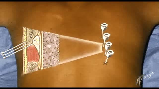
Intercostal Nerve Block
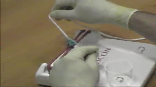
Surgical Knot
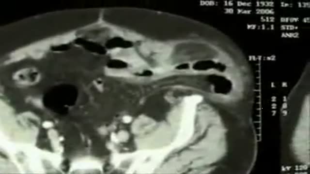
Giant spigelian stranguled hernia with small bowel loop and omental flap inside. The omentum required resection, the bowel appears vital. After the handle of hernia sac and his content has been done, a overlapped prolene repair will be done.

Watch that video of The Most Amazing Plastic Surgeries

Medical Examination of the cranial nerves

Video shows improvement of gait after a total knee replacement in the same patient. The sideways lurch has been abolished. This was possible by bone grafting and an advanced revision knee system.
Surgery performed at the MJRC, http://www.kneeindia.com/blog
http://www.kneeindia.com
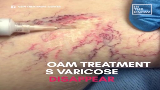
Sclerotherapy for varicose veins

This involves inserting a tube through the nasal passage, into the stomach

Shoulder Injection

Bronchiectasis is an abnormal dilation of the proximal and medium-sized bronchi (>2 mm in diameter) caused by weakening or destruction of the muscular and elastic components of the bronchial walls. Affected areas may show a variety of changes, including transmural inflammation, edema, scarring, and ulceration, among other findings. Distal lung parenchyma may also be damaged secondary to persistent microbial infection and frequent postobstructive pneumonia. Bronchiectasis can be congenital but is most often acquired.[9] Congenital bronchiectasis usually affects infants and children. These cases result from developmental arrest of the bronchial tree. Acquired forms occur in adults and older children and require an infectious insult, impairment of drainage, airway obstruction, and/or a defect in host defense. The tissue is also damaged in part by the host response of neutrophilic proteases, inflammatory cytokines, nitric oxide, and oxygen radicals. This results in damage to the muscular and elastic components of the bronchial wall. Additionally, peribronchial alveolar tissue may be damaged, resulting in diffuse peribronchial fibrosis.[12] The result is abnormal bronchial dilatation with bronchial wall destruction and transmural inflammation. The most important functional finding of altered airway anatomy is severely impaired clearance of secretions from the bronchial tree. Impaired clearance of secretions causes colonization and infection with pathogenic organisms, contributing to the purulent expectoration commonly observed in patients with bronchiectasis. The result is further bronchial damage and a vicious cycle of bronchial damage, bronchial dilation, impaired clearance of secretions, recurrent infection, and more bronchial damage
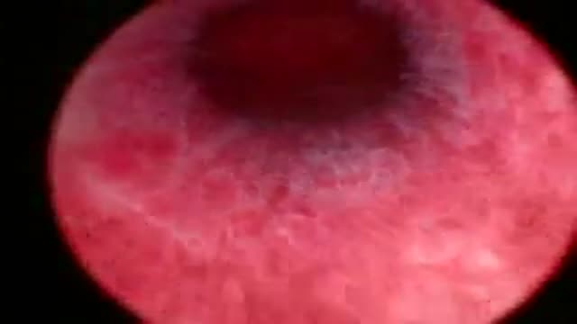
A hystroscopy showing a case of 2 intramural fibroids
