- Physical Examination
- Surgical Examination
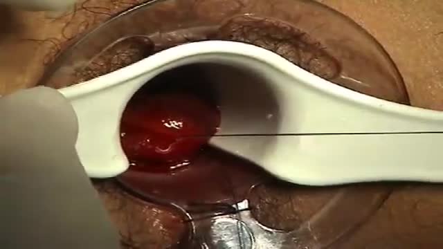
- Ophthalmology
- Clinical Skills
- Orthopedics
- Surgery Videos
- Laparoscopy
- Pediatrics
- Funny Videos
- Cardiothoracic Surgery
- Nursing Videos
- Plastic Surgery
- Otorhinolaryngology
- Histology and Histopathology
- Neurosurgery
- Dermatology
- Pediatric Surgery
- Urology
- Dentistry
- Oncology and Cancers
- Anatomy Videos
- Health and Fitness
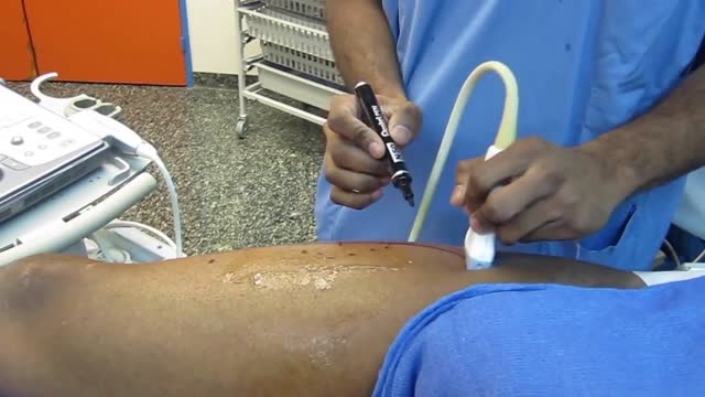
- Radiology
- Anaesthesia
- Physical Therapy
- Pharmacology
- Interventional Radiology
- Cardiology
- Endocrinology
- Gynecology
- Emergency Medicine
- Psychiatry and Psychology
- Childbirth Videos
- General Medical Videos
- Nephrology
- Physiology
- Diet and Food Health
- Diabetes Mellitus
- Neurology
- Women Health
- Osteoporosis
- Gastroenterology
- Pulmonology
- Hematology
- Rheumatology
- Toxicology
- Nuclear Medicine
- Infectious Diseases
- Vascular Disease
- Reproductive Health
- Burns and Wound Healing
- Other
Top videos

How to perform a Thyroid Gland Examination - Clinical Skills Revision
The thyroid examination is one of the first sessions of the clinical skills block for medical students at Warwick Medical School - largely as it touches lightly on to other clinical areas, such as the cardiac examination, and the peripheral neurological examination making it an excellent starting point for building further knowledge
This is a clinical examination of the thyroid gland is performed by Dr James Gill following the approach in Macleod’s Clinical examination.
------------------
Please note that there is no ABSOLUTE way to perform a clinical examination. Different institutions and even clinicians will have differing degrees of variations - the aim is the effectively identify medically relevant signs.
However, during OSCE assessments. Different medical schools, nursing colleges and other health professional courses will have their own preferred approach to a clinical evaluation - you should concentrate on THEIR marks schemes for your assessments.
The examination demonstrated here is derived from Macleods Clinical Examination - a recognised standard textbook for clinical skills.
Some people may experience an ASMR effect from watching this medical clinical examination
#ThyroidExamination #ClinicalSkills #DrGill #ASMR

plastic surgery cosmetic injections facial 3d medical animation company studio 3d visualization heal
High volume sinus irrigation!
Stories of breast implants exploding onboard airplanes are untrue - Silicone implants today are remarkably safe, and even when ruptured, they have a remarkable ability to retain its shape.
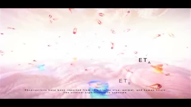
Pulmonary hypertension is a type of high blood pressure that affects the arteries in your lungs and the right side of your heart. In one form of pulmonary hypertension, tiny arteries in your lungs, called pulmonary arterioles, and capillaries become narrowed, blocked or destroyed. This makes it harder for blood to flow through your lungs, and raises pressure within your lungs' arteries. As the pressure builds, your heart's lower right chamber (right ventricle) must work harder to pump blood through your lungs, eventually causing your heart muscle to weaken and fail. Some forms of pulmonary hypertension are serious conditions that become progressively worse and are sometimes fatal. Although some forms of pulmonary hypertension aren't curable, treatment can help lessen symptoms and improve your quality of life. Pulmonary hypertension care at Mayo Clinic

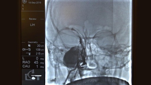
"Axillary Artery to Vein AV Graft for Dialysis Access"
Houston Methodist DeBakey Heart & Vascular Center, presents a cardiovascular procedure featuring Maham Rahimi, MD, M. Mujeeb Zubair, MD, and Louis Gomez, MD, as they demonstrate “Axillary Artery to Vein AV Graft for Dialysis Access".
Surgery: Maham Rahimi, MD, M. Mujeeb Zubair, MD, and Louis Gomez, MD
Narration: M. Mujeeb Zubair, MD
__________________________
FOR MORE INFORMATION
DeBakey CV Education: https://www.houstonmethodist.o....rg/education/medical
Follow Us:
Facebook: https://www.facebook.com/debakeycvedu
Twitter: https://twitter.com/DeBakeyCVedu
Livestream: https://livestream.com/debakey
Want concise, relevant reviews of the hottest topics in CV medicine? Subscribe for FREE to the Methodist DeBakey Cardiovascular Journal for quarterly, peer-reviewed issues delivered to your door.
https://journal.houstonmethodist.org/
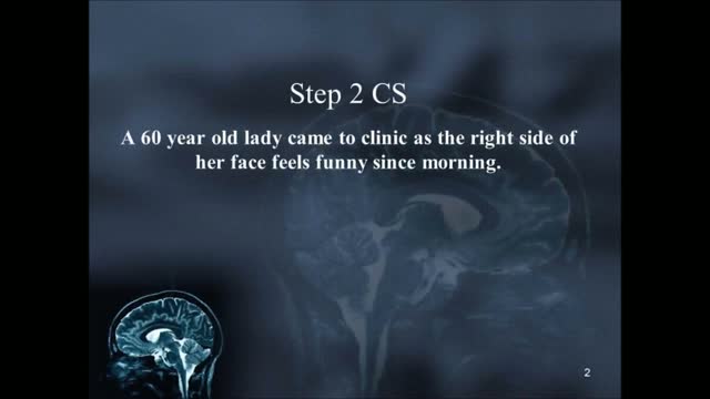
USMLE Step 2 CS - Numbness Weakness Full Video
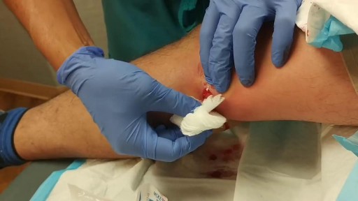
Popping and draining a leg abscess
Paracentesis is a procedure to take out fluid that has collected in the belly (peritoneal fluid). This fluid buildup is called ascites . Ascites may be caused by infection, inflammation, an injury, or other conditions, such as cirrhosis or cancer. The fluid is taken out using a long, thin needle put through the belly.
An unnamed Russian scientist has introduced the concept of a device that attaches to the wall of the artery. It would first stop blood flow to the area to prevent breakaway plaque. A drill would then scrape the plaque from the artery wall. The procedure of treating plaque buildup could include bypass surgery, stent replacement and balloon angioplasty. Since the plaques are of different types and locations in the body, the inventor proposed using different types of cutting mills.
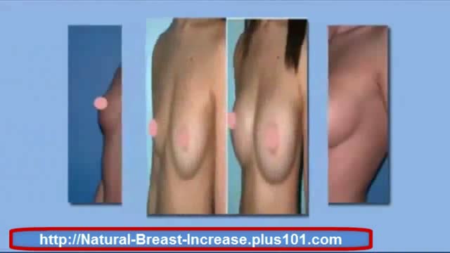
http://natural-breast-increase.plus101.com
---How To Make Bigger Breast. Can You Really Increase You Breast Size Naturally Without Surgery, Pills, Creams Etc Etc
Well a new website call natural-breast-increase.plus101.com says you can and thousands of women have already used there strategies.
And the best part about the this method is that you don't have to go through any expensive and painful surgery.
If you'd like to increase your own cup size then just visit the site below. I recommend the methods 100%
http://natural-breast-increase.plus101.com
How To Make Bigger Breast
http://www.youtube.com/watch?v=0cTQ3SZ4JIk
How To Make Bigger Breast,
can fenugreek increase breast size,
can i increase my breast size,
can i increase my breast size without surgery,
can i naturally increase breast size,
can we increase breast size naturally,
can women increase breast size naturally,
can you increase breast size,
can you increase breast size naturally,
can you make your breasts bigger,
cheap breast augmentation,
cheap breast implants,
cost of breast implants,
cream for breast enlargement,
cream to increase breast size,
curves breast enhancers,

Watch that video to know How to Increase Your Chances of Getting Pregnant?
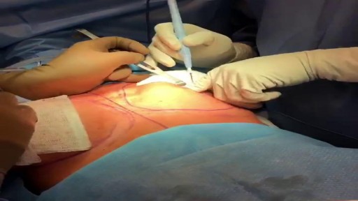
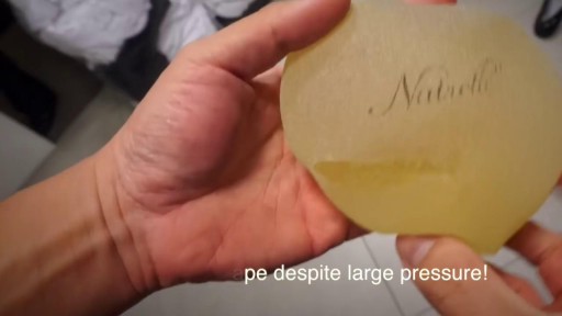
This anatomical implant was originally placed in 1997. Due to the dark yellow color inside the implant it is clear the implant has been ruptured for quite some time. When implants rupture, it is important to have them replaced as soon as possible to avoid excessive scarring in the breasts. If too much scar tissue has accumulated around the deflated implant, it becomes difficult to create a normal breast shape in the future. Therefor its important to know the signs of a ruptured implant such as, painful to touch, visible asymmetry or loss of integrity to the bag. Dr. Stuart Linder 9675 Brighton Way Suite 420 Beverly Hills, CA 90210
Psychotic Depression Information

Top 10 Shocking Before And After Drug Use Photos
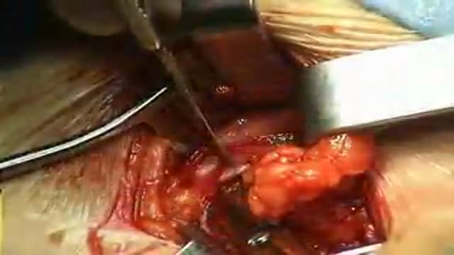
Femoral Hernia Repair with Prosthetic PHS repair placed on anterior way
minimally invasive procedure is the new gold standard for hemorrhoidectomy, according to American and European experts in the field. The procedure, known as PPH (procedure for prolapse and hemorrhoids) stapled hemorrhoidectomy, combines hemorrhoidal devascularization and repositioning to return the veins to the anal canal. “This year, this is the revolutionary new procedure in the United States,” Gary Hoffman, MD, clinical faculty member in general and colorectal surgery, Cedars-Sinai Medical Center, Los Angeles, told General Surgery News after moderating a live PPH telesurgery at the 2003 annual meeting of the Society of American Gastrointestinal Endoscopic Surgeons.
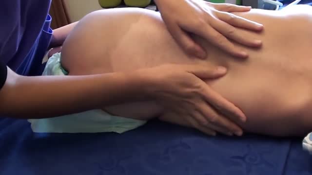
Pediatric Massage
it's the video of the OR during a dynamic reconstruction of the achilles tendon by a composite anterolateral perforator flap
