
- Physical Examination
- Surgical Examination
- Ophthalmology
- Clinical Skills
- Orthopedics
- Surgery Videos
- Laparoscopy
- Pediatrics
- Funny Videos
- Cardiothoracic Surgery
- Nursing Videos
- Plastic Surgery
- Otorhinolaryngology
- Histology and Histopathology
- Neurosurgery
- Dermatology
- Pediatric Surgery
- Urology
- Dentistry
- Oncology and Cancers
- Anatomy Videos
- Health and Fitness
- Radiology
- Anaesthesia
- Physical Therapy
- Pharmacology
- Interventional Radiology
- Cardiology
- Endocrinology
- Gynecology
- Emergency Medicine
- Psychiatry and Psychology
- Childbirth Videos
- General Medical Videos
- Nephrology
- Physiology
- Diet and Food Health
- Diabetes Mellitus
- Neurology
- Women Health
- Osteoporosis
- Gastroenterology
- Pulmonology
- Hematology
- Rheumatology
- Toxicology
- Nuclear Medicine
- Infectious Diseases
- Vascular Disease
- Reproductive Health
- Burns and Wound Healing
- Other
Top videos


All Solution of Male Disorder Male Infertility Diagnostic and Treatment Re-Slim Care Latest Technology in Pakistan Dr. Aslam Naveed is a well known sexologist in Pakistan. He has treated more than 1 Lac patients since last 30 years of clinical Practice in sexology, he knows how to help the people facing sexual disorders. Contact: 02134965050, 03432821919, 0345-8314663 http://www.sexologistpakistan.com/ https://www.facebook.com/menssexcareclinic/ https://www.youtube.com/channel/UCagSSgdEgQJWl_xfFM12BwA https://twitter.com/bettersexcare https://www.instagram.com/dr.aslamnaveed/ ADDRESS: Men’s Care Modern Hospital, Opposite, Safari Park, University Road, Karachi, Pakistan.

A recap of Mater Hospital patient Helen's story as she progressed from experiencing chronic knee pain due to osteoarthritis, through to knee replacement treatment and ultimately a new lease on life.
Dedicated to surgical excellence and patient-centred care, the Mater Hospital North Sydney is regarded as a leading orthopaedic hospital and the only Australian hospital to be accepted into the International Society of Orthopaedic Centres.
For more information, click here: https://bit.ly/3bvhY8G

STOP SMOKING
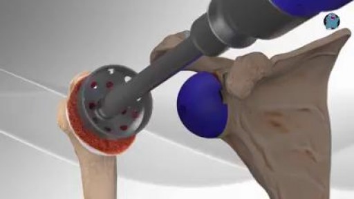
Every year, thousands of conventional total shoulder replacements are successfully done in the United States for patients with shoulder arthritis. This type of surgery, however, is not as beneficial for patients with large rotator cuff tears who have developed a complex type of shoulder arthritis called "cuff tear arthropathy." For these patients, conventional total shoulder replacement may result in pain and limited motion, and reverse total shoulder replacement is a better option.
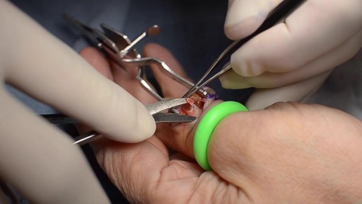
Giant cell tumors of the tendon sheath are common lesions and are the second most frequent tumors in the hand, after synovial cysts. They are diagnosed by means of clinical examination and complementary examinations (simple radiography and magnetic resonance). Erosion and invasion of the phalangeal bone affected may be seen on radiological examination. Magnetic resonance may show a “fluorescent or radiant effect” may be observed, caused by the high quantity of hemosiderin inside the tumor. Surgical treatment is the commonest practice, and complete excision is important for avoiding recurrence of the tumor, especially when bone invasion is observed on imaging examinations, which is generally related to greater tumor recurrence. In this paper, a case of a giant cell tumor of the tendon sheath in the middle phalanx of the third finger of a 45-year-old female patient is presented. This was successfully treated by means of surgery using a double access approach (dorsal and volar)
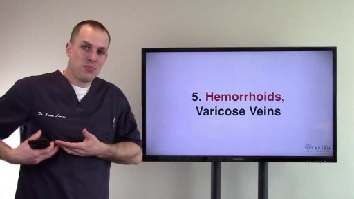
symptoms of liver dysfunction. Remember, the body doesn't work in isolation. Where there is dysfunction in one area of the body, be rest assured that dysfunction is happening throughout the body.

Erectile dysfunction (ED) is the inability to get or keep an erection firm enough for sexual function. It’s a common sexual problem, affecting as many as 30 million men in the United States. Most cases of ED have a physical cause, such as heart disease, diabetes, and obesity. Lifestyle choices like smoking and drinking excessive amounts of alcohol can also lead to ED. But for some men, psychological issues are the root of the problem.
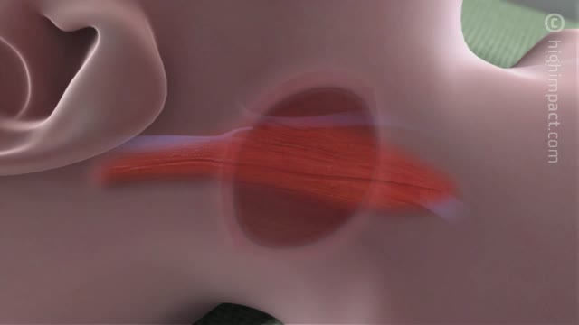
Central catheters provide dependable intravenous access and enable hemodynamic monitoring and blood sampling [1-3]. The jugular veins are one of the most popular sites for central venous access due to accessibility and overall low complication rates, and are the preferred site for temporary hemodialysis.

A Lecture Presented by Dr. Mostafa Yakoot to Vascular Surgery Congress. TITLE: SAFETY & EFFICACY OF A NEW HONEY OINTMENT (PEDYPHAR) FOR DIABETIC FOOT ULCERS. Based on the original article in JWC by: Yakoot M, Abdelatif M, Etman M.
Total thyroidectomy is the treatment of choice for all types of thyroid cancer(papillary, follicular, medular and anaplastic).
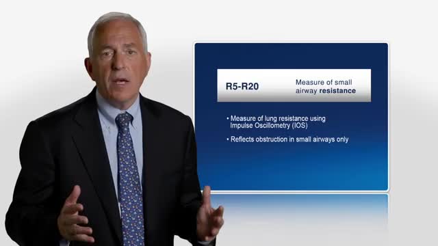
Asthma was originally described as an inflammatory disease that predominantly involves the central airways. Pathological and physiological evidence reported during the past few years suggests that the inflammatory process extends beyond the central airways to the peripheral airways and the lung parenchyma. The small airways are capable of producing T-helper-2 cytokines, as well as chemokines, and they have recently been recognized as a predominant site of airflow obstruction in asthmatic persons. The inflammation at this distal site has been described as more severe than large airway inflammation. These findings are of great clinical significance, and highlight the need to consider the peripheral airways as a target in any therapeutic strategy for treatment of asthma.
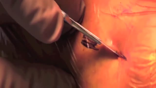
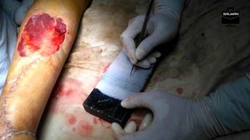
Removal of a Broken Intramedullary Nail and Exchange Nailing for Tibial Nonunion
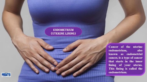
Obesity is the abnormal condition that causes a person to put on excessive amounts of weight due to accumulation of fat in their body. This extreme weight causes a variety of other disorders and diseases as complications associated with it. https://goo.gl/GgSAoY

Acromegaly is usually caused by a noncancerous tumor. Middle-aged adults are most commonly affected. Symptoms include enlargement of the face, hands, and feet. Prompt treatment is needed to avoid serious illness. Drugs can reduce the effects of growth hormone. If needed, surgery and radiation may be used to remove tumor cells.
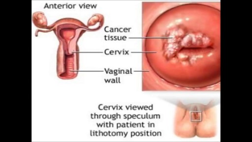
Menorrhagia is the medical term for menstrual periods with abnormally heavy or prolonged bleeding. Although heavy menstrual bleeding is a common concern, most women don't experience blood loss severe enough to be defined as menorrhagia. With menorrhagia, you can't maintain your usual activities when you have your period because you have so much blood loss and cramping. If you dread your period because you have such heavy menstrual bleeding, talk with your doctor. There are many effective treatments for menorrhagia.

Our surgeons take a compassionate, family-centered approach to both inpatient and outpatient care. We’re committed to making sure both you and your child understand our process. Told through a kid's eyes, this video tour reveals our caring approach.
To learn more about pediatric surgery at Stamford Hospital, visit: https://www.stamfordhealth.org..../care-treatment/pedi

