




Caesarean Section Childbirth Delivery Video

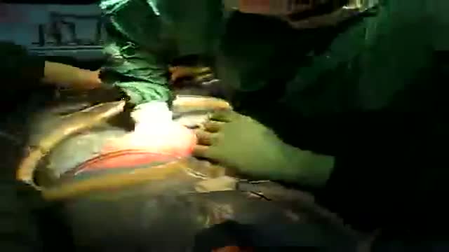
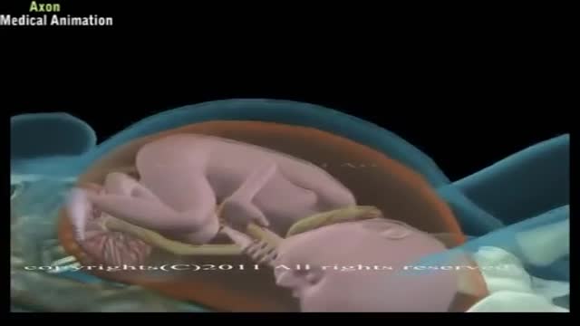
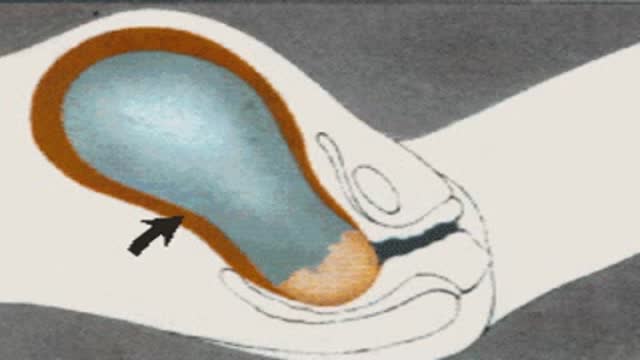
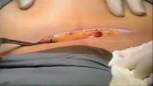
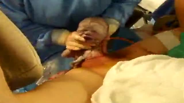
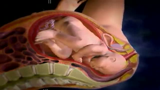
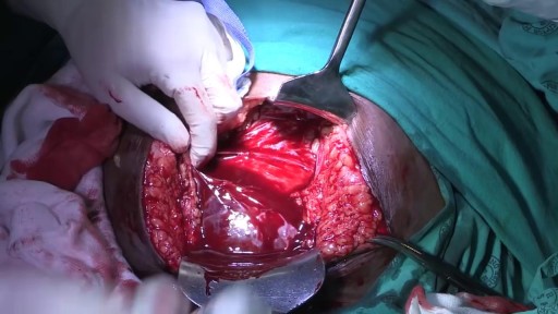
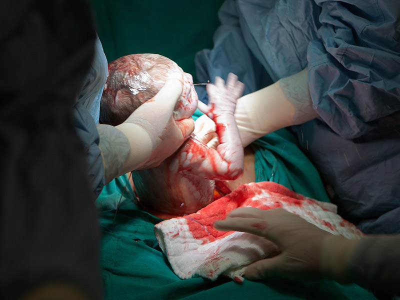
Not every woman undergoes a traditional vaginal delivery with the birth of her child. Under conditions of fetal or maternal distress, or in the case of breech presentation (when a baby is turned feet first at the time of delivery), or if the woman’s first baby was born by cesarean delivery, a procedure called a cesarean section may be required. During a cesarean, a doctor will make either a lateral incision in the skin just above the pubic hair line, or a vertical incision below the navel. As the incision is made, blood vessels are cauterized to slow bleeding. After cutting through the skin, fat, and muscle of the abdomen, the membrane that covers the internal organs is opened, exposing the bladder and uterus. At this time the physician will generally insert his or her hands into the pelvis in order to determine the position of the baby and the placenta. Next, an incision is made into the uterus and any remaining fluids are suctioned from the uterus. The doctor then enlarges the incision with his or her fingers. The baby’s head is then grasped and gently pulled with the rest of its body from the mother’s uterus. Finally, the abdominal layers are sewn together in the reverse order that they were cut. The mother is allowed to recover for approximately three to five days in the hospital. She will also be quite sore and restricted from activity for the following several weeks. There are several potential complications associated with this procedure that should be discussed with a doctor prior to surgery.
Video Transcript
A training film on cesarean section built explicitly around evidence from systematic reviews. It covers anesthesia choices, the Joel Cohen incision, blunt extension of the uterine incision, and trial findings on peritoneal closure and post-operative feeding, arguing that technique based on the best available evidence gives the lowest complication rates for this common operation.
Read the full transcript
Caesarean section is one of the most commonly performed major operations in the world. Often it's conducted as an emergency procedure by relatively junior doctors in peripheral hospitals. Like any other major surgery, it involves risks, including the risk of death for both the mother and her baby. It is especially important, therefore, that the technique used for this operation is based on the best available evidence of effectiveness and safety. The most reliable evidence of effectiveness of health care interventions comes from systematic reviews of randomized trials. Reviews of specific aspects of the Caesarean section technique are currently underway.
The technique presented in this video is based on the best currently available evidence for this operation. Begin by ensuring that pre-operative preparations are complete. Note, however, that this video does not cover these preparations. Before starting the operation, check that the baby's heartbeat is present and that the indication for Caesarean section is still valid. Check also the position of the baby and whether the woman has requested tubal ligation. Since general anesthesia carries particular risks in pregnant women, such as difficult endotracheal to intubation and aspiration of stomach contents, spinal anesthesia is recommended.
The main risk with spinal anesthesia is hypotension, which can be reduced by pre-loading the woman with prophylactic intravenous fluid, leg elevation and compression, and left lateral tilting. Only one randomized control trial is available on the relative advantages of vertical versus transverse abdominal incision for Caesarean section. Unfortunately, this trial was too small to yield conclusive results. Many physicians prefer the transverse incision developed by Joel Cohen for routine Caesarean section, owing to its effectiveness and good cosmetic results. In certain circumstances, when complications are anticipated or when the surgeon is less experienced, the vertical incision may be preferred as it has the advantage of speed, simplicity, and ease of upward extension to improve exposure of the uterus.
The vertical incision can also be performed using local analgesia, when general or regional analgesia are unavailable or deemed to be unsafe. Operations involving complications, such as placenta previa, transverse lye, and obstructed labor, require careful planning of the operation procedure in order to be prepared for potential problems. Check first that the spinal analgesia has taken effect. Then make a Joel Cohen's transverse abdominal incision about two centimeters below the line of the anterior superior iliac spines. Insize the subcutaneous tissues and the anterior rectus sheath in the midline, using only the scalpel.
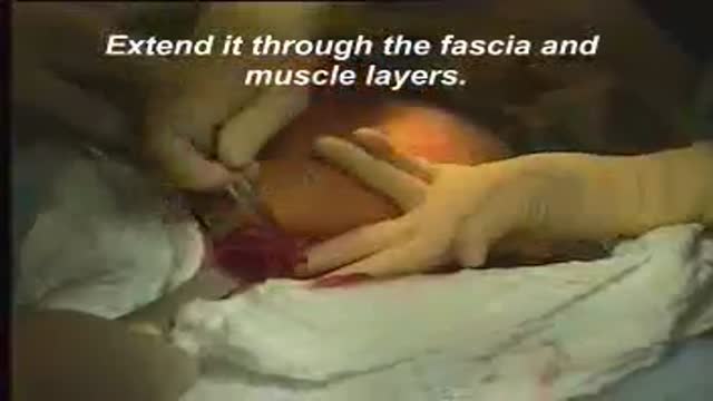
Extend the anterior rectus sheath incisions laterally beneath the subcutaneous tissues, with slightly opened scissor tips, or by stretching the tissue with your fingers. Separate the rectus muscles and open the periatal peritoneum, also by stretching it with your fingers. Stretch the abdominal opening laterally to separate the rectus muscles, and then stretch the subcutaneous tissues and the peritoneum. Randomized trials have found that, compared with Pannon-Steel's transverse incision, the Joel Cohen incision is quicker, carries less risk of morbidity, and leads to fewer subsequent adhesions.
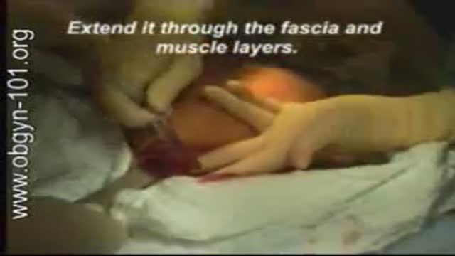
Open the loose visceral peritoneum over the lower uterine segment. In one randomized trial, not separating the peritoneum from the lower uterine segment had short-term benefits, but the numbers studied were too small to exclude the possibility of rare complications, such as bladder injury. Once the uterus is exposed, make a small transverse incision with a scalpel. Then, using both hands, stretch the opening laterally, incise the amniotic sac, and aspirate the amniotic fluid. In one large randomized trial, blunt lateral extension of the uterine incision resulted in less further wound extension and less bleeding.
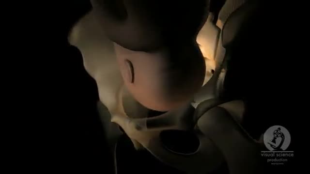
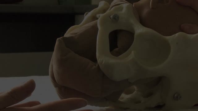
When the baby's head is deeply engaged in the pelvis, as seen in this case of obstructed labour, the head must be elevated carefully above the level of the uterine incision, and rotated to the occipito anterior position before it can be delivered through the incision. Deliver the placenta by applying steady, gentle traction on the cord. Randomized trials, comparing delivery of the placenta by cord traction and manual removal by inserting the hand inside the uterus to dislodge the placenta, show that the traction method results in less blood loss and less post-operative endometritis.
Removing the uterus from the abdominal cavity has no clear benefits or disadvantages over intra-abdominal repair. If the uterus is exteriorized to improve access, this should be done gently and carefully, as isolated cases of torn ovarian vessels have been reported.
Randomized trials have shown no clinically important differences between a single layer of locking sutures and the addition of a second layer. However, in a recent large non-randomized comparison, subsequent uterine rupture was less common when a second layer was used. Thus, at the present time there is inadequate evidence to recommend either approach with certainty. Non-closure of both the visceral and the perietal peritoneum have been shown in randomized trials to reduce operating time, post-operative fever, wound infection, analgesia use, and length of hospital stay. There is no evidence to justify closing either of the peritoneal layers.
The anterior rectus sheath is usually closed with a continuous absorbable suture. No clear benefits have been shown for routinely draining or suturing the subcutaneous tissues. But possible benefits have been reported when the subcutaneous layer is thicker than 2 cm. In one small randomized trial involving vertical skin incisions, an absorbable subcuticular skin suture gave better results than metal staples. A continuous subcutaneous suture is often used in transverse incisions. The misgav-ladakh technique developed by Stark and colleagues includes the use of the Joel Cohen abdominal incision, single layer closure of the uterus, non-closure of the peritoneum, and two or three mattress sutures to approximate the skin.
Tissue forceps are applied for about five minutes to the spaces between sutures and removed just before application of the wound dressing. Randomized trials comparing several of these modifications, for example in the misgav-ladakh technique, with traditional techniques have shown that the modified techniques reduce operating time and improve post-operative recovery. A benefit of particular relevance to resource-poor settings is that only two lengths of suture material are necessary. One such as chromic cat-gut on a round-bodied needle for the uterus, and the other such as polyglycolic acid on a cutting needle for the rectus sheath and skin.
Randomized trials have shown that it is not necessary to withhold oral fluids or food after a non-complicated caesarean section operation. When both the decision to perform the operation and the surgical technique used are based on the best available evidence of effectiveness and safety, it is possible to ensure the lowest rates of complications and mortality from this common but essential operation.
Transcript auto-generated from the video’s audio for accessibility and study. Educational content only — not medical advice.
Video Overview
From our Reproductive Health collection: Fertility, contraception, IVF and sexual health explained clearly by specialists. Browse all Reproductive Health videos →
SORT BY-
نظرات برتر
-
آخرین نظرات