



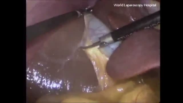
Transgastric Cholecystectomy

Surgery video of transgastric cholecystectomy
This video in 30 seconds
A research team narrates a transgastric cholecystectomy, an early natural-orifice operation removing the gallbladder through the stomach wall. The narration describes creating and closing the gastrotomy with a full-thickness suturing device, dissecting the gallbladder free endoscopically, removing it through the mouth, and reports the team's first five human cases under an ethics-approved protocol with a laparoscopic safety port.
Key points
- The endoscope first inspects the stomach and a site on the anterior stomach wall is selected for exit
- Full-thickness sutures are pre-placed to form a ridge that helps maintain insufflation and later closure
- A small gastrotomy is created and dilated to pass the operating endoscope into the peritoneal cavity
- The gallbladder is dissected free endoscopically, its duct and artery are ligated and divided, and it is drawn into the stomach
- The gastrotomy is closed with full-thickness sutures and the team reports its first five human cases under an approved protocol
Chapters
- 0:01 — Introduction to natural orifice surgery
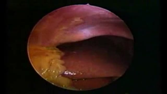
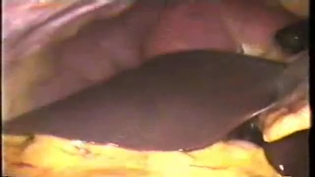
- 0:23 — Inspecting stomach and selecting gastrotomy site
- 1:34 — Creating and dilating the gastrotomy
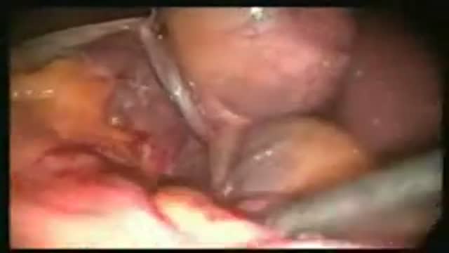
- 3:02 — Endoscopic dissection of the gallbladder
- 4:49 — Gallbladder drawn into stomach
- 5:19 — Closing the gastrotomy with sutures
- 7:11 — First human cases and results
Video Transcript
A research-team narration of a transgastric cholecystectomy — gallbladder removal through the stomach wall as an early natural-orifice (NOTES) operation. It describes creating and closing the gastrotomy with a full-thickness suturing device, dissecting the gallbladder free endoscopically, removing it through the mouth, and reports the team's first five human cases under an ethics-approved protocol with a laparoscopic safety port.
Read the full transcript
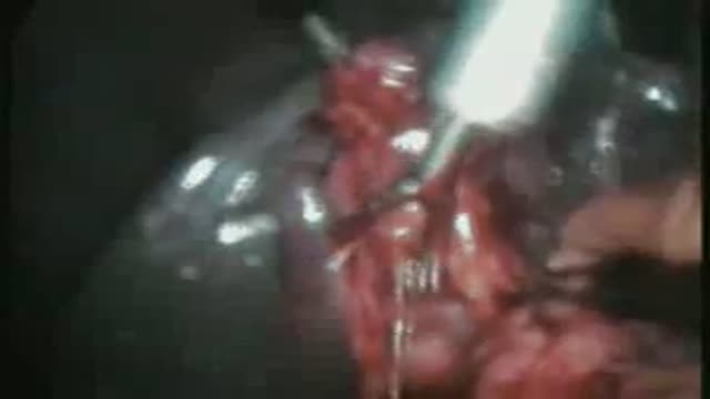
Natural orifice trans-illuminal endoscopic surgery has captured the interest of surgical endoscopists and may represent the next stage of evolution in minimally invasive surgery. We describe our technique of trans-gastric cholecystectomy in humans as developed during several years of laboratory work. Standard endoscope is first used to inspect the stomach and lavagia with an antibiotic solution. A critical step is the selection of the site for the gastrodomy. The ideal site to exit the stomach is the anterior mid-body which obviates the need for scope retreflection during the procedure. We pre-place full thickness sutures 2 cm apart to create an implicated ridge.
This will provide a valve to prevent loss of the pneumogastrium after removal of the endoscope. To create the ridge we use a novel full thickness suturing device which grabs the gastric wall and implicates it into the lumen. A needle is then fired transversely across the ridge and deposits a pledged permanent suture. Subsequently a second pledged is deposited on the other side and a cinching device used to tighten it down. A monopolar needle knife is used to create a 1 cm gastrodomy along the edge of the implicated ridge.
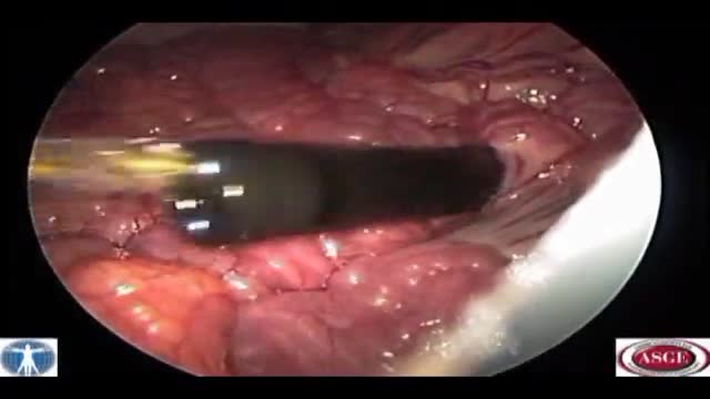
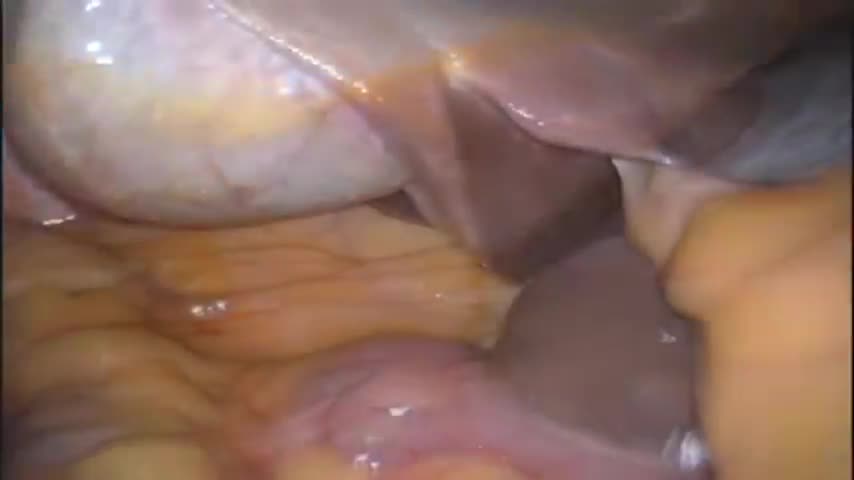
To minimize the resulting size of the gastrodomy a balloon dilator is then used to expand the hole to 2 cm. This easily allows passage of the 18 mm operating endoscope. Once the endoscope passes through the gastric wall into the peritoneal cavity, thorough exploration is performed. Our IRB protocol calls for laparoscopic observation for patient safety. A single 5 mm umbilical port is placed for monitoring. A laparoscopic monitor is positioned to not be visible to the endoscopic team. A percutaneous 2 mm suture passer or micro grasper is placed in the subziphoid position to retract the dome of the gallbladder.
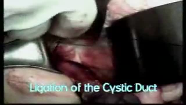
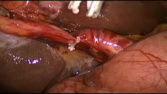
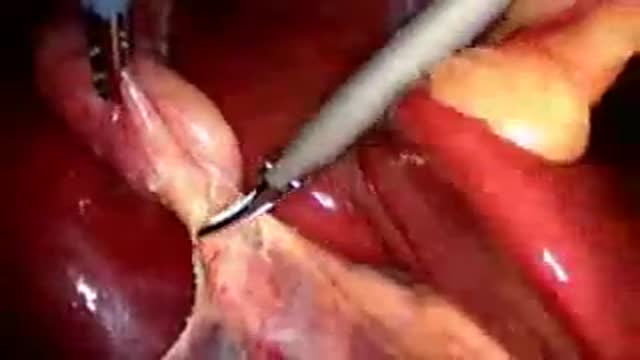
Endoscopic dissection is performed using a hook-cottery IT knife and follows the same precepts of safe dissection developed for laparoscopic surgery. Endoscopic dissection is performed using a dome down method completely freeing the gallbladder from the liver bed. Once it's completely freed, endoscopic endolubes are used to ligate both the cystic artery and cystic duct. After two endolubes are placed, endoscopic scissors are used to divide the cystic artery and cystic duct. The then completely mobilized gallbladder is with the torn into the gastric lumen. The previously created gastric fold allows the maintenance of pneumoparitoneum.
Once the specimen is inside the stomach, attention is turned to gastrodomy closure. We now see the value of the ridge or infolding created before gastrodomy. This allows CO2 pneumogastrian to be maintained and allows precise closure to be performed. The Geprox endoluminal suturing device is used to place full thickness sutures on both sides of the defect. The 5mm toothed grasper of the device is used to robustly grab the full thickness of the stomach wall. A needle is then fired perpendicularly across the tissue. Self-expanding tissue anchors are then deployed.
A one-way cinch is used to approximate the anchors and close the defect. As many sutures as are needed are deployed to tightly close the defect. Finally, the suture tails are trimmed. Finally, a toothed grasper is used to grasp the gallbladder and carefully withdraw it through the esophagus and out the mouth, completing the procedure. Our IRB approved protocol calls for Methylene Blue installation to check the integrity of our gastrodomy closure. To date, we have had no leaks in our human notes cholestestectomies. Our clinical experience began in May of 2007.
Under an IRB approved protocol, we performed transgastric cholestestectomy in five patients to date. There were four females and one male with an average operative time of 4.25 hours. As mentioned, our protocol calls for a minimum of one laparoscopic port for patient safety. In our experience, we've had to add two additional ports on several patients. Length of stay was 24 hours by protocol. Pain scores were recorded in the recovery room post-op day one and on post-op day two. In general, pain scores were slightly lower than seen in similar studies on laparoscopic cholestestectomy.
Transcript auto-generated from the video’s audio for accessibility and study. Educational content only — not medical advice.
Video Overview
From our Surgery Videos collection: Surgery videos: watch real operations step by step — general, laparoscopic and specialist surgery performed in the OR, explained for students and surgeons. Browse all Surgery Videos videos →
SORT BY-
শীর্ষ মন্তব্য
-
সর্বশেষ মন্তব্য