- Physical Examination
- Surgical Examination
- Ophthalmology
- Clinical Skills
- Orthopedics
- Surgery Videos
- Laparoscopy
- Pediatrics
- Funny Videos
- Cardiothoracic Surgery
- Nursing Videos
- Plastic Surgery
- Otorhinolaryngology
- Histology and Histopathology
- Neurosurgery
- Dermatology
- Pediatric Surgery
- Urology
- Dentistry
- Oncology and Cancers
- Anatomy Videos
- Health and Fitness
- Radiology
- Anaesthesia
- Physical Therapy
- Pharmacology
- Interventional Radiology
- Cardiology
- Endocrinology
- Gynecology
- Emergency Medicine
- Psychiatry and Psychology
- Childbirth Videos
- General Medical Videos
- Nephrology
- Physiology
- Diet and Food Health
- Diabetes Mellitus
- Neurology
- Women Health
- Osteoporosis
- Gastroenterology
- Pulmonology
- Hematology
- Rheumatology
- Toxicology
- Nuclear Medicine
- Infectious Diseases
- Vascular Disease
- Reproductive Health
- Burns and Wound Healing
- Other
Latest videos
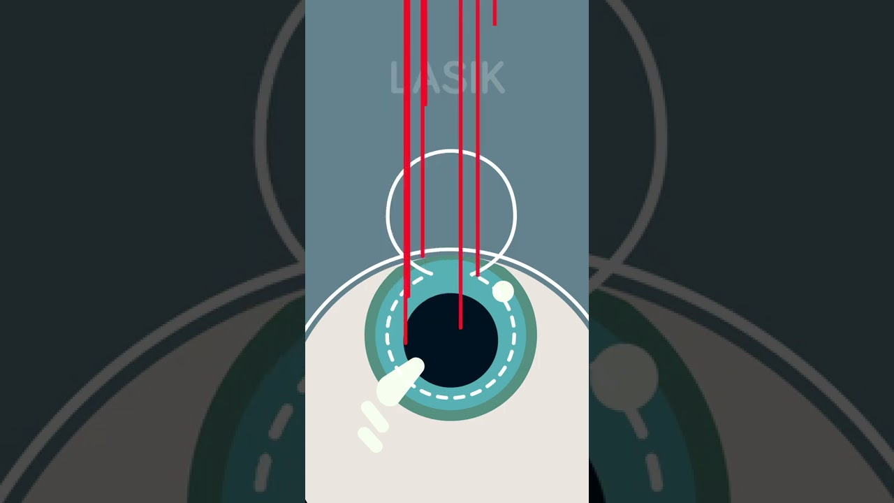
Ever considered getting laser eye surgery, but didn’t know how it worked? Allow us to help!
There are three different main types of laser eye surgery: LASIK, SMILE, and Surface Laser Treatments, and each can be explained pretty easily.
LASIK uses two lasers to open up a thin flap on the surface of the cornea, and then reshapes the cornea underneath. The flap is then placed back over the reshaped cornea, and heals independently with time.
SMILE uses one laser to reshape the cornea through a small, self-healing hole.
And Surface Eye Treatments remove the clear skin over the eye, to then reshape the cornea underneath with - you guessed it - a laser!

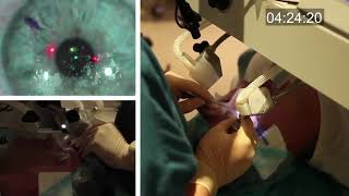
In this video, Professor Dan Reinstein performs a bilateral LASIK procedure filmed in real-time to demonstrate the full 8 and-a-half minute procedure from multiple angles. The superior design and experience of the Carl Zeiss Meditec Visumax femtosecond Laser for flap creation is seen, where the patient is only in contact with the device for about 30 seconds with extremely low contract force such that the patient feels effectively nothing, there are no red splodges (subconjunctival haemorages) left behind. From the surgeons' standpoint there is no device that is easier to use or faster for LASIK flap creation. The Carl Zeiss Meditec MEL80 excimer laser portion of the procedure is seamlessly integrated and incorporates all the features that make clinical outcomes so reproducible including the unique cone-for-controlled-atmosphere (CCA) and high efficiency, high sensitivity calibration test which can be performed for each individual patient to compensate for minor changes in energy that occur with excimer laser devices during the course of a day.
For reference to the clinical outcomes for LASIK with the MEL80 in presbyopia using PRESBYOND Laser Blended Vision see:
Reading glasses presbyopia (ageing eyes) only:
LASIK for presbyopia correction in emmetropic patients using aspheric ablation profiles and a micro-monovision protocol with the Carl Zeiss Meditec MEL 80 and VisuMax.
J Refract Surg. 2012 Aug;28(8):531-41. Reinstein DZ, Carp GI, Archer TJ, Gobbe M.
http://www.ncbi.nlm.nih.gov/pubmed/22869232
Short sighted, astigmatism and presbyopia (ageing eyes)
LASIK for Myopic Astigmatism and Presbyopia Using Non-Linear Aspheric Micro-Monovision with the Carl Zeiss Meditec MEL 80 Platform.
J Refract Surg. 2011 Jan;27(1):23-37. Epub 2010 Mar 1.
Reinstein DZ, Archer TJ, Gobbe M.
http://www.ncbi.nlm.nih.gov/pubmed/20205360
Long-sighted, astigmatism and presbyopia (ageing eyes)
LASIK for hyperopic astigmatism and presbyopia using micro-monovision with the Carl Zeiss Meditec MEL80 platform.
J Refract Surg. 2009 Jan;25(1):37-58. Reinstein DZ, Couch DG, Archer TJ.
http://www.ncbi.nlm.nih.gov/pubmed/19244952
For more information about laser eye surgery and PRESBYOND Laser Blended Vision, please contact the London Vision Clinic on 020 7224 1005.

Curious about LASIK eye surgery? NVISION's Dr. Richard Mauer talks risks, life-changing benefits, and outcomes (plus why he loves what he does!).
Want to start your journey to better vision? Schedule your complimentary consult today! https://bit.ly/3H2i0FU
NVISION: The Eye Doctors' #1 Choice in LASIK and Laser Cataract Surgery

Mitra Nejad, MD
Associate Physician Diplomate
UCLA Stein Eye Institute, Cataract and Refractive Surgery Division

LASIK is one of the most popular elective surgeries in the United States with 95% of patients walking away satisfied with their vision, according to one FDA study. But like with any surgery, there are risks.

See what it’s like to get LASIK eye surgery from Lisa Homsy’s perspective. Keep watching until the end to see the final results!

If you go to research LASIK eye surgery online, you may get conflicting messages. Some articles rave about it, but in some cases, others link it to severe pain or even suicide. 7 Action News' Carolyn Clifford sat down with one of the area's biggest providers of eye surgery to try and separate fact from fiction, so if you go under the laser, you know the risk.
An FDA survey has found some patients of Lasik eye surgery say the procedure ruined their sight.
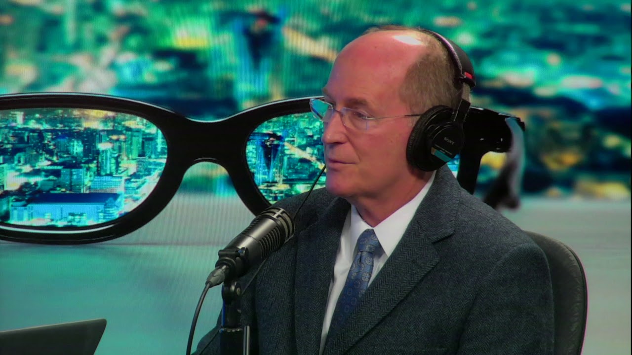
Dr. Leo Maguire, a Mayo Clinic ophthalmologist, explains how laser-assisted in situ keratomileusis (LASIK) eye surgery can correct common vision problems.
This interview originally aired Jan. 26, 2019.
To learn more about LASIK surgery, visit: https://www.mayoclinic.org/tests-procedures/lasik-eye-surgery/about/pac-20384774?mc_id=us&utm_source=newsnetwork&utm_medium=l&utm_content=content&utm_campaign=mayoclinic&geo=national&placementsite=enterprise&cauid=100721&_ga=2.112234244.1227307149.1547427243-1780934405.1469629163

An estimated 20 million LASIK procedures have been performed since 1998. The FDA website is filled with stories of complications, including pain, dizziness and detached retinas. CBS2's Chris Wragge reports.

Originally broadcast November 21, 2014.
They advertise low, low prices. But does anyone actually pay that rate? Erica Johnson investigates.
More from CBC Marketplace, Canada's top consumer affairs show:
Watch episodes online at http://cbc.ca/marketplace
Like us on Facebook: http://facebook.com/cbcmarketplace
Talk to us on Twitter: http://twitter.com/cbcmarketplace
Follow our hosts @cbctom and @cbcerica

Recovery Tips
LASIK eye surgery is the best known and most commonly performed laser refractive surgery to correct vision problems. The total recovery time is 1 to 2 weeks. However, vision may fluctuate slightly over the next 2 months.
Avoid watching television or reading for the first few days.
You can get back to work after 1 week.
Avoid applying pressure on the eyes for 7 days.
Avoid dust, smoke, yard and garden work, and eye make-up.
Wear eye shields at night given by a surgeon for 1 week.
Driving is allowed after 4 to 5 days.
Avoid swimming or using a hot tub for 2 weeks after surgery.
While using the computer, take frequent breaks and lubricate your eyes with artificial tears.
For treatment assistance in your country or abroad:
Email: hello@vaidam.com
Phone/WhatsApp/Viber: +91-9650001746
Website: www.vaidam.com
Vaidam is an ISO and NABH accredited medical assistance company. Patients from 100+ countries have used our services.
Useful Links:
India
Doctors: https://www.vaidam.com/doctors/opthalmology/lasik-procedure/india
Hospitals: https://www.vaidam.com/hospitals/opthalmology/lasik-procedure/india
Cost of Lasik Eye Surgery: https://www.vaidam.com/cost/lasik-procedure-cost-in-india
Turkey
Doctors: https://www.vaidam.com/doctors/opthalmology/lasik-procedure/turkey
Hospitals: https://www.vaidam.com/hospitals/opthalmology/lasik-procedure/turkey

Thinking about laser eye surgery? Watch our live stream and learn what the procedure involves.
Find out more about laser eye surgery
on our website: www.opticalexpress.co.uk
in our online magazine: www.opticalexpress.co.uk/magazine
on our Facebook page: https://www.facebook.com/opticalexpress/
or on our Instagram page: https://www.instagram.com/opticalexpressuk

His father, Dr. Joseph Dello Russo, helped turn Lasik eye surgery into the widespread procedure it is today. Now he explains a new technique and how it differs.

Dr. Ankur Gupta of the Virginia Eye Institute discusses LASIK eye surgery as a method of correcting refractive errors. LASIK was first performed in Virginia on an FDA-approved laser by a VEI surgeon in 1996. Today, Virginia Eye Institute offers both conventional LASIK and custom LASIK with the bladeless IntraLase laser to precisely sculpt your cornea to correct refractive errors.
For more information on the services and procedures offered at Virginia Eye Institute please visit: https://goo.gl/6nX4RZ
THE CONTENT IN THIS VIDEO IS GENERAL IN NATURE AND DOES NOT SUBSTITUTE PROFESSIONAL MEDICAL ADVICE. The content on our website including, but not limited to, text, images, and videos is for informational and educational purposes only. Although we work hard to provide accurate general information, it is not a substitute for professional medical advice or consultations with healthcare professionals, and does not establish any kind of provider-patient relationship. Our website information is not intended to make any promises about the results of our products and services. We are not liable for actions taken based on content found on our website. If you are seeking medical advice, diagnoses, or treatment, we encourage you to call 804-287-2020 to make an appointment with one of our providers for your individualized care plan.

I filmed my lasik eye surgery because it looks neat
See the full video here: https://youtu.be/wY_D5pMbEf0
Subscribe to my main channel: https://www.youtube.com/channe....l/UC1VLQPn9cYSqx8plb
#shorts
Purchase a license to download a non-watermarked copy of this video here: https://www.alilamedicalmedia.....com/-/galleries/all-
Voice by: Sue Stern.
©Alila Medical Media. All rights reserved.
Support us on Patreon and get FREE downloads and other great rewards: patreon.com/AlilaMedicalMedia
Perfect for patient education purposes.
All images/videos by Alila Medical Media are for information purposes ONLY and are NOT intended to replace professional medical advice, diagnosis or treatment. Always seek the advice of a qualified healthcare provider with any questions you may have regarding a medical condition.
LASIK, or "laser-assisted in situ keratomileusis," is the most commonly performed laser eye surgery to treat myopia, hyperopia and astigmatism. The goal of the treatment is to reshape the cornea to correct the refractive error of the eye.
The cornea is the transparent dome-shaped structure in front of the eye. The cornea refracts light and accounts for about two-thirds of the eye's total optical power. Altering the curvature of the cornea changes the way light rays enter the eye. As a result, the light rays can be focused properly onto the retina for clearer vision.
For nearsighted people, the laser is used to flatten the cornea. For farsighted people, the cornea is made steeper. For patients with astigmatism, the laser is used to smooth the irregularly-shaped cornea into a more regular shape.
The outer layer of the cornea - the epithelium – is capable of replacing itself within a few days after being damaged or removed. The deeper layer of the cornea – the stroma, on the contrary, is a permanent corneal tissue with very limited regenerative capacity. The stroma, if reshaped by a laser, will remain that way permanently.
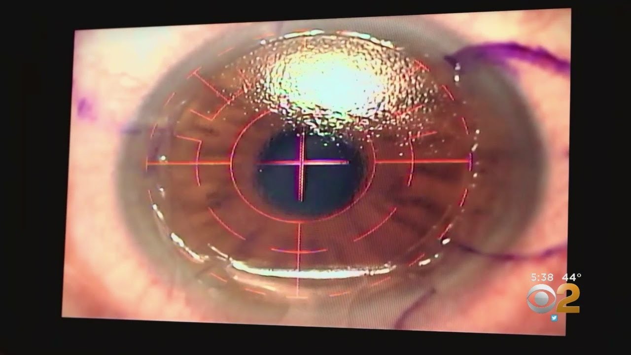
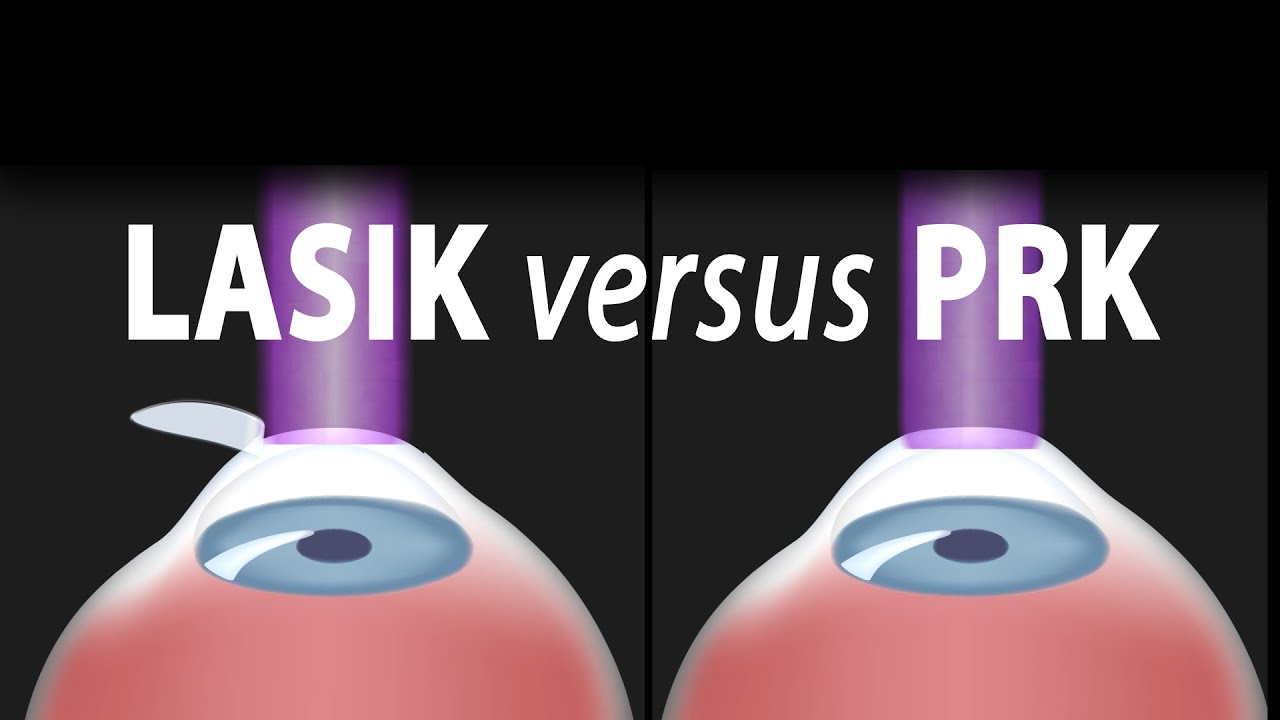
In this procedure, a thin, circular "FLAP" is created in the surface of the cornea to gain access to the permanent corneal tissue. This can be done with a mechanical cutting tool called a microkeratome, OR, for a blade-free experience, by a femtosecond laser. An excimer laser is then used to remove some corneal tissue to reshape the cornea. Excimer laser uses cool ultraviolet light beams to vaporize microscopic amounts of tissue in a precise manner to accurately reshape the cornea. The excimer laser is computer-controlled and is programmed based on the patient’s refractive error. The flap is then laid back in place and is allowed to heal.
LASIK eye surgery is mostly painless and can be completed within minutes. Improved vision can usually be seen overnight.
PRK, or photorefractive keratectomy, was the first type of laser eye surgery for vision correction and is the predecessor to the popular LASIK procedure. In PRK, NO flap is created. Rather, the epithelial cells on the eye surface are simply removed. An excimer laser is then used to reshape the cornea just like it does in LASIK.
The vision correction outcomes of PRK surgery are comparable to those of LASIK, but the recovery period is longer. This is because the epithelium is completely removed in PRK and it takes a few days to regenerate. PRK patients also have more discomfort and haziness of vision in the first few days after the surgery. Improved vision also takes longer to achieve.
PRK does, however, offer certain advantages. Because PRK does not involve creation of a flap, which contains both epithelial and deeper stromal tissue, the entire thickness of the stroma is available for treatment. The treatment range is therefore higher. This is particularly useful for patients with high levels of myopia or for those whose cornea is too thin for LASIK. PRK is also free of flap-related complication risks.

Laser-assisted in situ keratomileusis (LASIK) eye surgery can correct or improve your sight by using a laser to change the shape of the cornea. Find out more here: https://www.bupa.co.uk/health-....information/eyes-sig and https://www.bupa.co.uk/health-....information/eyes-sig/laser-eye-surgery
The content is intended for general information only and does not replace the need for personal advice from a qualified health professional.
