
- Procedures & Tests
- Symptoms A-Z
- Symptom Checker
- Kidney Diet
- First Aid
- Anatomy Atlas
- Ask MedicalVideos
- Drug Guide
- Lab Values
- Vital Signs
- Muscles A–Z
- Clinical Signs
- Medical Abbreviations
- Medical Terminology
- Physical Examination
- Surgical Examination
- Ophthalmology
- Clinical Skills
- Orthopedics
- Surgery Videos
- Laparoscopy
- Pediatrics
- Funny Videos
- Cardiothoracic Surgery
- Nursing Videos
- Plastic Surgery
- Otorhinolaryngology
- Histology and Histopathology
- Neurosurgery
- Dermatology
- Pediatric Surgery
- Urology
- Dentistry
- Oncology and Cancers
- Anatomy Videos
- Health and Fitness
- Radiology
- Anaesthesia
- Physical Therapy
- Pharmacology
- Interventional Radiology
- Cardiology
- Endocrinology
- Gynecology
- Emergency Medicine
- Psychiatry and Psychology
- Childbirth Videos
- General Medical Videos
- Nephrology
- Physiology
- Diet and Food Health
- Diabetes Mellitus
- Neurology
- Women Health
- Osteoporosis
- Gastroenterology
- Pulmonology
- Hematology
- Rheumatology
- Toxicology
- Nuclear Medicine
- Infectious Diseases
- Vascular Disease
- Reproductive Health
- Burns and Wound Healing
- Kidney Zone Podcast
- Other
Latest videos
The newest additions to the MedicalVideos.com library — freshly curated medical and surgery videos, explainers, and lectures from educators and institutions worldwide.
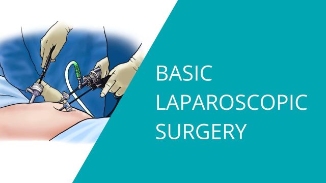

Learn Basic Laparoscopic Surgery, the components of a laparoscopic surgical setup, optimal positioning and ergonomics in laparoscopic surgery, and much more. Check out the full course for free here: https://www.incision.care/free-trial
What is Laparoscopic Surgery:
Laparoscopic surgery describes procedures performed using one or multiple small incisions in the abdominal wall in contrast to the larger, normally singular incision of laparotomy. The technique is based around principles of minimally invasive surgery (or minimal access surgery): a large group of modern surgical procedures carried out by entering the body with the smallest possible damage to tissues. In abdominopelvic surgery, minimally invasive surgery is generally treated as synonymous with laparoscopic surgery as are procedures not technically within the peritoneal cavity, such as totally extraperitoneal hernia repair, or extending beyond the abdomen, such as thoraco-laparoscopic esophagectomy. The term laparoscopy is sometimes used interchangeably, although this is often reserved to describe a visual examination of the peritoneal cavity or the purely scopic component of a laparoscopic procedure. The colloquial keyhole surgery is common in non-medical usage.
Surgical Objective of Laparoscopic Surgery:
The objective of a laparoscopic approach is to minimize surgical trauma when operating on abdominal or pelvic structures. When correctly indicated and performed, this can result in smaller scars, reduced postoperative morbidity, shorter inpatient durations, and a faster return to normal activity. For a number of abdominopelvic procedures, a laparoscopic approach is now generally considered to be the gold-standard treatment option.
Definitions
Developments of Laparoscopic Surgery:
Following a number of smaller-scale applications of minimally invasive techniques to abdominopelvic surgery, laparoscopic surgery became a major part of general surgical practice with the introduction of laparoscopic cholecystectomy in the 1980s and the subsequent pioneering of endoscopic camera technology. This led to the widespread adoption of the technique by the early- to mid-1990s. The portfolio of procedures that can be performed laparoscopically has rapidly expanded with improvements in instruments, imaging, techniques and training — forming a central component of modern surgical practice and cross-specialty curricula [2]. Techniques such as laparoscopically assisted surgery and hand-assisted laparoscopic surgery have allowed the application of laparoscopic techniques to a greater variety of pathology. Single-incision laparoscopic surgery, natural orifice transluminal endoscopic surgery, and minilaparoscopy-assisted natural orifice surgery continue to push forward the applications of minimally invasive abdominopelvic techniques; however, the widespread practice and specific indications for these remain to be fully established. More recently, robotic surgery has been able to build on laparoscopic principles through developments in visualization, ergonomics, and instrumentation.
This Basic Laparoscopic Surgery Course Will Teach You:
- Abdominal access techniques and the different ways of establishing a pneumoperitoneum
- Principles of port placement and organization of the operative field
- Key elements of laparoscopic suturing, basic knotting and clip application
Specific attention is paid to the following hazards you may encounter:
- Fire hazard and thermal injury
- Lens fogging
- Contamination of insufflation system
- Complications from trocar introduction
- Limitations of Veress needle technique
- Limitations of open introduction technique
- Complications of the pneumoperitoneum
- Gas embolism
- Mirroring and scaling of instrument movements
- Firing clip applier without a loaded clip
The following tips are designed to improve your understanding and performance:
- Anatomy of a laparoscope
- Checking for optic fiber damage
- "White balance" of camera
- Checking integrity of electrosurgical insulation
- Access at Palmer's point
- Lifting abdominal wall before introduction
- Confirming position of Veress needle
- Umbilical anatomy
- Identification of inferior epigastric vessels under direct vision
- Translumination of superficial epigastric vessels
- Selection of trocar size
- Aiming of trocar
- Working angles in laparoscopic surgery
- Choice of suture material
- Instruments for suturing
- Optimal ergonomics for suturing
- Extracorporeal needle positioning
- Optimal suture lengths
- "Backloading" needle
- Intracorporeal needle positioning
- Hand movements when suturing
- Optimal positioning of scissors
- Extracorporeal knot tying
- Visualization of clip applier around target structure
- Common clip configurations
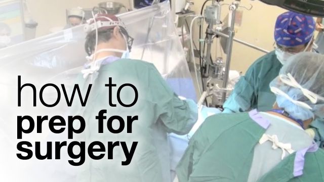
Thousands of Canadians undergo surgery every year, so how can you best prepare? The first step is having a dialogue, says Sunnybrook anesthesiologist Dr. Colin McCartney. Read the blog for more: http://sunnyview.sunnybrook.ca
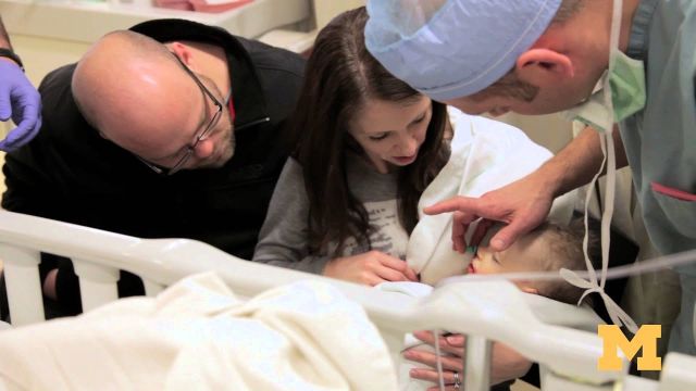
Cleft palate is among the most common birth defects affecting children in North America. The incomplete formation of the roof of the mouth can occur individually, or in addition to cleft lip. Cleft palate repair is a type of plastic surgery to correct this abnormal development both to restore function and a more normal appearance. This video explains what to expect for families scheduled for cleft palate surgery at the Craniofacial Anomalies Program at University of Michigan C.S. Mott Children's Hospital.
Learn more about our program at http://www.mottchildren.org/craniofacial
This video has been updated to include an alternate name for the internal thoracic arteries. View the updated video here: https://youtu.be/kxc22Fjd1NQ
For Employees of Hospitals, Schools, Universities and Libraries: Download 8 FREE medical animations from Nucleus by signing up for a free trial: http://nmal.nucleusmedicalmedi....a.com/free-trial-mem
Biology students: Subscribe to the Nucleus Biology channel to see new animations on biology and other science topics, plus short quizzes to ace your next exam: https://bit.ly/3lH1CzV
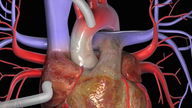
This video, created by Nucleus Medical Media, shows a coronary artery bypass graft (CABG) procedure used to combat coronary artery disease. Beginning with a midline sternal incision, the heart is connected to a perfusion machine which will take over the duties of the heart while the surgery takes place. Two different grafts are used to bypass the blocked coronary arteries: the internal thoracic artery from inside the chest wall, and the saphenous vein from the leg. After the procedure, the heart is shocked to restart its beating. A drainage tube is left at the incision site to drain away excess fluid. The animation continues to show two other types of approaches to a coronary artery bypass graft, off-pump bypass surgery and minimally invasive bypass surgery.
This is similar to the procedure performed on former president Bill Clinton and former California governor Arnold Schwarzenegger.
#HeartBypassSurgery #CABG #heart
ANCE00199
If a fetal lung lesion is causing heart failure, fetal surgery may be performed to remove the CCAM before birth. http://fetalsurgery.chop.edu
N. Scott Adzick, MD, Mark Johnson, MD, and Holly Hedrick, MD, experts from the Center for Fetal Diagnosis and Treatment at Children’s Hospital of Philadelphia, explain when fetal intervention for CCAM is recommended, the various approaches that may be used to treat the most complex fetal lung lesions before birth, and how these procedures are performed.
One concern with fetal lung lesions is that they take up space in the chest. If the lung mass grows and pushes the heart and other organs out of place, it can lead to complications such as fetal hydrops (heart failure in the fetus). If this happens, a fetal surgery procedure may be performed to remove the CCAM before birth.
In other cases, an EXIT procedure may be performed to partially deliver the baby, so the team can remove the mass before the baby is fully delivered.
In this video series, parents, nurses and doctors from Children’s Hospital of Philadelphia’s Center for Fetal Diagnosis and Treatment talk about the different types of fetal lung lesions like congenital cystic adenomatoid malformation (CCAM) and bronchopulmonary sequestration (BPS), the importance of accurate diagnosis and monitoring, and the most advanced treatment options currently available. They also discuss follow-up care and long-term outcomes for babies diagnosed with fetal lung lesions.
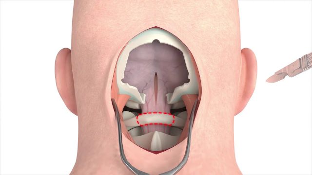
A brain surgery called a craniectomy is performed to remove a part of your skull in order to relieve pressure in an area when your brain swells from a traumatic brain injury. It is also performed to treat medical conditions that cause your brain to swell or bleed that can be caused by an aneurysm, brain tumor or other cancer.
This 3d animation shows how the surgical procedure decreases intracranial pressure (ICP), intracranial hypertension (ICHT), or heavy bleeding (also called hemorrhaging) inside your skull. If left untreated, pressure or bleeding can compress your brain and push it down onto the brain stem. This can be fatal or cause permanent brain damage.
Brain surgery is a very serious procedure under any circumstances, but a craniectomy is done when there is an immediate risk to the brain and neurological function due to severe brain injury or stroke.
For more information about custom 3D animation depicting surgery, please visit https://www.amerra.com/.
Watch additional medical animations:
Accessing an implantable port training - 3D animation: https://youtu.be/xSTpxjyv4O4
Open Suctioning with a Tracheostomy Tube - 3D animation: https://youtu.be/wamB7jpWCiQ
Ventriculostomy Brain Surgery - 3d animation: https://youtu.be/pUy0YDzVNzs
Suctioning the endotracheal tube - medical animation: https://youtu.be/pN6-EYoeh3g
Functional endoscopic sinus surgery (FESS) - 3D animation: https://youtu.be/qKTRyowwaLA
How to insert a nasogastric tube for NG intubation - 3d animation: https://youtu.be/Abf3Gd6AaZQ
Oral airway insertion - oropharyngeal airway technique - 3D animation: https://youtu.be/caxUdNwjt34
Nasotracheal suctioning (NTS) - 3D animation: https://youtu.be/979jWMsF62c
Learn about hemorrhoids with #3d #animation: https://youtu.be/R6NqlMpsiiY
LASIK eye surgery - 3D animation: https://youtu.be/Bb8bnjnEM00
CPR cardiopulmonary resuscitation - 3D animation: https://youtu.be/G87knTZnhks
What are warts (HPV)? - 3D animation: https://youtu.be/guJ1J7rRs1w
How Macular Degeneration Affects Your Vision - 3D animation: https://youtu.be/ozZQIZ_52YY
NeoGraft hair transplant procedure – animation: https://youtu.be/C-eTdH2UPXI
From UW Health's Neurosurgery Program: Learn more about the individual steps in the DBS surgery procedure. Visit uwhealth.org/dbs
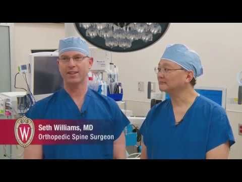
Orthopedic spine surgeons and vascular surgeons at UW Health in Madison, WI work together to perform minimally invasive anterior lumbar interbody fusion (Mini-ALIF). With this type of spinal fusion surgery, patients have smaller incisions, usually spend less time in the hospital and typically return to daily activities more quickly. Learn more https://www.uwhealth.org/ALIF
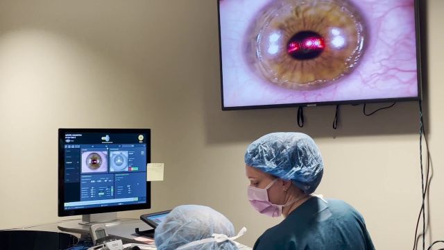
For those that aren't good LASIK candidates, PRK is a procedure that offers the same great results! Watch Dr. Ferguson explain each step during Samantha's procedure. Her reaction at the end is incredible!
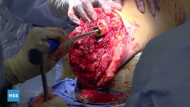
In this video, Dr. Robert Rozbruch, chief of Limb Lengthening and Complex Reconstruction at Hospital for Special Surgery performs an osseointegration after a primary amputation. The patient, a 40 year old woman, had chronic nerve pain and compromised function of her residual limb.
For more information, visit: https://www.limblengthening.com/
https://www.hss.edu/limblengthening
https://www.hss.edu/LSARC
https://www.facebook.com/limblengtheningNYC
https://www.instagram.com/limblengthening
https://www.twitter.com/limblengthen
https://www.youtube.com/channe....l/UC-JL_X6ALjZXiXtcP
key words: Osseointegration, Amputee, Amputation, Limb Replacement, Tibia, Osseointegration
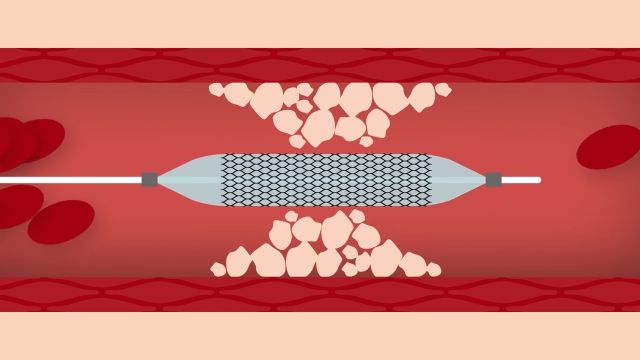
Watch to learn more about what happens during a stent procedure.
More information about this procedure and other heart care at BJC: https://www.bjc.org/Services/M....edical-Services/angi
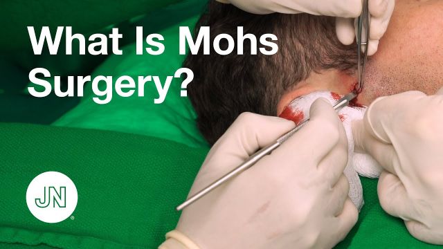
Mohs surgery is a procedure used to remove skin cancers (most commonly basal and squamous cell carcinoma and melanoma) in a way that preserves a maximum amount of healthy tissue. It is useful for skin cancers when:
(1) the location of the cancer is near sensitive areas, like the fingers or face;
(2) earlier treatments have not worked;
(3) a skin cancer is large; and
(4) regular surgery is less likely to remove the cancer.
This procedure video illustrates the procedure on 2 patients with basal cell carcinoma. Click https://ja.ma/3b4scuY to learn more.
0:00 Disclaimer
0:07 Introduction
0:28 What this video will cover
0:43 Mohs "stage" steps
1:09 Marking surgical sites
1:27 Stage 1: skin layer resection (patient 1)
1:59 Maintaining skin layer orientation
2:55 Stage 1: processing layer onto slides (patient 1)
3:53 Stage 1: histology review for cancer (patient 1)
4:31 Discussion on wound closure
5:10 Wound closure (patient 1)
5:47 Stage 1: histology review for cancer (patient 2)
6:24 Stage 2: skin layer resection (patient 2)
6:38 Stage 2: histology review for cancer (patient 2)
6:56 Stage 3: skin layer resection (patient 2)
7:07 Stage 3: histology review for cancer (patient 2)
7:20 Wound closure (patient 2)
7:57 1-week follow-up before-and-after wound healing
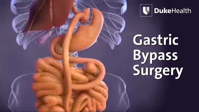
Gastric bypass, also called Roux-en-Y gastric bypass surgery, is considered a “metabolic” procedure because it changes how your body absorbs fat, calories and nutrients. This metabolic change occurs because your gastrointestinal tract is altered when your gastric bypass surgeon attaches the smaller section of your stomach directly to your small intestine. As a result, your appetite changes and you feel full faster.
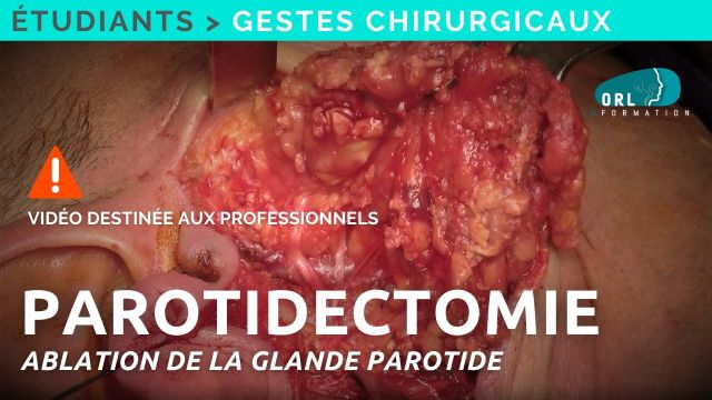
How to perform a parotidectomy gland resection? In this video we take you step by step through the protid gland resection surgical technique. This video is intended for ENT residents and Head and Neck Surgery Surgeons. It is part of the ORL-Information's Head and Neck surgery Masterclass in collaboration with the University Hospital of Nîmes. Surgeons Editors: Pr. Benjamin LALLEMANT, MD, PhD - Dr. Camille GALY, MD Head and and Neck Department, University Hospital of Nîmes, France Official video | www.orl-information.fr
Cette vidéo présentent la technique de la parotidectomie avec dissection du nerf facial. Elle illustre les différents temps de l'intervention notamment le temps de repérage du nerf facial.
--------------------------------------
@Prévention médecine | Comment préserver sa santé ?
✔Abonnez-vous à la chaîne ORL-Information : https://www.youtube.com/c/ORLINFORMATION
✔Retrouvez-nous sur internet: https://www.orl-information.fr
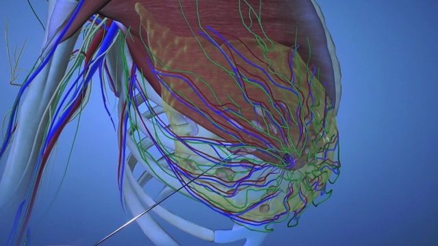
Lumpectomy means that a focal area of cancer is going to be removed. A lot of patients with a lumpectomy don’t need any specific breast reconstruction, explains Dr. Miguel Angel Medina, Director of Microsurgery with Miami Cancer Institute.
Al the end of surgical treatment, all those patients go on to need radiation therapy. For patients who have large breasts, physicians have to take a larger lumpectomy than normal.
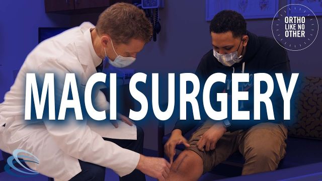
Lattrell Wells was a perfect candidate for the MACI procedure. Dr. Michael O'Malley is a sports medicine surgeon at Carilion Clinic, "It’s a two stage procedure. So what we do is we actually harvest a small portion of the patient's cartilage and bone cells and we send it to a lab where the lab then that grows additional cartilage cells. It comes back to us in a little sheet and six weeks after that initial surgery, we re-implant the cartilage in a second surgery where we implant that sheet depending on the size of lesion right where his defect. This the only option where there’s virtually no risk of any kind of graft rejection or anything of that nature.
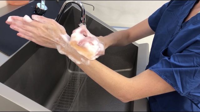
Surgical site infections (SSIs) remain a prevalent threat to patient safety. Proper surgical hand scrub or rub techniques are essential to decreasing the incidence of SSIs. This video provides instructions on the anatomical surgical hand scrub procedure using the brushstroke method. Learn more from the Department of Hospital Epidemiology and Infection Control (HEIC) at The Johns Hopkins Hospital: http://www.hopkinsmedicine.org/heic
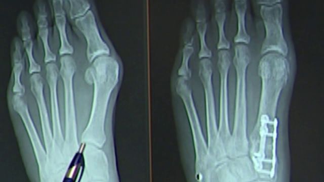
A new procedure helps patients with unattractive bunions and hammertoes.
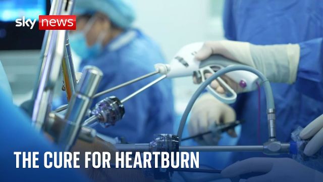
Surgeons at St Mary's Hospital, part of Imperial College Healthcare have come up with a new surgical procedure that cures heartburn with a device called RefluxStop.
Mr Ahmed Ahmed, a consultant surgeon, says surgery should now be seen as an alternative to life-long drug treatment - as Sky's Thomas Moore reports.
Read more: https://news.sky.com/story/new....-nhs-heartburn-surge
#heartburncure #surgery #skynews
SUBSCRIBE to our YouTube channel for more videos: http://www.youtube.com/skynews
Follow us on Twitter: https://twitter.com/skynews
Like us on Facebook: https://www.facebook.com/skynews
Follow us on Instagram: https://www.instagram.com/skynews
Follow us on TikTok: https://www.tiktok.com/@skynews
For more content go to http://news.sky.com and download our apps: Apple https://itunes.apple.com/gb/ap....p/sky-news/id3163919 Android https://play.google.com/store/apps/details?id=com.bskyb.skynews.android&hl=en_GB
Sky News Daily podcast is available for free here: https://podfollow.com/skynewsdaily/
Sky News videos are now available in Spanish here/Los video de Sky News están disponibles en español aquí: https://www.youtube.com/channe....l/UCzG5BnqHO8oNlrPDW
To enquire about licensing Sky News content, you can find more information here: https://news.sky.com/info/library-sales
This video is available for instant download licensing here: https://www.alilamedicalmedia.....com/-/galleries/all-
©Alila Medical Media. All rights reserved.
Support us on Patreon and get FREE downloads and other great rewards: patreon.com/AlilaMedicalMedia
All images/videos by Alila Medical Media are for information purposes ONLY and are NOT intended to replace professional medical advice, diagnosis or treatment. Always seek the advice of a qualified healthcare provider with any questions you may have regarding a medical condition.
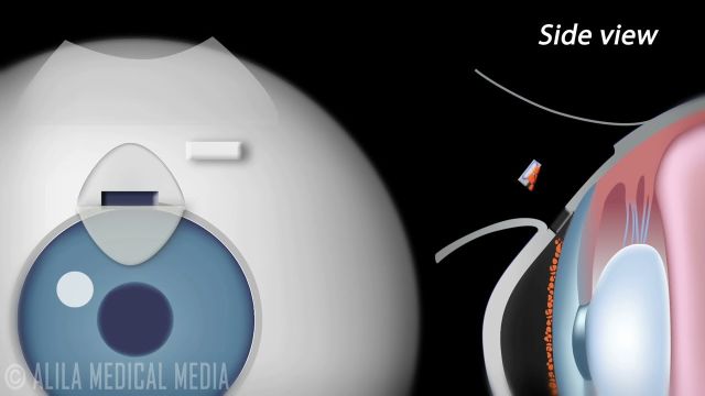
Trabeculectomy, also called Filtration Surgery, is a surgical procedure performed for treatment of glaucoma. The treatment involves removing part of the trabecular meshwork and creating a new escape route for the aqueous humor. When successful, it allows the aqueous fluid to drain from the eye into an area underneath the conjunctiva where it is subsequently absorbed by the body's circulatory system or filtered into tears.
In this procedure:
- A conjunctival pocket is created and maybe treated with Mitomycin or other antimetabolites for a few minutes. These drugs are used to prevent scarring of the operation site. Scarring, if occurs, may clog the new drainage canal, and is therefore the major reason the procedure may fail.
- A half thickness flap is then made in the sclera and is dissected all the way to the clear cornea.
- A block of scleral tissue including part of the trabecular meshwork and Schlemm's canal is then removed to make a hole into the anterior chamber of the eye.
- As the iris may plug up this hole from the inside, a piece of the iris maybe removed at this time. This is called iridectomy.
- The scleral flap is then sutured loosely back in place. These sutures can be released gradually during a couple of weeks after surgery. This allows adjustment of the aqueous flow in order to achieve target pressure and to avoid the complication of having a too low intraocular pressure.
- The conjunctiva is sewn back in place to cover the area.
After surgery, aqueous humor drains into a filtering area called a "bleb" under the conjunctiva. Since the surgery is usually performed near the top of the eye, the bleb can easily be concealed behind the upper eyelid.
Medical Videos
Free medical and surgery videos for patients, students and professionals — trusted since 2007.
Founded by Dr. Mohamed Ibrahim, MD, PhD — physician-scientist.
Follow us on Facebook · Instagram