Video teratas

General Examination - Clinical Skills OSCE - Dr Gill
The general examination is one of those early exams, which is essentially used to start medical students off with their clinical skills studies.
In the real world, it's mainly used with regard to gaining an overview of a patient, such as for a medical check up, or a baseline examination, for example, a health report.
They have been a couple of comments about the pulse monitor used in the video. For those who are interested. I’ve reached out to the manufacturer, and they’ve requested that the following code is provided to viewers, in order to get 20% off, if they decide on themselves.
Product model number: Vibeat SP20
Official Website: https://vibeatstore.com/produc....ts/sp20-handheld-pul
Special 20% OFF code: JAMES
--------------
Different medical schools, nursing colleges and other health professional courses will have their own preferred approach to a clinical assessment - you should concentrate on THEIR marks schemes for your assessments.
Some people watching this video may experience an ASMR effect
#DrGill #Asmr #Clinicalskills
#drgill #clinicalskills #asmr

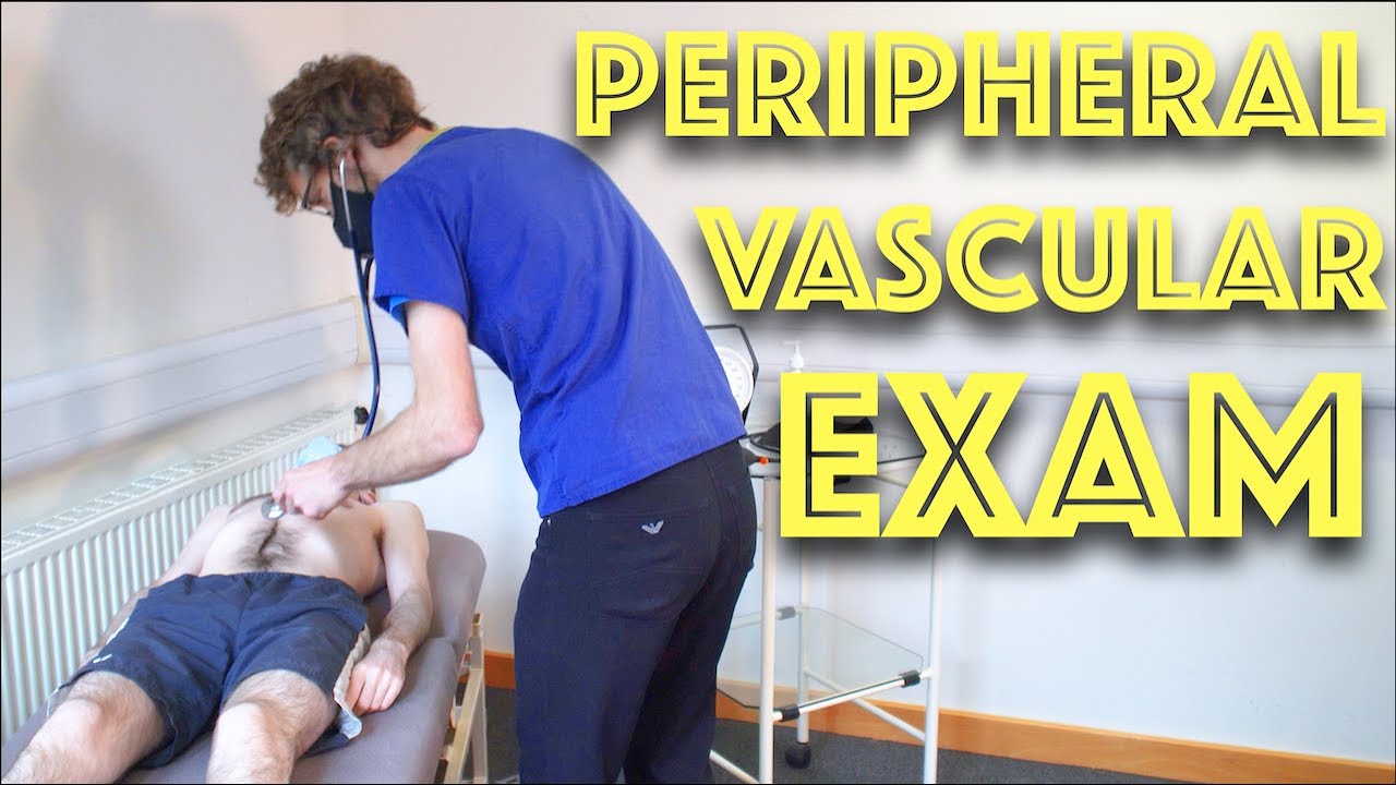
Examination of Peripheral Vascular System - Clinical Skills OSCE Revision - Dr Gill
In this video, we demonstrate the peripheral vascular examination - a less common examination, but still vitally important, particularly amongst the older population
Starting with the examination of the hands looking for clinical signs of vascular compromise, we then check the pulses of the major arteries of the upper body - the radial, brachial and carotid arteries, before moving down to assess for an abdominal aortic aneurysm.
At this point, I feel it's a practical step to check the femoral pulses before doing the overview of the legs.
After visually assessing we must examine the major vascular areas of leg.- namely the popliteal pulses, before wrapping up around the ankle with the posterior tibial and dorsalis pedis pulses
For completeness, the cardiovascular examination is demonstrated here
https://www.youtube.com/watch?v=ECs9O5zl6XQ&t=2s
#PeripheralVascular #ClinicalSkills #DrGill
Talley + O'Connor's essential video guide to Abdominal Examination is here! Brush up on your skills and be sure to ace your OSCEs!

Elbow Exam - Orthopaedic OSCE - Clinical Skills - Dr Gill
The elbow examination is a core skill - in this video, we demonstrate how to perform an elbow EXAM for an Orthopaedic Clinical Skills OSCE, which should be one of the more accessible examination stations for medical students.
For a passing grade in your Clinical Skills OSCE, an elbow assessment should follow the LOOK, FEEL, MOVE approach
Initially looking for erythema, scars, swelling and position
Palpating the elbow - specifically the olecranon, medial and lateral epicondyles, and radial head for heat, oedema and crepitus
Finally assess range of movement with flexion and extension at the elbow, before determining for tennis and golfers' elbows
Watch further orthopaedic examinations for your OSCE revision:
The Elbow - Deep Dive
https://youtu.be/SX5buhtCVDw
The Spine Examination:
https://youtu.be/pJxMHa6SCgU
The Knee examination
https://youtu.be/oyKH4EYfJDM
The Hip examination
https://youtu.be/JC9GKq5nSdQ
The GALS examination
https://youtu.be/5qJaf7gW-B0 - Gait, Arms, Legs, Spine - GALS screen
------------
Please note that there is no ABSOLUTE way to perform a clinical examination. Different institutions and even clinicians will have differing degrees of variations - the aim is the effectively identify medically relevant signs.
However during OSCE assessments. Different medical schools, nursing colleges and other health professional courses will have their own preferred approach to a clinical assessment - you should concentrate on THEIR marks schemes for your assessments.
The examination demonstrated here is derived from Macleods Clinical Examination - a recognised standard textbook for clinical skills.
Some people viewing this medical examination video may experience an ASMR effect
#clinicalskills #Elbow #DrGill

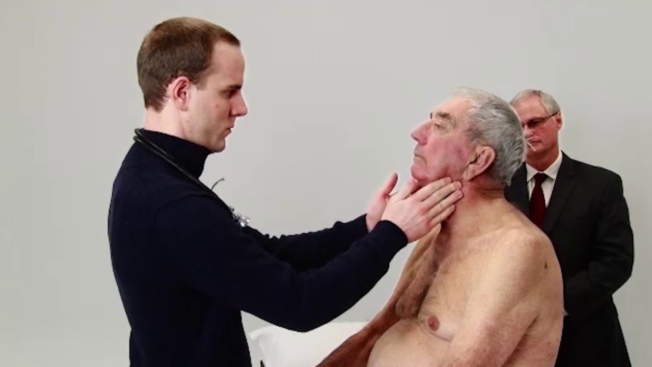
How to perform a Thyroid Gland Examination - Clinical Skills Revision
The thyroid examination is one of the first sessions of the clinical skills block for medical students at Warwick Medical School - largely as it touches lightly on to other clinical areas, such as the cardiac examination, and the peripheral neurological examination making it an excellent starting point for building further knowledge
This is a clinical examination of the thyroid gland is performed by Dr James Gill following the approach in Macleod’s Clinical examination.
------------------
Please note that there is no ABSOLUTE way to perform a clinical examination. Different institutions and even clinicians will have differing degrees of variations - the aim is the effectively identify medically relevant signs.
However, during OSCE assessments. Different medical schools, nursing colleges and other health professional courses will have their own preferred approach to a clinical evaluation - you should concentrate on THEIR marks schemes for your assessments.
The examination demonstrated here is derived from Macleods Clinical Examination - a recognised standard textbook for clinical skills.
Some people may experience an ASMR effect from watching this medical clinical examination
#ThyroidExamination #ClinicalSkills #DrGill #ASMR

Cardiovascular Examination Clinical skills - Medical School Revision - Dr Gill
The cardiac exam is one of the clinical skills that medical students learn completely, as more often than not, patients will consult regularly about chest pain, and it is important to be able to identify key cardiovascular signs
As a junior doctor, the examination of the cardiovascular system can be almost a dreaded examination, as cardiac murmurs can literally take years of exposure in order to gain confidence with their identification through cardiac auscultation.
This video demonstrates not merely the examination of the heart, but the complete cardiovascular system including its peripheries.
I hope these clinical skill revision videos are helpful, please like and subscribe and join the community so that we can create more effective videos to help with your journey through medical school
#ClinicalExamination #ASMR #drgill
Some people have found this video useful for ASMR

The most reliable clinical sign to detect ascites is checking for bilateral flank dullness. If a patient with ascites is lying supine, fluid accumulates in the flank regions, leading to dullness on percussion. At the same time, the air-filled bowel loops are forced upwards by the free fluid due to buoyancy, resulting in tympanitic percussion. To locate specifically where dullness shifts to tympany, or the air-fluid level, percussion should be performed from the sides towards the middle. To confirm that the dullness is caused by ascites, ask the patient to switch to a lateral decubitus position. If ascites is present, the air-filled bowel loops will shift accordingly and remain at the surface of the fluid. As a result, the air-fluid level will shift as well. This is known as shifting dullness.
Subscribe to AMBOSS YouTube for the latest clinical examination videos, medical student interviews, study tips and tricks, and live webinars!
Free 5 Day Trial: https://go.amboss.com/amboss-YT
Instagram: https://www.instagram.com/amboss_med/
Facebook: https://www.facebook.com/AMBOSS.Med/
Twitter: https://twitter.com/ambossmed
Blog: https://blog.amboss.com/us
#AMBOSSMed #ClinicalExamination
Peripheral Vascular Examination OSCE - Clinical Skills - Dr Gill
In the cardiovascular examination, particularly in the case of an OSCE station, we conclude the examination often by stating that the examiner would want to perform:
- An ECG
- Check full blood count
- and "do a peripheral vascular examination
In this video, we demonstrate that oft-talked about, but comparatively less common examination.
Starting off, with the examination of the hands, the radial, brachial and carotid pulses. before moving down to assess for a AAA, checking the femoral and popliteal pulses, before wrapping up around the ankle with the posterior tibial and dorsalis pedis pulses
For completeness, the cardiovascular examination is demonstrated here
https://www.youtube.com/watch?v=ECs9O5zl6XQ&t=2s
#PeripheralVascular #ClinicalSkills #DrGill
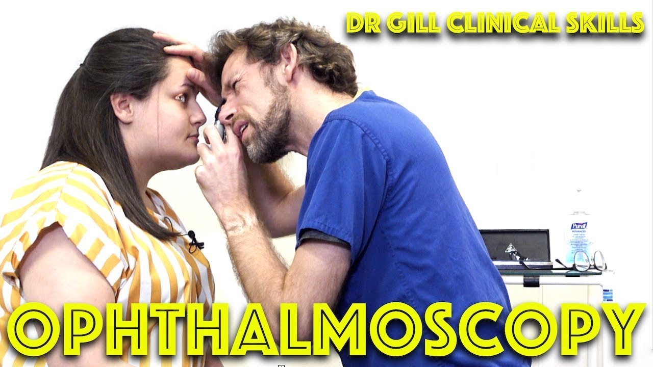
Ophthalmoscopy - Eye Clinical Examination - OSCE - Dr Gill
Direct Ophthalmoscopy use of the eyes is a very challenging clinical skill, incorporating both the examiner's knowledge of the retina, but also understanding the use of the ophthalmoscope
In this clinical skills tutorial, we look at the use of the direct ophthalmoscope as part of an ophthalmic examination
it should be noted that in the ideal circumstances, the room lights will be dimmed during the examination, and dilating eye drops used to improve the visualisation of the fundus
Some people may notice an ASMR effect from this clinical examination
#DrGill #Ophthalmoscopy #ClinicalSkills #EyeExam

Are you worried about getting a sports hernia exam? In this video, we'll show you exactly what to expect when you get your hernia exam.
We'll take you through the various steps that are taken during the hernia exam, so you can have a more comfortable and informative experience. After watching this video, you'll have a better idea of what to expect and be prepared for your hernia exam!
#sportshernia #groinpain #california

Learn with Dr. Wahdan 2
You can download the lecture from this link
https://docdro.id/5ni1FFZ

Sports Hernia Self Test (TRY IT)
714-502-4243 | Costa Mesa, CA | http://www.p2sportscare.com
[FREE GIFT] Audio Download
#sportshernia #hernia #hippain
Sports Hernia Diagnosis
What Is A Sports Hernia?
A sports hernia is tearing of the transversalis fascia of the lower abdominal or groin region. A common misconception is that a sports hernia is the same as a traditional hernia. The mechanism of injury is rapid twisting and change of direction within sports, such as football, basketball, soccer and hockey.
The term “sports hernia” is becoming mainstream with more professional athletes being diagnosed. The following are just to name a few:
Torii Hunter
Tom Brady
Ryan Getzlaf
Julio Jones
Jeremy Shockey
If you follow any of these professional athletes, they all seem to have the same thing in common: Lingering groin pain. If you play fantasy sports, this is a major headache since it seems so minor, but it can land a player on Injury Reserve on a moments notice. In real life, it is a very frustrating condition to say the least. It is hard to pin point, goes away with rest and comes back after activity, but is hardly painful enough to make you want to stop. It lingers and is always on your mind. And if you’re looking for my step-by-step sports hernia rehab video course here it is.
One the best definitions of Sport hernias is the following by Harmon:
The phenomena of chronic activity–related groin pain that it is unresponsive to conservative therapy and significantly improves with surgical repair.”
This is truly how sports hernias behave in a clinical setting. It is not uncommon for a sports hernia to be unrecognized for months and even years. Unlike your typical sports injury, most sports medicine offices have only seen a handful of cases. It’s just not on most doctors’ radar. The purpose of this article is not only to bring awareness about sports hernias, but also to educate.
Will you find quick fixes in this article for sports hernia rehab?
Nope. There is no quick fix for this condition, and if someone is trying to sell you one, they are blowing smoke up your you-know-what.
Is there a way to decrease the pain related to sports hernias?
Yes. Proper rehab and avoidance of activity for a certain period of time will assist greatly, but this will not always stop it from coming back. Pain is the first thing to go and last thing to come. Do not be fooled when you become pain-free by resting it. Pain is only one measure of improvement in your rehab. Strength, change of direction, balance and power (just to name a few) are important, since you obviously desire to play your sport again. If you wanted to be a couch potato, you would be feeling better in no time. Watching Sports Center doesn’t require any movement.
Why is this article so long?
There is a lot of information on sports hernias available to you on the web. However, much of the information is spread out all over the internet and hard for athletes to digest due to complicated terminology. This article lays out the foundational terminology you will need to understand what options you have with your injury. We will go over anatomy, biomechanics, rehab, surgery, and even the fun facts. The information I am using is from the last ten years of medical research, up until 2016. We will be making updates overtime when something new is found as well. So link to this page and share with friends. This is the best source for information on sports hernias you will find.
Common Names (or Aliases?) for Sports Hernias
Sportsman’s Hernia
Athletic Pubalgia
Gilmore’s Groin
How Do You Know If You Have A Sports Hernia?
Typical athlete characteristics:
Male, age mid-20s
Common sports: soccer, hockey, tennis, football, field hockey
Motions involved: cutting, pivoting, kicking and sharp turns
Gradual onset
How A Sports Hernia Develops
Chronic groin pain typically happens over time, which is why with sports hernias, we do not hear many stories of feeling a “pop” or a specific moment of injury. It is the result of “overuse” mechanics stemming from a combination of inadequate strength and endurance, lack of dynamic control, movement pattern abnormalities, and discoordination of motion in the groin area.
There is a lot going on in the groin area. There are many muscles, tendons, and fascia pulling in different directions. These contracting structures need to coordinate together for any athletic motion. This perspective is also known as the injury prevention model.

MRCPCH Clinical Revision - more videos at http://mrcpch.paediatrics.co.uk
Revise for your MRCPCH Clinical exam, with videos and high quality content created by the London Paediatrics Trainees Committee.
Examiner: Jonathan Round
Candidate: Amitav Parida
Filming: Mary Chesshyre, Huey Miin Lee, Chris Kelly
Thank you to the Evelina Children's Hospital for allowing us to film during their MRCPCH Revision Course (https://www.guysandstthomaseve....nts.co.uk/mrcpch-cli

The typical radiograph is of a well-defined, rounded, retrocardiac opacity with an air-fluid level. In this image, the radiolucent gas is highlighted in blue, while the gastric contents are highlighted in the green. In many cases of hiatal hernia, there will not be an air bubble below the left hemidiaphragm. This is a relatively expected finding considering that the stomach is no longer in its usual position. The anatomical position of the herniated organ can be further elucidated on the lateral radiograph. Here we can see that the stomach is in the middle mediastinum posterior to the heart and above the diaphragm. Hiatal hernias can look similar to a retrocardiac lung abscess or another cavitary lesion, but it will change in size and shape between radiographs. Large hernias can shift the mediastinum to the right and result in a widening of the carinal angle. They can even give the appearance of cardiomegaly. In this radiograph, the cardiac silhouette is distinctly visible within the confines of the hiatal hernia. To review, a hiatal hernia on an AP chest radiograph typically appears as a round retrocardiac opacity with an air-fluid level.
🌐 Check out our website for more video lectures
https://www.med4vl.com
📺 Subscribe To My Channel and Get More Great Quizzes and Tutorials
https://www.youtube.com/channe....l/UC95TzSH1B_2EjaZMg
#FOAMrad #MedEd #radiology
Disclaimer: All the information provided by Medical Education for Visual Learners and associated videos are strictly for informational purposes only. It is not intended as a substitute for medical advice from your health care provider or physician. It should not be used to overrule the advice of a qualified healthcare provider, nor to provide advice for emergency medical treatment. If you think that you or someone that you know may be suffering from a medical condition, then please consult your physician or seek immediate medical attention.

The anatomy of the direct and indirect inguinal hernia.
Music:
Berries and Lime by Gregory David
https://www.epidemicsound.com/track/z6iCiiyCPm/

This is how Paraumbilical hernia looks like and how it is examined although it looks very simple but in exam it can be very difficult to perform all steps in small amount of time. This can be short case or even long of #cpsp #fcps #mbbs #medicalstudent #mbbsexams #plab2 #plab #plab1 and MS #genernalknowledge #surgery exams
#para-umbilical hernia
#umbilical hernia #paraumbilical #hernia repair#laparoscopic paraumbilical hernia repair. #umbilical defect, #vetral hernia surgery. #herniatreatment #herniatreatment #ventral hernia hernia,#laparoscopic ventral hernia repair,umbilicus,carl lowe jr,hernia repair,training,north carolina,hernia repair surgery,charlotte,operation,laparoscopic,bulge,surgery,surgeon,dr. lowe,ipom repair,live surgery,mesh,
#mesh #ipom repair

Endoscopy in Hiatal Hernia.
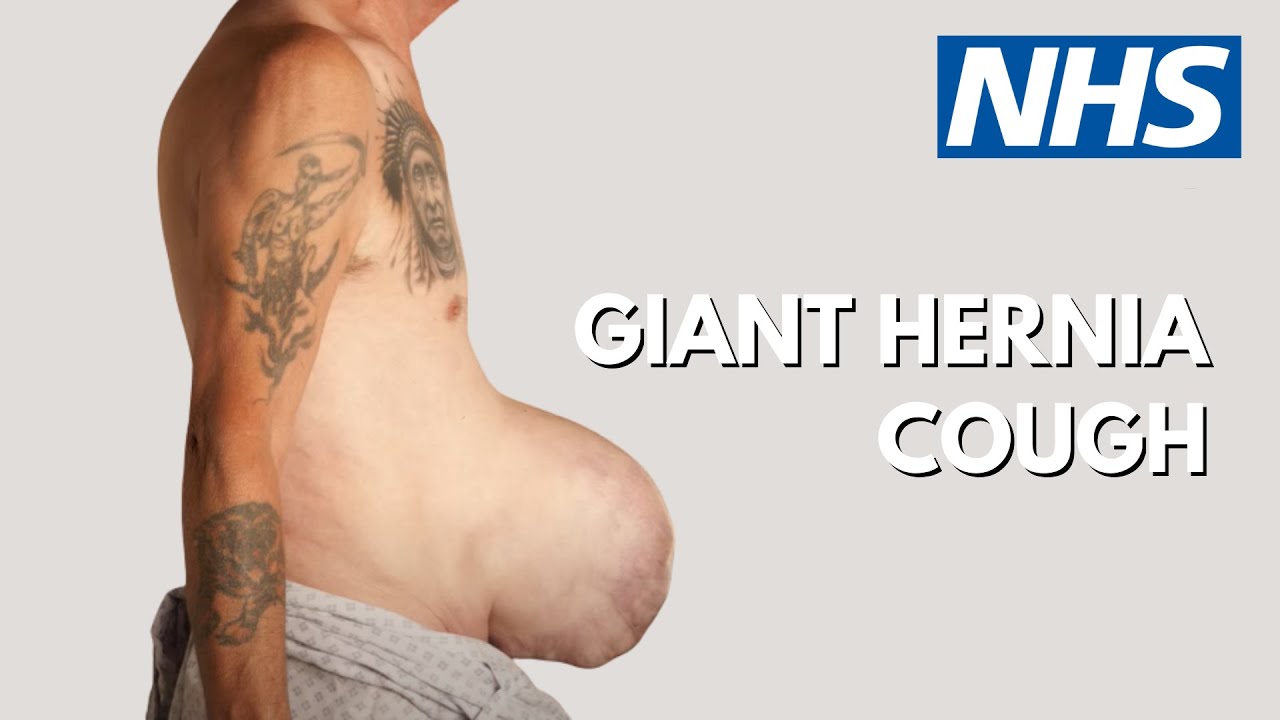
Patient Glenn Williams had a hernia measuring 20cm x 30cm. Consultant Graham Offer has performed ground breaking surgery to help Glenn.
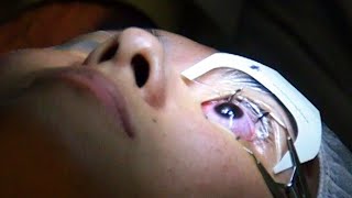
#lasik #eyesurgery #BusinessInsiderIndia #lasereyesurgery
Business Insider's Michelle Yan has been nearsighted since she was 9 years old. After laser eye surgery, she has 20/20. She walks us through the pre-surgery steps, the actual surgery which the doctor referred to as a spa for eyes, as well as the recovery process.
Don't miss the suggestions and recommendations she has for all those who are planning to undergo laser eye surgery for themselves, towards the end.
-----------------------------------------------
Business Insider India features the country's business heavyweights, ranging from start-ups to veterans across industries. It offers amazing synergy between India and its foreign counterpart - developed, analyzed and presented in hallmark BI Style. It covers latest news & trends on Tech, Business, Careers, Startups and Finance.
For more such content, Visit us at: https://www.businessinsider.in/
Subscribe at: https://www.youtube.com/channe....l/UC62AhAC25Ukuscqux
BI on Facebook: https://www.facebook.com/BusinessInsiderIndia/
BI on Twitter:https://twitter.com/BiIndia
BI on Instagram: https://www.instagram.com/businessinsiderin/
BI on Tiktok: https://vm.tiktok.com/GNpHXg/
What It's Like To Get Laser Eye Surgery
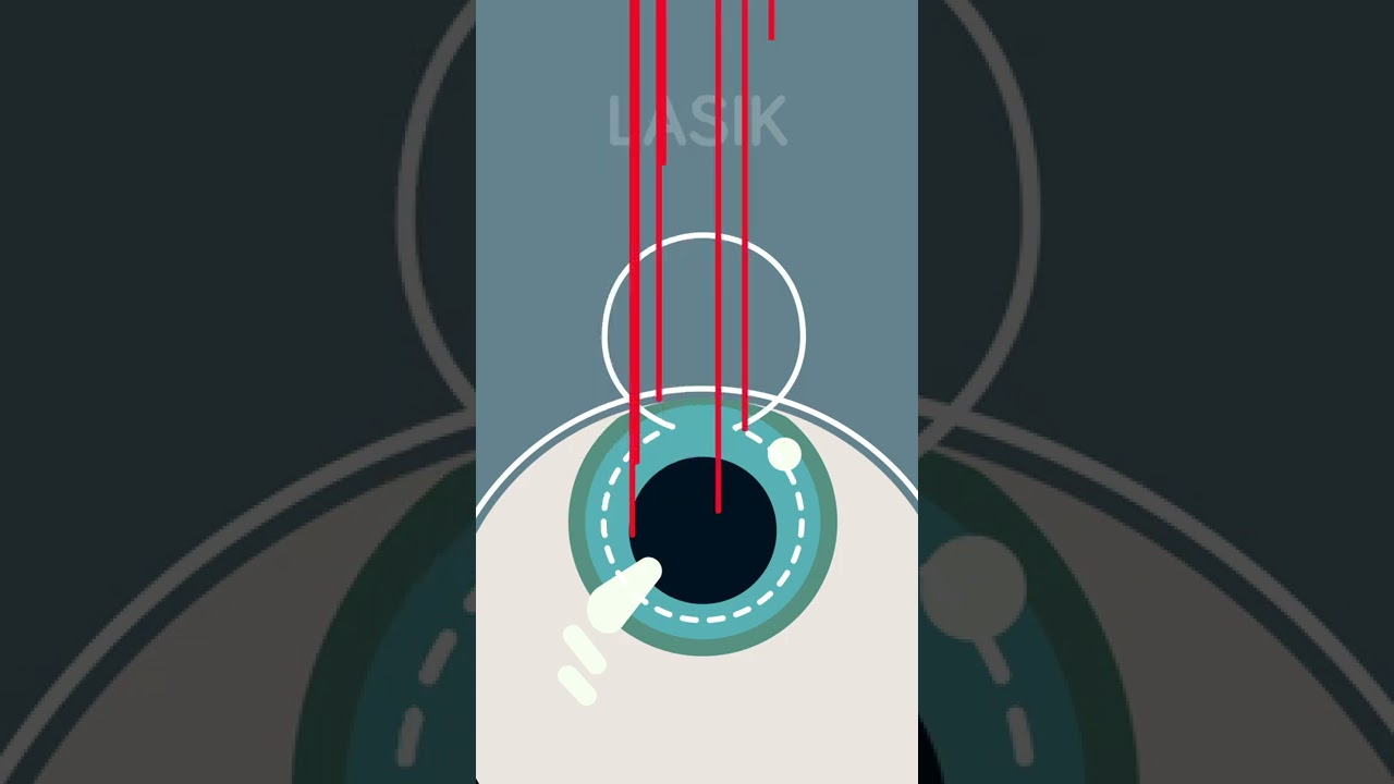
Ever considered getting laser eye surgery, but didn’t know how it worked? Allow us to help!
There are three different main types of laser eye surgery: LASIK, SMILE, and Surface Laser Treatments, and each can be explained pretty easily.
LASIK uses two lasers to open up a thin flap on the surface of the cornea, and then reshapes the cornea underneath. The flap is then placed back over the reshaped cornea, and heals independently with time.
SMILE uses one laser to reshape the cornea through a small, self-healing hole.
And Surface Eye Treatments remove the clear skin over the eye, to then reshape the cornea underneath with - you guessed it - a laser!
