- Physical Examination
- Surgical Examination
- Ophthalmology
- Clinical Skills
- Orthopedics
- Surgery Videos
- Laparoscopy
- Pediatrics
- Funny Videos
- Cardiothoracic Surgery
- Nursing Videos
- Plastic Surgery
- Otorhinolaryngology
- Histology and Histopathology
- Neurosurgery
- Dermatology
- Pediatric Surgery
- Urology
- Dentistry
- Oncology and Cancers
- Anatomy Videos
- Health and Fitness
- Radiology
- Anaesthesia
- Physical Therapy
- Pharmacology
- Interventional Radiology
- Cardiology
- Endocrinology
- Gynecology
- Emergency Medicine
- Psychiatry and Psychology
- Childbirth Videos
- General Medical Videos
- Nephrology
- Physiology
- Diet and Food Health
- Diabetes Mellitus
- Neurology
- Women Health
- Osteoporosis
- Gastroenterology
- Pulmonology
- Hematology
- Rheumatology
- Toxicology
- Nuclear Medicine
- Infectious Diseases
- Vascular Disease
- Reproductive Health
- Burns and Wound Healing
- Other
Top videos
Dilation and Curettage D and C
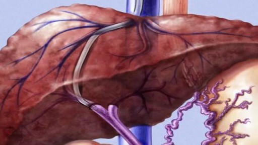
ransjugular intrahepatic portosystemic shunt (TIPS) is a procedure to create new connections between two blood vessels in your liver. You may need this procedure if you have severe liver problems.
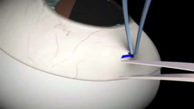
Glaucoma Surgery 3D Animation
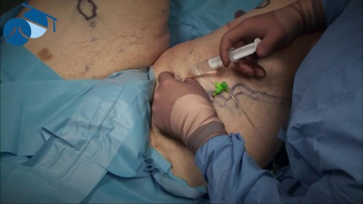
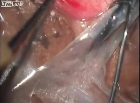
Local anaesthetic injection prior to tumescence ready for varicose vein surgery

Real Story: Youngest Mother In History (5 years old) Pregnant FIVE YEAR OLD! Youngest Mother In The World, Lina Medina's True Story!
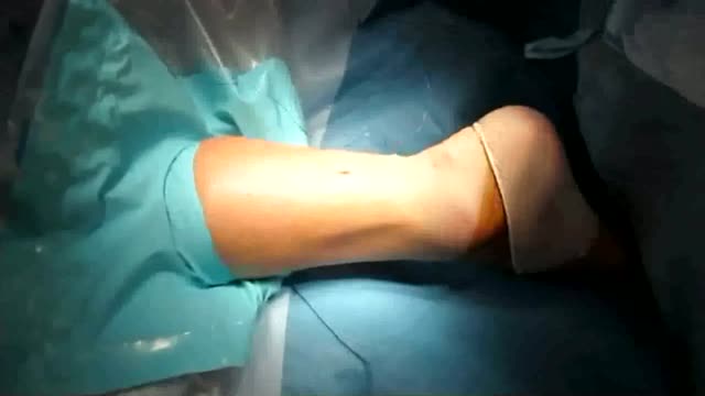
an incision made on the back of the lower leg starting just above the heel bone. After the surgeon finds the two ends of the ruptured tendon, these ends are sewn together with sutures. The incision is then closed. Another repair method makes a small incision on the back of the lower leg at the site of the rupture.
Transradial Cardiac Catheterization

New Drugs Improve Osteoporosis Treatment

Tinnitus (TIN-ih-tus) is the perception of noise or ringing in the ears. A common problem, tinnitus affects about 1 in 5 people. Tinnitus isn't a condition itself — it's a symptom of an underlying condition, such as age-related hearing loss, ear injury or a circulatory system disorder
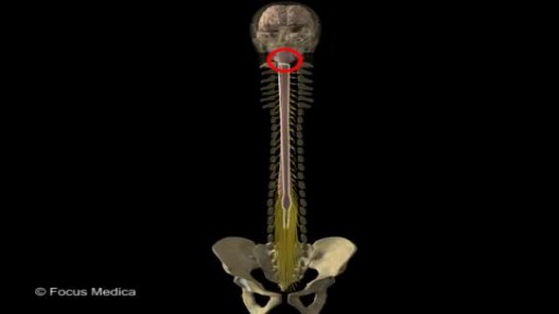
Back and Spinal cord Anatomy

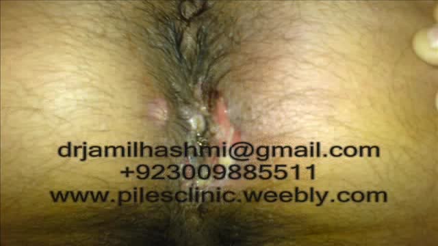
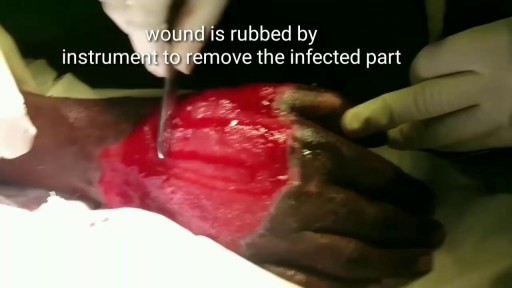
Treatment for Piles,Fistula,hemorrhoids, Hydrocele Without Operation or surgery in pakistan Dr Jamil Ahmad Hashmi ( haripur hazar pakistan )... +923009885511 --- drjamil79@gmail.com
Treatment for Piles,Fistula,Hydrocele Without Operation piles treatment with 60 days Quickly! pain free treatment full life Piles Medicine dr jamil ahmad hashmi ( haripur hazar pakistan ) drjamil79@yahoo.com +923009885511 piles treatment with 60 days Quickly! pain free treatment full life Piles Medicine dr jamil ahmad hashmi...
Skin grafting is a type of medical grafting involving the transplantation of skin. The transplanted tissue is called a skin graft. Skin grafting is often used to treat: Extensive wounding or trauma Burns Areas of extensive skin loss due to infection such as necrotizing fasciitis or purpura fulminans Specific surgeries that may require skin grafts for healing to occur – most commonly removal of skin cancers. Skin grafts are often employed after serious injuries when some of the body’s skin is damaged. Surgical removal (excision or debridement) of the damaged skin is followed by skin grafting. The grafting serves two purposes: it can reduce the course of treatment needed (and time in the hospital), and it can improve the function and appearance of the area of the body which receives the skin graft. There are two types of skin grafts, the more common type is where a thin layer is removed from a healthy part of the body (the donor section), like peeling a potato, or a full thickness skin graft, which involves pitching and cutting skin away from the donor section. A full thickness skin graft is more risky, in terms of the body accepting the skin, yet it leaves only a scar line on the donor section, similar to a Cesarean section scar. For full thickness skin grafts, the donor section will often heal much more quickly than the injury and is less painful than a partial thickness skin graft.
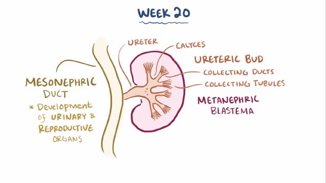
Renal agenesis is a condition in which a newborn is missing one or both kidneys. Unilateral renal agenesis (URA) is the absence of one kidney. Bilateral renal agenesis (BRA) is the absence of both kidneys. Both types of renal agenesis occur in fewer than 1 percent of births annually, according to the March of Dimes. Fewer than 1 in every 1,000 newborns has URA. BRA is much rarer, occurring in about 1 in every 3,000 births.
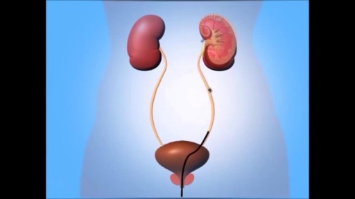
A ureteroscopy is an examination or procedure using a ureteroscope. A ureteroscope, like a cystoscope, is an instrument for examining the inside of the urinary tract. The urologist can insert small instruments through the cystoscope to treat problems in the urethra and bladder or perform a biopsy. For a ureteroscopy, the urologist passes the ureteroscope through the bladder and into a ureter.

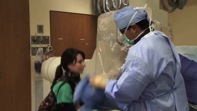
As you consider Fort HealthCare and our Pediatric Surgical Services, here is a quick tour to give you and your child an idea of what to expect.
We look forward to helping you.
To find out more information, please visit forthealthcare.com/PediatricSurgery
Video production by Highlights Media, LLC
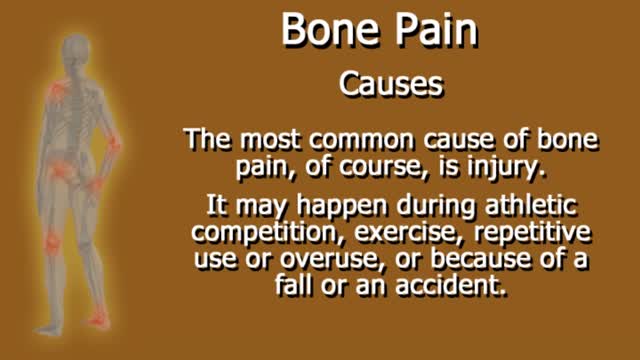
Bone pain: Pain is the most common sign of bone cancer, and may become more noticeable as the tumor grows. Bone pain can cause a dull or deep ache in a bone or bone region (e.g., back, pelvis, legs, ribs, arms). Early on, the pain may only occur at night, or when you are active.
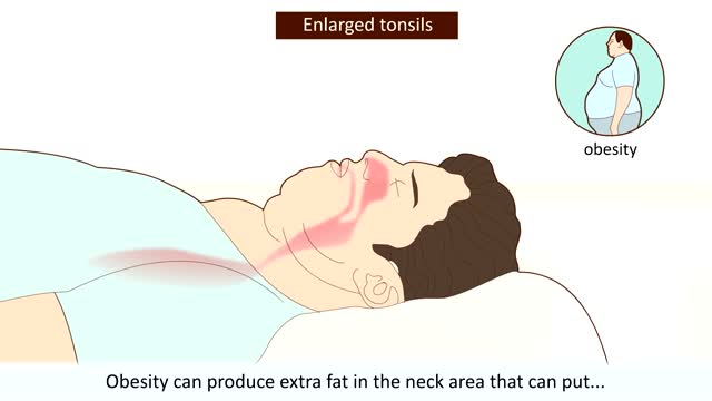
What Is Sleep Apnea? When you have this condition, your breath can become very shallow or you may even stop breathing -- briefly -- while you sleep. It can happen many times a night in some people. Obstructive sleep apnea happens when something partly or completely blocks your upper airway during shut-eye. That makes your diaphragm and chest muscles work harder to open the obstructed airway and pull air into the lungs. Breathing usually resumes with a loud gasp, snort, or body jerk. You may not sleep well, but you probably won't be aware that this is happening.
Loa loa filariasis (also known as loiasis, loaiasis, Calabar swellings, Fugitive swelling, Tropical swelling and African eyeworm) is a skin and eye disease caused by the nematode worm, loa loa. Humans contract this disease through the bite of a Deer fly or Mango fly (Chrysops spp), the vectors for Loa loa. The adult Loa loa filarial worm migrates throughout the subcutaneous tissues of humans, occasionally crossing into subconjunctival tissues of the eye where it can be easily observed. Loa loa does not normally affect one's vision but can be painful when moving about the eyeball or across the bridge of the nose.The disease can cause red itchy swellings below the skin called "Calabar swellings". The disease is treated with the drug diethylcarbamazine (DEC), and when appropriate, surgical methods may be employed to remove adult worms from the conjunctiva.
