- Physical Examination
- Surgical Examination
- Ophthalmology
- Clinical Skills
- Orthopedics
- Surgery Videos
- Laparoscopy
- Pediatrics
- Funny Videos
- Cardiothoracic Surgery
- Nursing Videos
- Plastic Surgery
- Otorhinolaryngology
- Histology and Histopathology
- Neurosurgery
- Dermatology
- Pediatric Surgery
- Urology
- Dentistry
- Oncology and Cancers
- Anatomy Videos
- Health and Fitness
- Radiology
- Anaesthesia
- Physical Therapy
- Pharmacology
- Interventional Radiology
- Cardiology
- Endocrinology
- Gynecology
- Emergency Medicine
- Psychiatry and Psychology
- Childbirth Videos
- General Medical Videos
- Nephrology
- Physiology
- Diet and Food Health
- Diabetes Mellitus
- Neurology
- Women Health
- Osteoporosis
- Gastroenterology
- Pulmonology
- Hematology
- Rheumatology
- Toxicology
- Nuclear Medicine
- Infectious Diseases
- Vascular Disease
- Reproductive Health
- Burns and Wound Healing
- Other
Top videos
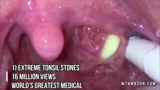
Tonsil Stone Removal Techniques

Sober Living Facility @ http://soberliving.ca/guide-to-sober-living/
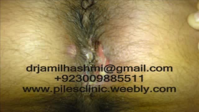
Treatment for Piles,Fistula,hemorrhoids, Hydrocele Without Operation or surgery in pakistan Dr Jamil Ahmad Hashmi ( haripur hazar pakistan )... +923009885511 --- drjamil79@gmail.com
Treatment for Piles,Fistula,Hydrocele Without Operation piles treatment with 60 days Quickly! pain free treatment full life Piles Medicine dr jamil ahmad hashmi ( haripur hazar pakistan ) drjamil79@yahoo.com +923009885511 piles treatment with 60 days Quickly! pain free treatment full life Piles Medicine dr jamil ahmad hashmi...
Before Dr. Benjamin Carson became the first person to successfully separate twins conjoined at the head, before he had a TV movie made about his life, before he became known for his "gifted hands" and before he became head of pediatric neurosurgery at Johns Hopkins, Ben Carson was headed down the wrong path in life.
Symptoms of Acute Angle-Closure Glaucoma Hazy or blurred vision. The appearance of rainbow-colored circles around bright lights. Severe eye and head pain. Nausea or vomiting (accompanying severe eye pain) Sudden sight loss.
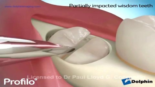
our dentist says it's time to remove your wisdom teeth. He may refer you to an oral surgeon, who will do the procedure in his office. It should only take a few days for you to heal and feel back to normal.

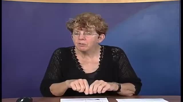
This one goes out to all the student, resident and fellows trying to clarify what their bosses are trying to say to the patient

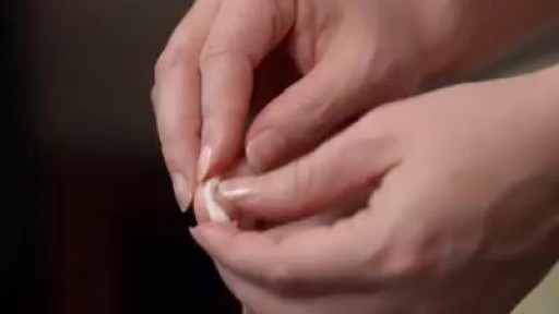
An ingrown toenail may be painful, but most you can treat at home. Here's how -- and when to call a doctor:
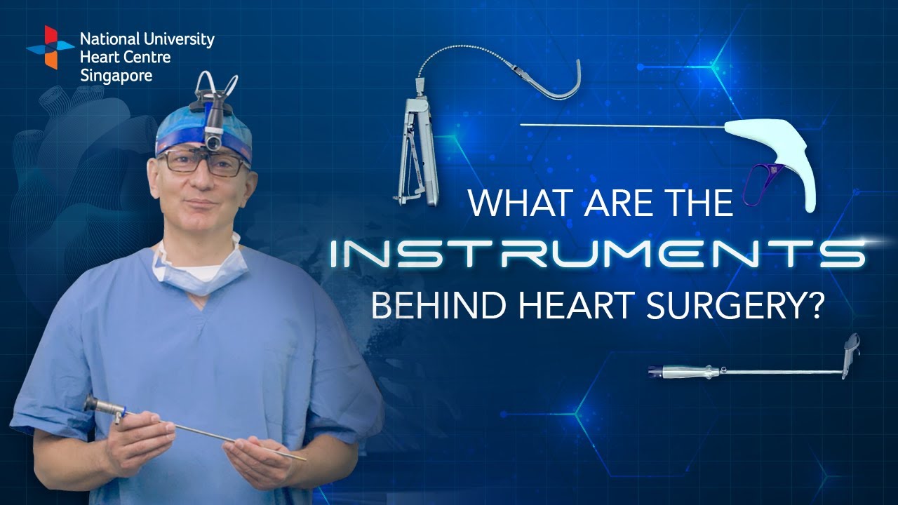
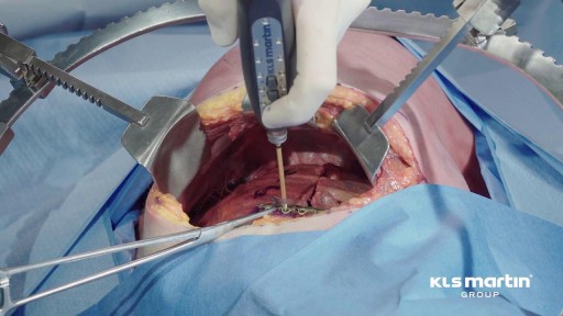
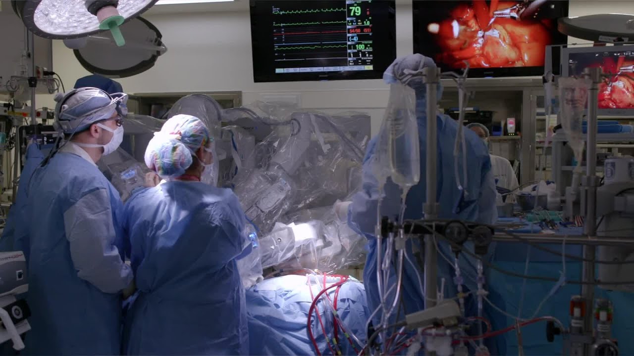
Instruments at work, innovation at play. 🔍
Watch on to discover the behind-the-scenes instruments utilised by our NUHCS cardiac surgery expert, A/Prof Theodoros Kofidis, Head of NUHCS' Department of Cardiac, Thoracic & Vascular Surgery (CTVS), for keyhole heart operations. 🔑
To find out more about Minimally Invasive Heart Surgery @ NUHCS, visit: https://[a]www.nuhcs.com.sg%2FOur-Services%2FSpecialties%2FPages%2FMinimally-Invasive-Cardiac-Surgery-Programme.aspx[/a]
Connect with us:
Instagram: @nuhcsofficial
Facebook: www.facebook.com/nuhcs
Website: www.nuhcs.com.sg
LinkedIn: www.linkedin.com/company/nuhcs
To make an appointment with the NUHCS Heart Clinic, email us at appointment@nuhs.edu.sg
#NUHCS #cardiacsurgery #heartsurgery #keyholesurgery #minimallyinvasive
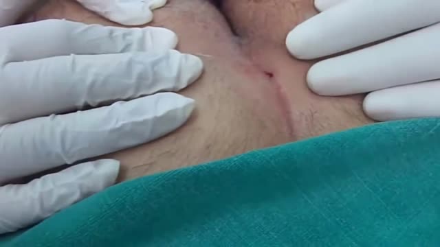
A pilonidal sinus (PNS) is a small cyst or abscess that occurs in the cleft at the top of the buttocks. A PNS usually contains hair, dirt, and debris. It can cause severe pain and can often become infected. If it becomes infected, it may ooze pus and blood and have a foul odor. A PNS is a condition that mostly affects men and is also common in young adults. It’s also more common in people who sit a lot, like cab drivers.
A pelvic mass is a general term for any growth or tumor on the ovary or in the pelvis. A pelvic mass can be cystic (cystadenoma), solid (fibroma), or both (dermoid). A pelvic mass can be benign or malignant.
A fractured rib is usually a result of a fall or accident. Prolonged coughing and sports with repetitive movement, such as golf, also can cause a rib fracture. Symptoms include pain when taking a deep breath, pressing on the injured area, or bending or twisting the body. In most cases, fractured ribs usually heal on their own in one or two months. Pain relievers can make it easier to breathe deeply.
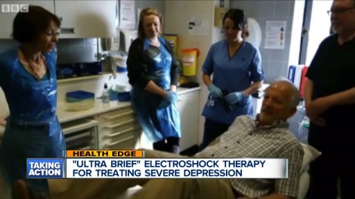
With ECT, electrodes are placed on the patient's scalp and a finely controlled electric current is applied while the patient is under general anesthesia. The current causes a brief seizure in the brain. ECT is one of the fastest ways to relieve symptoms in severely depressed or suicidal patients.
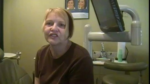
We have just enhanced the smile of another wonderful patient! She just received 6 mini dental implants place by DR. Jue www.sugarlanddentalspa.com.
To learn more about robotically assisted heart surgery, please visit https://cle.clinic/2Y6aHXH
Robotically assisted heart surgery is a minimally invasive option most often used for mitral valve repair. Cleveland Clinic cardiothoracic surgeons explain how it works and what to expect.
To learn more about our cardiothoracic experts, please visit
Marc Gillinov, MD - https://cle.clinic/2ZtNM7b
Daniel Burns, MD - https://cle.clinic/2W1MdxI
If you liked the video hit like and subscribe for more!
#clevelandclinic #heartsurgery #roboticsurgery #heartcare #cardiothoracic
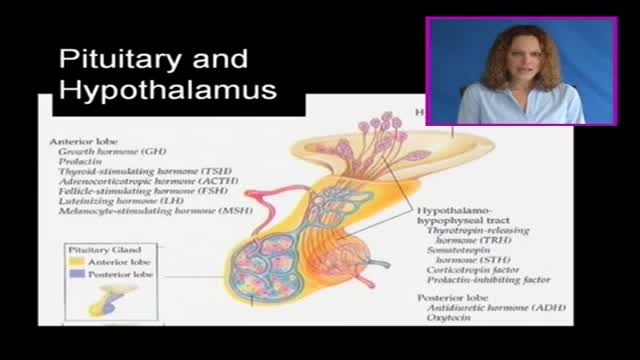
The pituitary gland is often portrayed as the "master gland" of the body. Such praise is justified in the sense that the anterior and posterior pituitary secrete a battery of hormones that collectively influence all cells and affect virtually all physiologic processes. The pituitary gland may be king, but the power behind the throne is clearly the hypothalamus. As alluded to in the last section, some of the neurons within the hypothalamus - neurosecretory neurons - secrete hormones that strictly control secretion of hormones from the anterior pituitary. The hypothalamic hormones are referred to as releasing hormones and inhibiting hormones, reflecting their influence on anterior pituitary hormones.
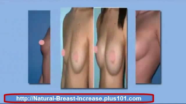
http://natural-breast-increase.plus101.com
---How To Make Bigger Breast. Can You Really Increase You Breast Size Naturally Without Surgery, Pills, Creams Etc Etc
Well a new website call natural-breast-increase.plus101.com says you can and thousands of women have already used there strategies.
And the best part about the this method is that you don't have to go through any expensive and painful surgery.
If you'd like to increase your own cup size then just visit the site below. I recommend the methods 100%
http://natural-breast-increase.plus101.com
How To Make Bigger Breast
http://www.youtube.com/watch?v=0cTQ3SZ4JIk
How To Make Bigger Breast,
can fenugreek increase breast size,
can i increase my breast size,
can i increase my breast size without surgery,
can i naturally increase breast size,
can we increase breast size naturally,
can women increase breast size naturally,
can you increase breast size,
can you increase breast size naturally,
can you make your breasts bigger,
cheap breast augmentation,
cheap breast implants,
cost of breast implants,
cream for breast enlargement,
cream to increase breast size,
curves breast enhancers,
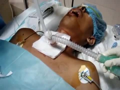
we use a single, tapered PDT dilator and kit . All the equipment and supplies listed must be present at the bed-side, because there is no time to go looking for supplies if an airway emergency occurs during the procedure. Two teams are used simultaneously. One team manages the endo-tracheal tube, and the other manages the placement of the tracheostomy tube

In this video, Professor Dan Reinstein performs a bilateral LASIK procedure filmed in real-time to demonstrate the full 8 and-a-half minute procedure from multiple angles. The superior design and experience of the Carl Zeiss Meditec Visumax femtosecond Laser for flap creation is seen, where the patient is only in contact with the device for about 30 seconds with extremely low contract force such that the patient feels effectively nothing, there are no red splodges (subconjunctival haemorages) left behind. From the surgeons' standpoint there is no device that is easier to use or faster for LASIK flap creation. The Carl Zeiss Meditec MEL80 excimer laser portion of the procedure is seamlessly integrated and incorporates all the features that make clinical outcomes so reproducible including the unique cone-for-controlled-atmosphere (CCA) and high efficiency, high sensitivity calibration test which can be performed for each individual patient to compensate for minor changes in energy that occur with excimer laser devices during the course of a day.
For reference to the clinical outcomes for LASIK with the MEL80 in presbyopia using PRESBYOND Laser Blended Vision see:
Reading glasses presbyopia (ageing eyes) only:
LASIK for presbyopia correction in emmetropic patients using aspheric ablation profiles and a micro-monovision protocol with the Carl Zeiss Meditec MEL 80 and VisuMax.
J Refract Surg. 2012 Aug;28(8):531-41. Reinstein DZ, Carp GI, Archer TJ, Gobbe M.
http://www.ncbi.nlm.nih.gov/pubmed/22869232
Short sighted, astigmatism and presbyopia (ageing eyes)
LASIK for Myopic Astigmatism and Presbyopia Using Non-Linear Aspheric Micro-Monovision with the Carl Zeiss Meditec MEL 80 Platform.
J Refract Surg. 2011 Jan;27(1):23-37. Epub 2010 Mar 1.
Reinstein DZ, Archer TJ, Gobbe M.
http://www.ncbi.nlm.nih.gov/pubmed/20205360
Long-sighted, astigmatism and presbyopia (ageing eyes)
LASIK for hyperopic astigmatism and presbyopia using micro-monovision with the Carl Zeiss Meditec MEL80 platform.
J Refract Surg. 2009 Jan;25(1):37-58. Reinstein DZ, Couch DG, Archer TJ.
http://www.ncbi.nlm.nih.gov/pubmed/19244952
For more information about laser eye surgery and PRESBYOND Laser Blended Vision, please contact the London Vision Clinic on 020 7224 1005.
