
- Physical Examination
- Surgical Examination
- Ophthalmology
- Clinical Skills
- Orthopedics
- Surgery Videos
- Laparoscopy
- Pediatrics
- Funny Videos
- Cardiothoracic Surgery
- Nursing Videos
- Plastic Surgery
- Otorhinolaryngology
- Histology and Histopathology
- Neurosurgery
- Dermatology
- Pediatric Surgery
- Urology
- Dentistry
- Oncology and Cancers
- Anatomy Videos
- Health and Fitness
- Radiology
- Anaesthesia
- Physical Therapy
- Pharmacology
- Interventional Radiology
- Cardiology
- Endocrinology
- Gynecology
- Emergency Medicine
- Psychiatry and Psychology
- Childbirth Videos
- General Medical Videos
- Nephrology
- Physiology
- Diet and Food Health
- Diabetes Mellitus
- Neurology
- Women Health
- Osteoporosis
- Gastroenterology
- Pulmonology
- Hematology
- Rheumatology
- Toxicology
- Nuclear Medicine
- Infectious Diseases
- Vascular Disease
- Reproductive Health
- Burns and Wound Healing
- Other
Top videos


Watch that video to know the Symptoms and Causes Of Female Genital Infections

UCLA Hand Transplant Procedure
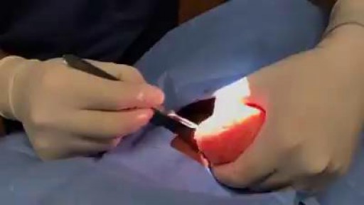
Lipomas are slow-growing soft tissue tumours that rarely reach a size larger than 2 cm. Lesions larger than 5 cm, so-called giant lipomas, can occur anywhere in the body but are seldom found in the upper extremities. The authors present their experiences with eight patients having giant lipomas of the upper extremity. In addition, a review of the literature, and a discussion of the appropriate evaluation and management are included.
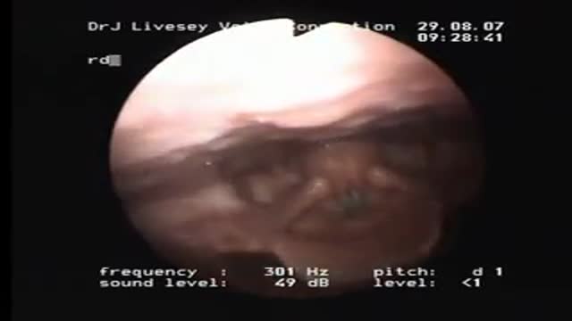
A check up at the Ear Nose & Throat doctor to make sure Genie's Opera singing vocal chords are working properly.
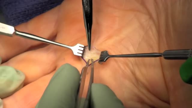
Trigger finger, also known as stenosing tenosynovitis (stuh-NO-sing ten-o-sin-o-VIE-tis), is a condition in which one of your fingers gets stuck in a bent position. Your finger may straighten with a snap — like a trigger being pulled and released. Trigger finger occurs when inflammation narrows the space within the sheath that surrounds the tendon in the affected finger. If trigger finger is severe, your finger may become locked in a bent position. People whose work or hobbies require repetitive gripping actions are at higher risk of developing trigger finger. The condition is also more common in women and in anyone with diabetes. Treatment of trigger finger varies depending on the severity.
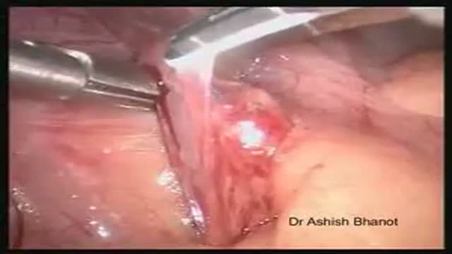
Laparoscopic Appendectomy for Acute Appendicitis Post-illeal Appendix
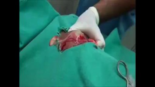
Different types of Abscess- Drainage and Aspiration of Pus.

Patient 65-year-old of age who comes to the medical consultation with pain moderated pain in the right hypochondrium of “several years of evolution” but that it increased one week ago. Also, she shows pain in the umbilical region of “many years of evolution”, that is supported according to the patient - in a constant way.rnTo the examination, we observe an umbilical hernia, apparently divided into two parts. The hernia of the external region measures 25.1 centimeters x 18.0 centimeters and the one that occupies the average region measures 12.0 centimeters x 10.0 centimeters.rnPatient who comes to the medical consultation with moderated pain in the right hypochondrium of one year of evolution but it increased one week ago after eat duck.rnIn the ultrasound scan of the region of the right hypochondrium (patient came having breakfast, that is to say, without previous preparation ) we can observe the liver of 123.8 millimeters high, as well as the porta vein with a diameter of 7.3 millimeters.rnOn having observed the Gallbladder, we think that a side wall is increased in 2.7 mm (hyperechogenic) with several “echogenics points” in the interior (”Biliary Mud”).
The measurements of the gallbladder were: 39.0 x 17.4 millimeters.rnWe can appreciates an echogenic image in the interior that it would make think about stone. The stones are identified as echogenic foci casting acoustic shadowing but but this image did not appear and a re-evaluation is decided in 15 days.
Acalculous cholecystopathy which means disease or condition of the gallbladder without the presence of gallstones. You might also call it functional gallbladder disorder or impaired gallbladder emptying. Some causes may be chronic inflammation, a problem with the smooth muscles of the gallbladder or the muscle of the Sphincter of Oddi being too tight.
REMEMBER:
Umbilical hernia is a congenital malformation, especially common in infants of African descent, and more frequent in boys. An Acquired umbilical hernia directly results from increased intra-abdominal pressure and are most commonly seen in obese individuals.
Presentation:A hernia is present at the site of the umbilicus (commonly called a navel, or belly button) in the newborn; although sometimes quite large, these hernias tend to resolve without any treatment by around the age of 5 years. Obstruction and strangulation of the hernia is rare because the underlying defect in the abdominal wall is larger than in an inguinal hernia of the newborn. The size of the base of the herniated tissued is inversely correlated with risk of strangulation (i.e. narrow base is more likely to strangulate).
Babies are prone to this malformation because of the process during fetal development by which the abdominal organs form outside the abdominal cavity, later returning into it through an opening which will become the umbilicus.
Differential diagnosisrnImportantly this type of hernia must be distinguished from a paraumbilical hernia which occurs in adults and involves a defect in the midline near to the umbilicus, and from omphalocele.

Most intact aortic aneurysms do not produce symptoms. As they enlarge, symptoms such as abdominal pain and back pain may develop. Compression of nerve roots may cause leg pain or numbness. Untreated, aneurysms tend to become progressively larger, although the rate of enlargement is unpredictable for any individual. Rarely, clotted blood which lines most aortic aneurysms can break off and result in an embolus. They may be found on physical examination. Medical imaging is necessary to confirm the diagnosis. Symptoms may include: anxiety or feeling of stress; nausea and vomiting; clammy skin; rapid heart rate. In patients presenting with aneurysm of the arch of the aorta, a common symptom is a hoarse voice as the left recurrent laryngeal nerve (a branch of the vagus nerve) is stretched. This is due to the recurrent laryngeal nerve winding around the arch of the aorta. If an aneurysm occurs in this location, the arch of the aorta will swell, hence stretching the left recurrent laryngeal nerve. The patient therefore has a hoarse voice as the recurrent laryngeal nerve allows function and sensation in the voicebox. Abdominal aortic aneurysms, hereafter referred to as AAAs, are the most common type of aortic aneurysm. One reason for this is that elastin, the principal load-bearing protein present in the wall of the aorta, is reduced in the abdominal aorta as compared to the thoracic aorta (nearer the heart). Another is that the abdominal aorta does not possess vasa vasorum, hindering repair. Most are true aneurysms that involve all three layers (tunica intima, tunica media and tunica adventitia), and are generally asymptomatic before rupture. The most common sign for the aortic aneuysm is the Erythema nodosum also known as leg lesions typically found near the ankle area. The prevalence of AAAs increases with age, with an average age of 65–70 at the time of diagnosis. AAAs have been attributed to atherosclerosis, though other factors are involved in their formation. An AAA may remain asymptomatic indefinitely. There is a large risk of rupture once the size has reached 5 cm, though some AAAs may swell to over 15 cm in diameter before rupturing. Before rupture, an AAA may present as a large, pulsatile mass above the umbilicus. A bruit may be heard from the turbulent flow in a severe atherosclerotic aneurysm or if thrombosis occurs. Unfortunately, however, rupture is usually the first hint of AAA. Once an aneurysm has ruptured, it presents with a classic pain-hypotension-mass triad. The pain is classically reported in the abdomen, back or flank. It is usually acute, severe and constant, and may radiate through the abdomen to the back. The diagnosis of an abdominal aortic aneurysm can be confirmed at the bedside by the use of ultrasound. Rupture could be indicated by the presence of free fluid in potential abdominal spaces, such as Morison's pouch, the splenorenal space (between the spleen and left kidney), subdiaphragmatic spaces (underneath the diaphragm) and peri-vesical spaces. A contrast-enhanced abdominal CT scan is needed for confirmation. Only 10–25% of patients survive rupture due to large pre- and post-operative mortality. Annual mortality from ruptured abdominal aneurysms in the United States alone is about 15,000. Another important complication of AAA is formation of a thrombus in the aneurysm.
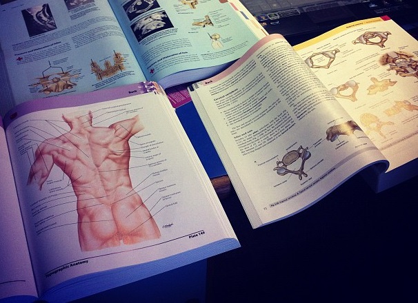
Watch that video to learn How to Study The Human Anatomy

Watch that video to know the Natural Ways to Whiten Teeth at Home
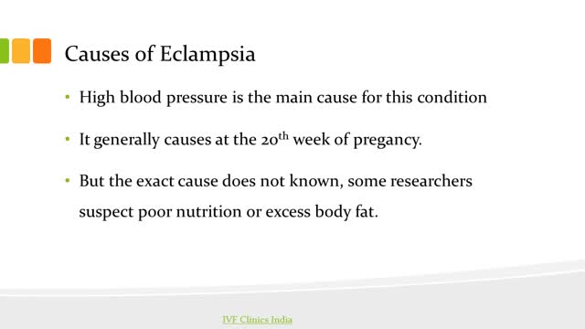
Ten percent of all pregnancies are complicated by hypertension. Eclampsia and preeclampsia account for about half of these cases worldwide, and these conditions have been recognized and described for years despite the general lack of understanding of the disease. [1] In the fifth century, Hippocrates noted that headaches, convulsions, and drowsiness were ominous signs associated with pregnancy. In 1619, Varandaeus coined the term eclampsia in a treatise on gynecology. [2, 3]
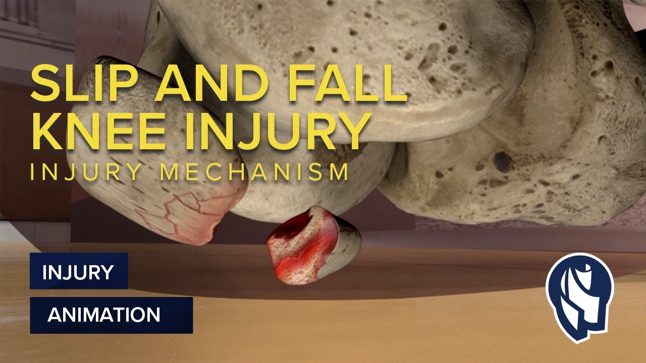
MotionLit can help you multiply the value of your case by portraying the mechanisms of injuries in a 3D Accident Reconstruction Animation. Call (855)850-0650 or visit motionlit.com to learn & earn more for your client!
MotionLit is a one-stop-shop for litigators, offering complete trial services from video production, animation, litigation support, exhibit design, trial technician, and video depositions. Our visuals have proven to help attorneys obtain record-breaking verdicts with our persuasive trial presentations, settlement documentaries, and demonstratives.
Contact Us At:
www.motionlit.com
(213) 291 9141
info@motionlit.com
Follow Us On:
Twitter: @motionlit
Instagram: https://www.instagram.com/motionlit/
Facebook: https://www.facebook.com/MotionLit/

ROTIGS medical device by Honolulu inventor Dr. Brad NaPier makes tracheal intubations easier for medical professionals. For more info, visit www.rotigs.com
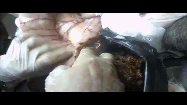
Megacolon, as well as megarectum, is a descriptive term. It denotes dilatation of the colon that is not caused by mechanical obstruction.[1, 2] Although the definition of megacolon has varied in the literature, most researchers use the measurement of greater than 12 cm for the cecum as the standard. Because the diameter of the large intestine varies, the following definitions would also be considered: greater than 6.5 cm in the rectosigmoid region and greater than 8 cm for the ascending colon. Megacolon can be divided into the following 3 categories: Acute megacolon ( pseudo-obstruction) Chronic megacolon, which includes congenital, acquired, and idiopathic causes Toxic megacolon
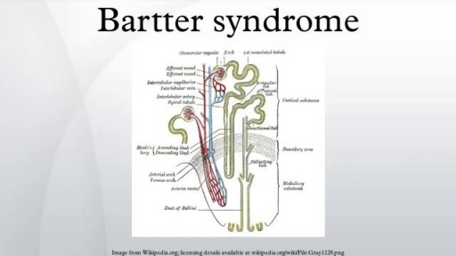
Bartter syndrome is a rare inherited defect in the thick ascending limb of the loop of Henle. It is characterized by low potassium levels (hypokalemia), increased blood pH (alkalosis), and normal to low blood pressure. There are two types of Bartter syndrome: neonatal and classic
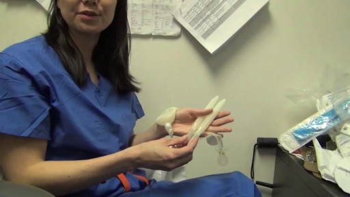
Implant comparison: Coloplast vs AMS for Penile Implants
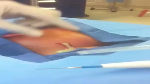
Huge pus in the neck after a bad cold and sore throat!
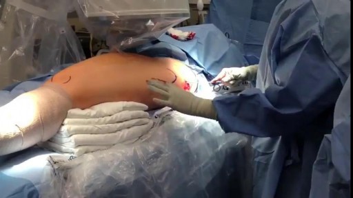
This is an example of a surgery to fix a femur (thigh bone) fracture utilizing an intramedullary nail. This is a minimally invasive way of fixing this surgical problem and allows for immediate range of motion and full weight-bearing.
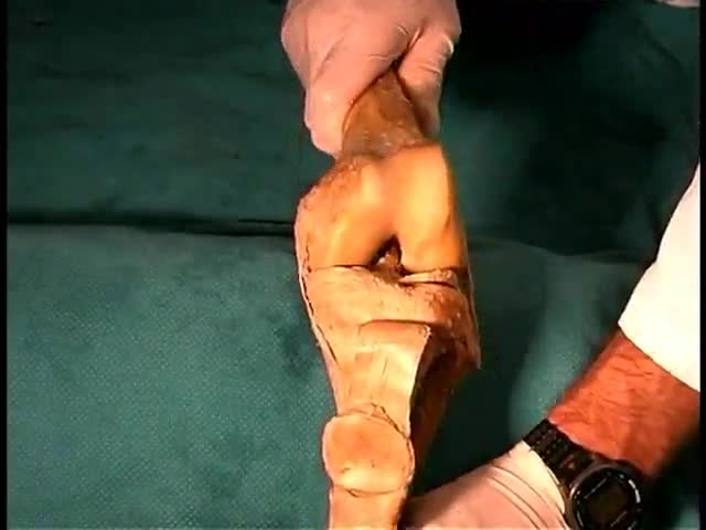
Anatomy of The Lower Limb Joints
