- Physical Examination
- Surgical Examination
- Ophthalmology
- Clinical Skills
- Orthopedics
- Surgery Videos
- Laparoscopy
- Pediatrics
- Funny Videos
- Cardiothoracic Surgery
- Nursing Videos
- Plastic Surgery
- Otorhinolaryngology
- Histology and Histopathology
- Neurosurgery
- Dermatology
- Pediatric Surgery
- Urology
- Dentistry
- Oncology and Cancers
- Anatomy Videos
- Health and Fitness
- Radiology
- Anaesthesia
- Physical Therapy
- Pharmacology
- Interventional Radiology
- Cardiology
- Endocrinology
- Gynecology
- Emergency Medicine
- Psychiatry and Psychology
- Childbirth Videos
- General Medical Videos
- Nephrology
- Physiology
- Diet and Food Health
- Diabetes Mellitus
- Neurology
- Women Health
- Osteoporosis
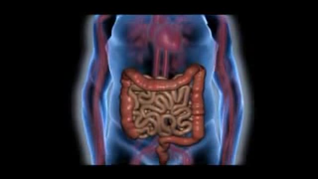
- Gastroenterology
- Pulmonology
- Hematology
- Rheumatology
- Toxicology
- Nuclear Medicine
- Infectious Diseases
- Vascular Disease
- Reproductive Health
- Burns and Wound Healing
- Other
Top videos
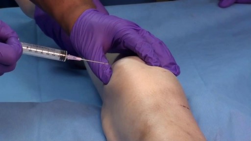
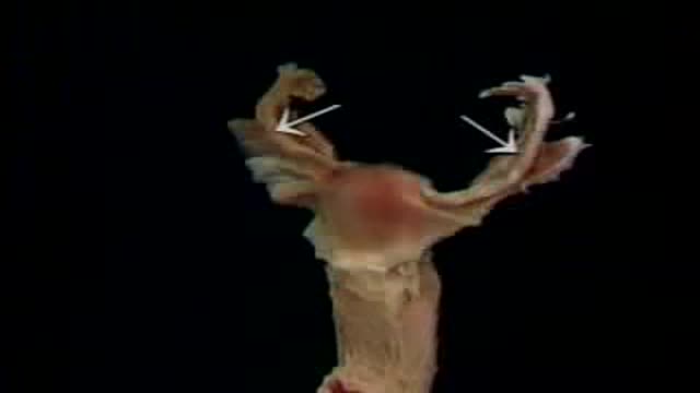
The clinician performing the procedure should be familiar with the anatomy of the specific joint and cognizant of the relevant landmarks in order to avoid puncture of tendons, blood vessels, and nerves (see the images below).
Amazing Surgery: Bilateral Nephrectomy for polycystic kidneys and cholecystectomy.
Genital warts are growths on the skin of the genital area and around the anus. They are caused by certain types of the human papilloma virus (HPV). There are more than 100 types of HPV. Some types of HPV produce warts on different parts of the body, like plantar warts on the feet and common hand warts. There is no specific treatment for HPV, but there are treatments for health problems caused by HPV. Genital warts can be treated by your healthcare provider, or with prescription medication. HPV-related cancers are more treatable when diagnosed and treated promptly. For more information, visit www.cancer.org.
Implantation of a long-lasting implant for diabetic macular edema (DME)- steroidal implants
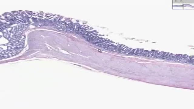
Histology of GastroDuodenal Junction
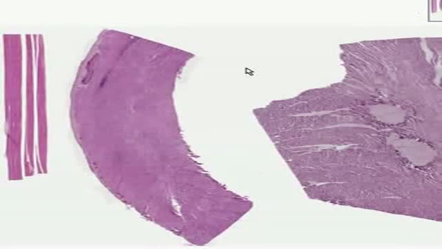
Histology of Mucles Skeletal Smooth Cardiac

Atrial flutter is a type of abnormal heart rate, or arrhythmia. It occurs when the upper chambers of your heart beat too fast. When the chambers in the top of your heart (atria) beat faster than the bottom ones (ventricles), it complicates your heart rhythm
Cell Adhesion Molecule Inhibition Animation
Worlds Most Amazing Medical Case

There are four major blood groups determined by the presence or absence of two antigens – A and B – on the surface of red blood cells: Group A – has only the A antigen on red cells (and B antibody in the plasma) Group B – has only the B antigen on red cells (and A antibody in the plasma) Group AB – has both A and B antigens on red cells (but neither A nor B antibody in the plasma) Group O – has neither A nor B antigens on red cells (but both A and B antibody are in the plasma)
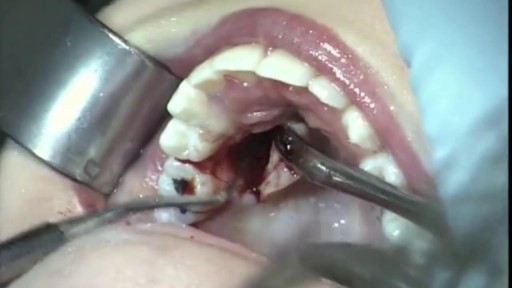
wisdom teeth removal - surgery,extraction

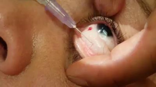
Business Insider's Michelle Yan has been nearsighted since she was 9 years old. After laser eye surgery, she has 20/20. She walks us through the pre-surgery steps, the actual surgery, as well as the recovery process.
MORE MEDICAL TECH:
8 Medical Procedures That Are Improving Lives
https://www.youtube.com/watch?v=kTMMrAP6DNI
13 Medical Procedures Changing The Health World
https://www.youtube.com/watch?v=VAR44vnxWis
Lifelike Medical Robot Actually Bleeds
https://www.youtube.com/watch?v=IjnhmcCQLsc
------------------------------------------------------
#Lasik #Surgery #TechInsider
Tech Insider tells you all you need to know about tech: gadgets, how-to's, gaming, science, digital culture, and more.
Visit us at: https://www.businessinsider.com
TI on Facebook: https://www.facebook.com/techinsider
TI on Instagram: https://www.instagram.com/tech_insider/
TI on Twitter: https://twitter.com/techinsider
TI on Amazon Prime: http://read.bi/PrimeVideo
INSIDER on Snapchat: https://insder.co/2KJLtVo
------------------------------------------------------
What It's Like To Get Laser Eye Surgery

Nursing skills lab procedure for IV insertion.

Ellis demonstrates the need to drop the tip of the needle when withdrawing medication from a vial.
#NCLEX #NewGrad #ClinicalSkills #HESI #Kaplan #ATI #NursingSchool #NursingStudent #Nurse #RN #PN #Education #LPN #NurseEducator #lvn
🚨 Reminder: shipping deadlines are looming 👀
🎁 Regular Shipping: Order by Friday, December 15
🚀 Expedited Shipping: Order by Monday, December 18
🔍 Still searching for last-minute gifts? Consider a Level Up RN Gift Card! 💌 It’s not only a thoughtful present but also the perfect way to share treasures like Pharmacology Flashcards OR digital treasures like Flashables Digital Nursing Flashcards & the Level Up RN membership. Give the gift of knowledge this holiday season! 🧠⚡️💖 bit.ly/LevelUpRNGC
🚪 Access our Cram Courses, Quizzes and Videos all in one ad free space with Level Up RN Membership https://bit.ly/LevelUpRNMembership
Want more ways to MASTER Clinical Skills? Check out our flashcards & videos!
👇👇👇👇👇👇👇👇👇👇
👉 https://bit.ly/clinicalnursingskills 👈
☝️👆☝️👆☝️👆☝️👆☝️👆
This is your one-stop-shop for materials to help you LEARN & REVIEW so you can PASS Nursing School.
🤔🤔🤔 DO YOU WANT TO PASS your classes, proctored exams and the NCLEX? 🤔🤔🤔 Our resources are the best you can buy. They are built with a single goal: help you pass with no fluff. Everything you need, and nothing you don’t. Don’t take our word for it, though! Check out our hundreds of ⭐️⭐️⭐️⭐️⭐️ reviews from nurses who passed their exams and the NCLEX with Level Up RN.
🗂️ Our Ultimate Nursing School Survival kit is your number 1 resource to get through nursing school and to pass the NCLEX. Whether you're just starting school or you’re already prepping for the NCLEX, this bundle of flashcards is the best you can buy. It covers all the information you need to know to pass all your exams and it has FREE shipping!
➡️ https://bit.ly/TUNSSK ⬅️
L👀king for EVEN MORE resources to survive Nursing School? Make your Nursing School experience your own! Life’s difficult enough—learning shouldn’t be.
🪅 Games https://nursesquad.com
💻 Digital resources https://bit.ly/NursingStudyCourses
📅 Organizational tools https://bit.ly/OrganizingSchool
✨Want perks? Join our channel!
https://youtube.com/leveluprn/join
🏷 Head to https://leveluprn.com/specials for all our latest deals!🥳️
📧 LOOKING FOR FREE RESOURCES TO HELP WITH YOUR EXAMS? Get exclusive tips, latest video releases and more delivered to your email!
➡️ https://leveluprn.com/signup ⬅️
⚕ 👩 LEVEL UP NURSE SQUAD 👩⚕️
All of the nurses at Level Up RN are here to help! Cathy Parkes started helping her fellow classmates back when she was in nursing school, tutoring so they could pass their exams and graduate. After she got her BSN and started working as an RN at Scripps Encinitas Hospital, she started this YouTube channel to help nursing students around the world. Since then she has built a team of top-notch dedicated nurses and nurse educators who are focused on improving nursing education and supporting career advancement for nurses everywhere. With flashcards, videos, courses, organizational tools and more, we are singularly focused on helping students and nurses Level Up on their exams and nursing careers.

How to Improve Sexual Health or Stamina part 4 All Solution of Male Disorder Male Infertility Diagnostic and Treatment Re-Slim Care Latest Technology in Pakistan Dr. Aslam Naveed is a well known sexologist in Pakistan. He has treated more than 1 Lac patients since last 30 years of clinical Practice in sexology, he knows how to help the people facing sexual disorders. Contact: 02134965050, 03432821919 www.sexologistpakistan.com https://www.facebook.com/menssexcareclinic/ https://youtu.be/_fRbtwWtLoE Part 1 https://youtu.be/S17bCnwCLuI Part 2 https://youtu.be/CPAXxkdz7mM Part 3 https://youtu.be/YlsdBZJ4prg Part 4 https://youtu.be/fylxbK4azvs Part 5 https://youtu.be/Zb8TcdgJ7Io Part 6 https://youtu.be/0wbDDNAwsmo Part 7 https://youtu.be/gHDmwfsMgTw Part 8 https://youtu.be/IasXoRKUlV4 Part 9 ADDRESS: Men’s Care Modern Hospital, Opposite, Safari Park, University Road, Karachi, Pakistan.
Breath sounds can be either normal or abnormal. These sounds come from the lungs when you breathe in or out. These sounds can be heard using a stethoscope or simply when breathing. Abnormal breath sounds can indicate a lung problem, such as: an obstruction inflammation an infection fluid in the lungs asthma Listening to breath sounds is an important part of diagnosing many different medical conditions.
Female Reproductive System Anatomy

Foreceps Delivery Birth Video
