शीर्ष वीडियो
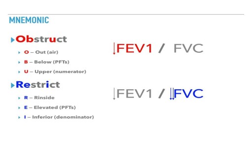
Obstructive lung diseases include conditions that make it hard to exhale all the air in the lungs. People with restrictive lung disease have difficulty fully expanding their lungs with air. Obstructive and restrictive lung disease share the same main symptom: shortness of breath with exertion.
Anterior Cruciate Ligament Reconstruction
Thoracic Epidural Placement Paramedian Approach
The term dermoid cyst does not appear to be restricted to a single kind of lesion nor is it used in only a single medical discipline. The term dermoid cyst can be found in the vocabulary of dermatologists, dermatopathologists, general pathologists, gynecologists, neurosurgeons, or pediatricians. If asked, all of these clinicians would most probably define and describe dermoid cysts differently. For example, gynecologists and general pathologists might say that a dermoid cyst is a cystic tumor of the ovary. In contrast, neurosurgeons tend to view a dermoid cyst is associated with a congenital cyst of the spine or an intracranial congenital cyst. For pediatricians and dermatologists, dermoid cyst means subcutaneous cysts, which are usually congenital.[1]

Panic attacks involve sudden feelings of terror that strike without warning. These episodes can occur at any time, even during sleep. People experiencing a panic attack may believe they are having a heart attack or they are dying or going crazy. The fear and terror that a person experiences during a panic attack are not in proportion to the true situation and may be unrelated to what is happening around them. Most people with panic attacks experience several of the following symptoms:
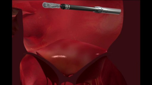
Certain high-surgical-risk patients with severe degenerative mitral regurgitation (DMR) now have a minimally invasive treatment option. MitraClip® therapy is a minimally-invasive transcatheter mitral valve repair (TMVr) procedure that has been established as a proven option with demonstrated safety and clinically important improvements. Used in more than 25,000 patients worldwide, MitraClip® is a well-established therapy. The MitraClip® device received CE Mark approval in Europe in 2008 and U.S. FDA approval in 2013, and has been approved for commercial use in 50 countries throughout the world.

How to Improve Sexual Health or Stamina part 4 All Solution of Male Disorder Male Infertility Diagnostic and Treatment Re-Slim Care Latest Technology in Pakistan Dr. Aslam Naveed is a well known sexologist in Pakistan. He has treated more than 1 Lac patients since last 30 years of clinical Practice in sexology, he knows how to help the people facing sexual disorders. Contact: 02134965050, 03432821919 www.sexologistpakistan.com https://www.facebook.com/menssexcareclinic/ https://youtu.be/_fRbtwWtLoE Part 1 https://youtu.be/S17bCnwCLuI Part 2 https://youtu.be/CPAXxkdz7mM Part 3 https://youtu.be/YlsdBZJ4prg Part 4 https://youtu.be/fylxbK4azvs Part 5 https://youtu.be/Zb8TcdgJ7Io Part 6 https://youtu.be/0wbDDNAwsmo Part 7 https://youtu.be/gHDmwfsMgTw Part 8 https://youtu.be/IasXoRKUlV4 Part 9 ADDRESS: Men’s Care Modern Hospital, Opposite, Safari Park, University Road, Karachi, Pakistan.
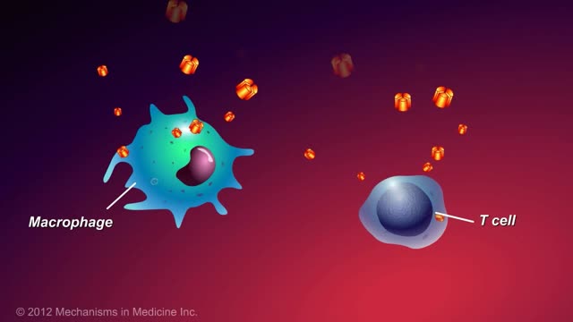
This animation describes what anti-TNF-alpha therapies are, how they work, and how patients with inflammatory bowel disease (IBD) can benefit.

A video discussing Causes of Itching in the Vulva
A subdural hematoma is a collection of blood outside the brain. Subdural hematomas are usually caused by severe head injuries. The bleeding and increased pressure on the brain from a subdural hematoma can be life-threatening.
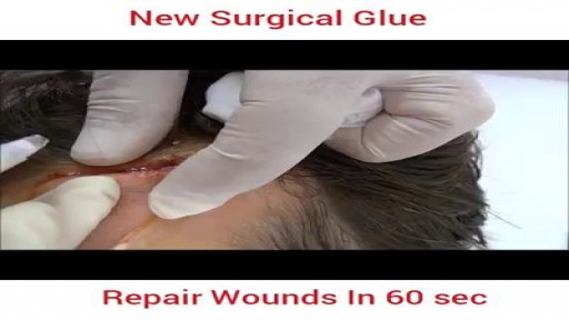
Wow Must Watch & Share wound Repair in 60 seconds

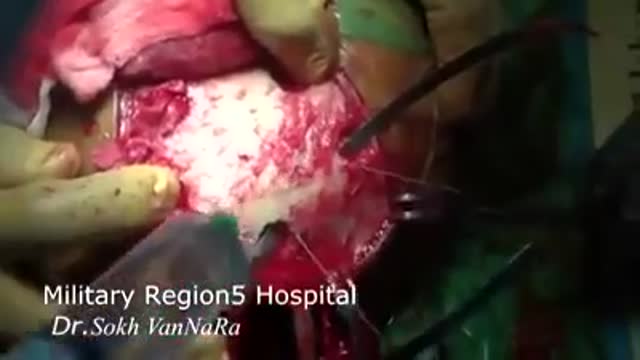
A 28 years old man lost his right arm with a conveyor device in 2014. The video is taken 2 years after replantation. You can see another videos in my site: https://drliaghatclinic.com, https://instagram.com/liaghatclinic, https://t.me/liaghatclinic
Breath sounds can be either normal or abnormal. These sounds come from the lungs when you breathe in or out. These sounds can be heard using a stethoscope or simply when breathing. Abnormal breath sounds can indicate a lung problem, such as: an obstruction inflammation an infection fluid in the lungs asthma Listening to breath sounds is an important part of diagnosing many different medical conditions.
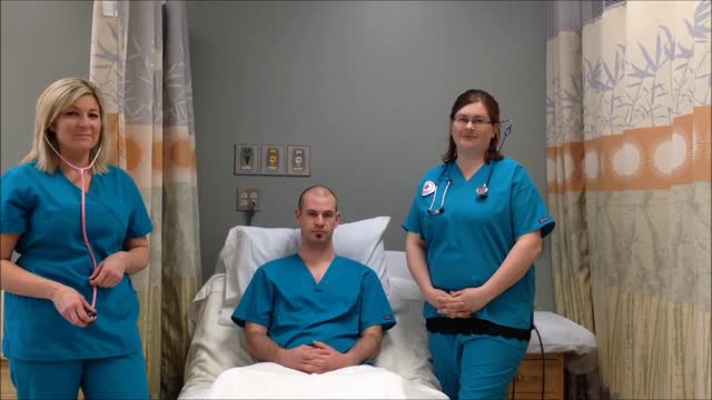
USMLE Step 2 CS - Pain Seeking This is just preview video. To get full access please visit our website : www.usmletutoring.com
The epididymis is a long coiled tube that lies above and behind each testicle. The epididymis collects and transports sperm from the testis to the vas deferens (tubes that transport sperm to the urethra). An epididymal cyst is a cyst-like mass in the epididymis that contains clear fluid. Typically, epididymal cysts and spermatoceles do not cause symptoms. When discovered, the epididymal cyst is usually about the size of a pea and feels separate from the top of the testis. Spermatoceles typically arise from the head of the epididymis, and are felt on the top portion of the testicle. Epididymal cysts and spermatoceles are often incidental findings on testicular self-examination or routine physical examination. It is important that any mass noted in the scrotum be examined by a urologist in order to obtain an accurate diagnosis, especially a mass on the testicle itself. Our team in the Division of Urology will typically be able to confirm the diagnosis on physical exam. However, a scrotal ultrasound may also be used in order to rule out other conditions.
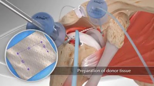
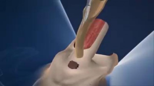
Superior capsule reconstruction (SCR) is a promising alternative treatment for irreparable posterosuperior rotator cuff tears (Figure 1). It utilizes a graft from the superior glenoid to the greater tuberosity to stabilize the humeral head. In a study by Mihata and colleagues of 23 patients who underwent SCR with a fascia lata autograft at a minimum of 2 years follow-up, the American Shoulder and Elbow Surgeons (ASES) score improved significantly from 23.5 preoperatively to 92.9. Postoperative MRI showed 83% of patients had intact reconstructions with no progression of muscle atrophy.

Craniectomy is neurosurgical procedure that involves removing a portion of the skull in order to relieve pressure on the underlying brain. This procedure is typically done in cases where a patient has experienced a very severe brain injury that involves significant amounts of bleeding around the brain or excessive swelling of the brain.
Male To Female Gender Reassignment Surgery
Female Reproductive System Anatomy

Foreceps Delivery Birth Video
