
- Physical Examination
- Surgical Examination
- Ophthalmology
- Clinical Skills
- Orthopedics
- Surgery Videos
- Laparoscopy
- Pediatrics
- Funny Videos
- Cardiothoracic Surgery
- Nursing Videos
- Plastic Surgery
- Otorhinolaryngology
- Histology and Histopathology
- Neurosurgery
- Dermatology
- Pediatric Surgery
- Urology
- Dentistry
- Oncology and Cancers
- Anatomy Videos
- Health and Fitness
- Radiology
- Anaesthesia
- Physical Therapy
- Pharmacology
- Interventional Radiology
- Cardiology
- Endocrinology
- Gynecology
- Emergency Medicine
- Psychiatry and Psychology
- Childbirth Videos
- General Medical Videos
- Nephrology
- Physiology
- Diet and Food Health
- Diabetes Mellitus
- Neurology
- Women Health
- Osteoporosis
- Gastroenterology
- Pulmonology
- Hematology
- Rheumatology
- Toxicology
- Nuclear Medicine
- Infectious Diseases
- Vascular Disease
- Reproductive Health
- Burns and Wound Healing
- Other
Top videos


Watch that video to know How to get Pregnant Fast
How To Breastfeed - Deep Latch Technique
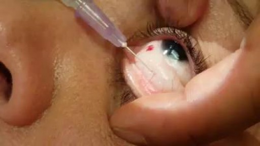
Implantation of a long-lasting implant for diabetic macular edema (DME)- steroidal implants
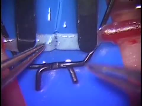
Simple microinstruments and a medical school laboratory microscope were used for anastomosis training. Chicken blood vessels were used as a material for this study. A long segment of blood vessel from the proximal brachial artery to the distal radial artery was used for training. End-to-side anastomosis was practiced first, and the training continued with end-to-end anastomosis of the appropriate segments.

Have you ever wondered if your toothbrush bristles are too hard or soft or whether or not it even mattered? Contrary to popular belief, the extra soft or soft bristle option is the best choice you can make when it comes to selecting your bristle. The soft bristles are gentler on the gums and because of their delicate nature, it’s easier for them to glide in between teeth. Using hard or medium bristles increases your risk of receding gums and also heightens tooth sensitivity, since you are wearing down the enamel on your teeth with the rough nature. Schedule a dental appointment now to learn more! visit : https://www.urbndental.com/
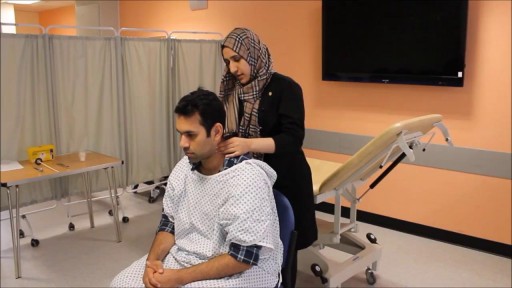

USMLE Step 2 CS - LOC This is just preview video. To get full access please visit our website : www.usmletutoring.com
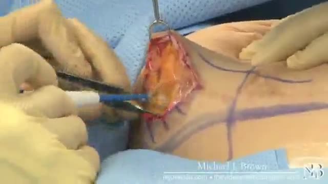
METHODS:
Previously existing methods are characterized by unpleasant scars that, despite surgeons promises, remain for life.
Incisions are:
- around the areola (Round block) leading to a flat areola, often unpleasant hypertophic skars, skin rippling.
- inverted T (around the areola, vertically down and in the fold under the breast).
- Vertical (around the areola and vertically down). Due to the extess skin, incisions often turn into inverted L or T. Rearrangement of glandular tissue and skin changes the shape of the breasts and may be different from expectations. Scars worry patients and sometimes cause disturbances in the relationship with their partner.
- No scars. The "Serdev Suture" lifting technique for breast lifting without scars (only points - needle perforations in the skin) is created by the Bulgarian cosmetic surgeon Prof. Dr. Nikolay Serdev. It is a novelty that had changed the cosmetic surgery world in the last 10-14 years for young patients. The technique is especially important in Asia and Latin America, for Asians, African-Americans, Indians, and others who form keloids and lumpy scars after operations.
The Serdev suture method can achieve lift upto and over 14 centimeters and is most suitable for the following types of breasts:
- not very heavy full breasts.
- in the presence of subpectoral implants with subsequent drooping of the breasts after childbirth and lactation.
- empty and loose breasts after childbirth and breastfeeding. In such cases this technique is combined with subpectoral implants. In sagging breasts implants should not be placed in the skin over the pectoral muscles, because thus will lead to even more drooping. Therefore, breast lift requires breast fixation to the level of the pectoral muscle (the normal position in young women), and then placement of appropriate implants under the muscle, to hold them in the appropriated position.
- in drooping breasts after subglandular augmentation (over the muscle). In such cases, patients should not wait until the skin elongation becomes visible. The implants should be removed, the capsule removed - a difficult but a necessary operation, preventing postop seromas and infection. Implants should be placed under the pectoralis muscle to wear them. Patients should orient the cosmetic surgeon at what level they want the nipples - in the middle of the implant, higher or lower.
Implants should be generally replaced - below the muscle implants should be smooth, move naturally without hurting the muscle.
Because of modern anesthetics and new methods without trauma, pain and swelling after surgery are not significant. In 3-4 days, patients can return to social life, even the next day, but it is preferable to rest for 2-3 days.
Exercises with the arms and weight lifting is prohibited for a month and a half.
Due to lack of scars, the breast lift using the Serdev sutures can be repeated to maintain the aesthetic appearence of the breasts even in advanced age.
Gigantomastia i.e. very large, very heavy and drooping breasts can not be operated in this manner, because of gravity and overskin.
Early mastopexy using Serdev sutures is recommended before too much changes in the tissues. If late, more and more complex interventions are required.
"A lot of people are opting for various breast procedures and one of the most common among them is “mastopexy”. This is the surgery that involved uplifting of sagging breasts and, in certain cases, repositioning of the nipple and areola in order to restore normality and beauty. The excess skin is removed and firmness is provided to the breasts. Though mastopexy can be done as a stand alone surgery, many people combine it with breast augmentation which involves inserting implants inside the b

Central Venous Catheter Placement & Pulmonary Artery Catheter Video

The spleen is the largest lymphoid organ. It receives blood from the splenic artery and is the only lymphoid organ that primarily filters blood instead of lymph.
Find our full video library only on Osmosis Prime: http://osms.it/more.
Join over 3 million current & future clinicians who learn by Osmosis, and over 130 universities around the world who partner with us to make medical and health education more engaging and efficient. We have unparalleled tools and materials to prepare you to succeed in school, on board exams, and as a future clinician. Sign up for a free trial at http://osms.it/more. If you're interested in exploring an institutional partnership, visit osmosis.org/educators to request a personalized demo.
Follow us on social:
Facebook: http://osms.it/facebook
Twitter: http://osms.it/twitter
Instagram for med: http://osms.it/instagram
Instagram for nursing: https://osms.it/ignursing
Linkedin: https://osms.it/linkedin
Our Vision: Everyone who cares for someone will learn by Osmosis.
Our Mission: To empower the world’s clinicians and caregivers with the best learning experience possible. Learn more here: http://osms.it/mission
Medical disclaimer: Knowledge Diffusion Inc (DBA Osmosis) does not provide medical advice. Osmosis and the content available on Osmosis's properties (Osmosis.org, YouTube, and other channels) do not provide a diagnosis or other recommendation for treatment and are not a substitute for the professional judgment of a healthcare professional in diagnosis and treatment of any person or animal. The determination of the need for medical services and the types of healthcare to be provided to a patient are decisions that should be made only by a physician or other licensed health care provider. Always seek the advice of a physician or other qualified healthcare provider with any questions you have regarding a medical condition. © 2023 Elsevier. All rights reserved.

To get started, you need to find your pelvic floor muscles by stopping urination in midstream. If you succeed, you have located the right muscles. Once you have located your pelvic floor muscles, tighten the contraction for about 5 seconds, before relaxing for another 5 seconds.

Sometimes we live in different worlds...
mply put, relapses, also known as flare ups, or (MS) attacks are new or worsening MS symptoms. But there is a concrete definition used by healthcare providers to identify MS attacks. To be considered an MS relapse: Old symptoms of MS must have become worse or new symptoms appeared.

SEX DURING PREGNANCY & 7 Tips For A Healthy Pregnancy
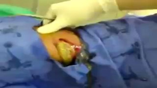
The largest amount of pus I have ever seen!!
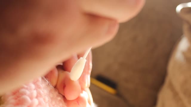
Description: Use warm water and sea salt. Soak the wart for 10 to 15 minutes in warm salt water to moisten the skin. Scrape the dead skin layers off the wart using a nail file, pumice stone or mild sandpaper. You could also use your fingers, but wash them thoroughly before and after, as warts can easily spread.

UCLA Hand Transplant Procedure

A Cryptic Pregnancy is a pregnancy, where the feotus is hidden or camouflaged in its natural environment; due to no HcG hormone being produced by the placenta and/or by difficulty in finding with ultrasound devices.
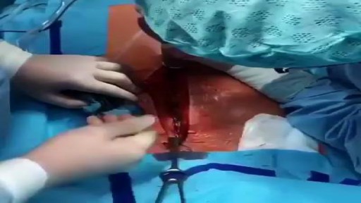
Median Sternotomy performed before open heart surgery !

Psychological testing refers to the administration of psychological tests. A psychological test is "an objective and standardized measure of a sample of behavior" (p. 4). The term sample of behavior refers to an individual's performance on tasks that have usually been prescribed beforehand.
