
- Physical Examination
- Surgical Examination
- Ophthalmology
- Clinical Skills
- Orthopedics
- Surgery Videos
- Laparoscopy
- Pediatrics
- Funny Videos
- Cardiothoracic Surgery
- Nursing Videos
- Plastic Surgery
- Otorhinolaryngology
- Histology and Histopathology
- Neurosurgery
- Dermatology
- Pediatric Surgery
- Urology
- Dentistry
- Oncology and Cancers
- Anatomy Videos
- Health and Fitness
- Radiology
- Anaesthesia
- Physical Therapy
- Pharmacology
- Interventional Radiology
- Cardiology
- Endocrinology
- Gynecology
- Emergency Medicine
- Psychiatry and Psychology
- Childbirth Videos
- General Medical Videos
- Nephrology
- Physiology
- Diet and Food Health
- Diabetes Mellitus
- Neurology
- Women Health
- Osteoporosis
- Gastroenterology
- Pulmonology
- Hematology
- Rheumatology
- Toxicology
- Nuclear Medicine
- Infectious Diseases
- Vascular Disease
- Reproductive Health
- Burns and Wound Healing
- Other
Top videos
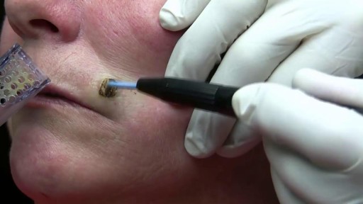

Watch that video of a Very Large Mole Removal Surgery
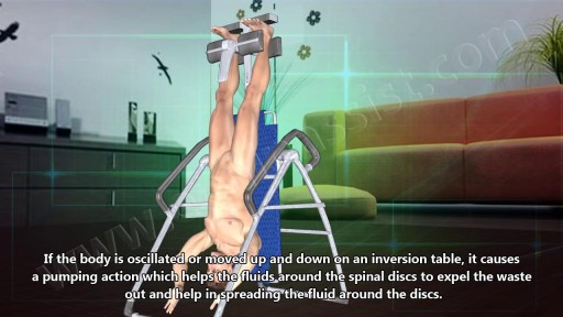
Many people report that inversion table therapy is a great way to stretch muscles and ligaments, reduce muscle spasms, and improve circulation. Stretching stimulates the lymph glands to increase the flow of lymphatic fluids; part of the body's waste disposal system. Similarly, cellular health depends on good blood circulation to deliver nourishment and remove waste. Inversion table therapy also helps to relieve motion sickness and stress. In addition, the body becomes more aware of its spatial orientation and balance when the inner ear is stimulated during inversion. Plus, it is not necessary for the body to be positioned completely upside down to gain benefits from inversion therapy! Unlike antigravity boots used with an inversion rack, an adjustable inversion table offers the flexibility to choose the most comfortable angle. Dialogue with Your Doctor Like anything that can affect your health, talk to your doctor before you start using an inversion table. This is important because certain medications and health conditions may make using an inversion table unsafe. Your doctor may recommend against inversion table therapy if you have obesity, a detached retina, fracture, glaucoma, heart condition (circulatory problem), hernia, implanted device, middle ear or eye infection, osteoporosis, are pregnant, or have a spinal injury. There may be other medical conditions not listed that your doctor may view as a contraindication.

Home hemodialysis gives you the flexibility to fit treatment into your lifestyle and may help you feel better with fewer medications and dietary restrictions. Watch this video to find out about how home hemodialysis could be right for you, and visit our website to learn more: www.FreseniusKidneyCare.com/HHD. #HomeDialysis #DialysisTreatment #KidneyDialysis
Subscribe to Our YouTube Channel
Subscribe Here: https://www.youtube.com/user/FMSMarketing
Find Fresenius Kidney Care Online at:
Website: https://www.freseniuskidneycare.com/
Facebook:@FreseniusKidneyCare
https://www.facebook.com/FreseniusKidneyCare/
Pinterest: Fresenius Kidney Care
https://www.pinterest.com/FreseniusKidneyCarePins/
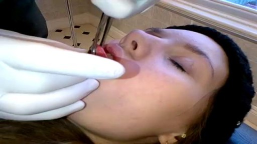
This video demonstrates tongue piercing procedure done OUTSIDE a clinical setting

M. D. Anderson Cancer Center provides a basic education on cancer.

Most healthy children are inattentive, hyperactive or impulsive at one time or another. It’s normal for preschoolers to have short attention spans and be unable to stick with one activity for long. Even in older children and teenagers, attention span often depends on the level of interest. The same is true of hyperactivity. Young children are naturally energetic — they often are still full of energy long after they’ve worn their parents out. In addition, some children just naturally have a higher activity level than others do. Children should never be classified as having ADHD just because they’re different from their friends or siblings. Children who have problems in school but get along well at home or with friends are likely struggling with something other than ADHD. The same is true of children who are hyperactive or inattentive at home, but whose schoolwork and friendships remain unaffected.
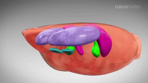
The mainstay of treatment is usually medication, talk therapy, or a combination of the two. Increasingly, research suggests these treatments may normalize brain changes associated with depression.
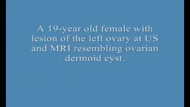
Laparoscopic Resection of Ovary Dermoid Cyst
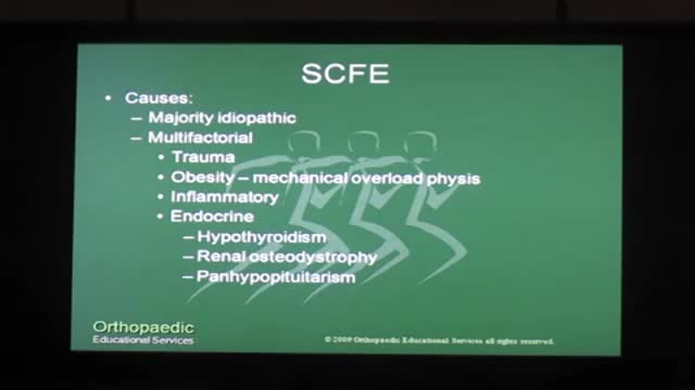
There is a strong association with obesity. In children younger than 10 years, it is associated with metabolic endocrine disorders {hypothyroidism, panhypopituitarism, hypogonadism, renal osteodystrophy, growth hormone abnormalities). SCFE is considered chronic if it has been present more than 3 weeks and acute if it has been present for 3 weeks or less. It is called "stable" if the patient can bear weight and "unstable" if the patient cannot ambulate. Unstable SCFE is associated with more complications, including avascular necrosis of the femoral head (AVN). SCFE is diagnosed by x-ray of the pelvis and bilateral hips. The underlying cause is a widened epiphyseal growth plate, due to abnormal cartilage maturation and endochondral ossification. The treatment is surgical, requiring immediate internal fixation with a single screw. Delay in treatment {> 24 hours) leads to increased AVN, SCFE progression from stable to unstable, and high risk of future degenerative arthritis. Prophylactic contralateral fixation of the unaffected hip is not routinely done in the U.S., except in patients with endocrine abnormalities.

How to Start an IV Like a Pro (Nursing Skills)
Get the full lesson here: https://nursing.com/lesson/ski....lls-02-01-starting-a
FREE Nursing School Cheat Sheets at: http://www.NURSING.com
Welcome to the NURSING Family, we call it the most supportive nursing cohort on the planet.
At NURSING.com, we want to help you remove the stress and overwhelm of nursing school so that you can focus on becoming an amazing nurse.
Check out our freebies and learn more at: (http://www.nursing.com)
In our Nursing Skills course, we show you the most common and most important skills you will use as a nurse! We included everything from bed baths, to inserting a foley, to advanced skills like chest tube management.
How to Start an IV Like a Pro (Nursing Skills):
This video covers the nursing skill of starting an IV. Here are some tips and tricks to hit that vein every time!
Bookmarks:
0:07 Introduction to starting an IV
0:32 First steps/ Locating a good vein
1:03 Preparing supplies
1:59 Tourniquet replacement
2:11 Cleaning the site
2:26 Inspecting the angiocath
2:46 How to insert the angiocath
3:19 Stabilizing the catheter
3:53 Dressing the catheter
4:19 Labeling the dressing
4:25 Sharps and trash disposal
4:34 Closing words of inspiration
Visit us at http://www.nursing.com/medical-inform... for disclaimer information.
NCLEX®, NCLEX-RN® are registered trademarks of the National Council of State Boards of Nursing, INC. and hold no affiliation with NURSING.com.
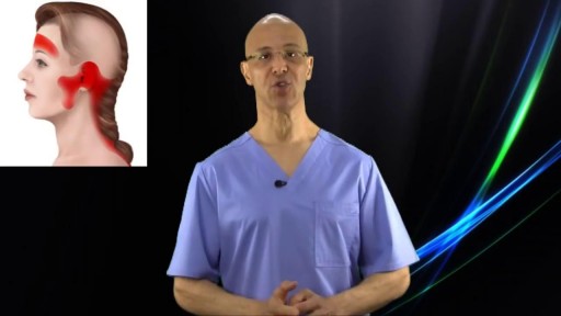
The majority of all headaches are tension related headaches. The blockage of blood circulation along with contraction/shortening of muscles is what causes this condition. This simple technique can take away most tension related headaches in seconds.

Endoscopic crushing of a bladder stone
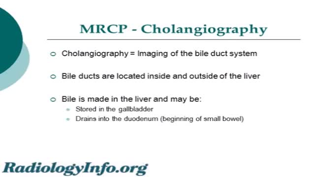
An MRCP scan is a scan that uses magnetic resonance imaging (MRI) to produce pictures of the liver, bile ducts, gallbladder and pancreas. Note: the information below is a general guide only. The arrangements,and the way tests are performed, may vary between different hospitals.

The abortion pill is a medicine called mifepristone that ends an early pregnancy.In general, it's used up to 70 days — 10 weeks — after the first day of a woman's last period. Patients over this mark can have an in-clinic abortion procedure.
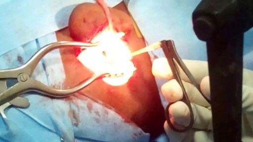
ow does a perforation of the eardrum occur? There are many ways an eardrum perforation can occur. An infection behind the eardrum in the middle ear may cause a rupture of the eardrum. Trauma to the ear may result from an object entering the ear canal and puncturing the eardrum. A traumatic blow to the ear with a cupped hand can rupture the eardrum. Hot welding slag can burn a hole through the eardrum. After a ventilation tube has been extruded or is removed, the opening usually closes; in some cases a permanent opening of the eardrum may occur. Chronic ear problems such as deep retraction pockets and cholesteatoma can weaken and erode the eardrum, resulting in a defect or perforation.
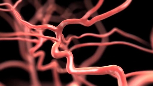
Narrated animation of stroke intervention. Video supplied by Covidien, showing the Solitaire mechanical thrombectomy device, which was the first FDA-approved device for such an indication.

Home dialysis treatment, including for both peritoneal and hemodialysis, has been a slowly developing trend in recent years. Between 2017 and 2018, the number of patients receiving treatment for peritoneal and hemodialysis jumped 7.7% and 8.8%, respectively, according to the United States Renal Data System 2020 Annual Data Report (ADR).
#KidneyPatients #WION #WorldNews
About Channel:
WION -The World is One News, examines global issues with in-depth analysis. We provide much more than the news of the day. Our aim to empower people to explore their world. With our Global headquarters in New Delhi, we bring you news on the hour, by the hour. We deliver information that is not biased. We are journalists who are neutral to the core and non-partisan when it comes to the politics of the world. People are tired of biased reportage and we stand for a globalised united world. So for us the World is truly One.
Please keep discussions on this channel clean and respectful and refrain from using racist or sexist slurs as well as personal insults.
Subscribe to our channel at https://goo.gl/JfY3NI
Check out our website: http://www.wionews.com
Connect with us on our social media handles:
Facebook: https://www.facebook.com/WIONews
Twitter: https://twitter.com/WIONews
Follow us on Google News for latest updates
Zee News:- https://bit.ly/2Ac5G60
Zee Bussiness:- https://bit.ly/36vI2xa
DNA India:- https://bit.ly/2ZDuLRY
WION: https://bit.ly/3gnDb5J
Zee News Apps : https://bit.ly/ZeeNewsApps
METHODS:
Previously existing methods are characterized by unpleasant scars that, despite surgeons promises, remain for life.
Incisions are:
- around the areola (Round block) leading to a flat areola, often unpleasant hypertophic skars, skin rippling.
- inverted T (around the areola, vertically down and in the fold under the breast).
- Vertical (around the areola and vertically down). Due to the extess skin, incisions often turn into inverted L or T. Rearrangement of glandular tissue and skin changes the shape of the breasts and may be different from expectations. Scars worry patients and sometimes cause disturbances in the relationship with their partner.
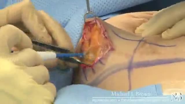
- No scars. The "Serdev Suture" lifting technique for breast lifting without scars (only points - needle perforations in the skin) is created by the Bulgarian cosmetic surgeon Prof. Dr. Nikolay Serdev. It is a novelty that had changed the cosmetic surgery world in the last 10-14 years for young patients. The technique is especially important in Asia and Latin America, for Asians, African-Americans, Indians, and others who form keloids and lumpy scars after operations.
The Serdev suture method can achieve lift upto and over 14 centimeters and is most suitable for the following types of breasts:
- not very heavy full breasts.
- in the presence of subpectoral implants with subsequent drooping of the breasts after childbirth and lactation.
- empty and loose breasts after childbirth and breastfeeding. In such cases this technique is combined with subpectoral implants. In sagging breasts implants should not be placed in the skin over the pectoral muscles, because thus will lead to even more drooping. Therefore, breast lift requires breast fixation to the level of the pectoral muscle (the normal position in young women), and then placement of appropriate implants under the muscle, to hold them in the appropriated position.
- in drooping breasts after subglandular augmentation (over the muscle). In such cases, patients should not wait until the skin elongation becomes visible. The implants should be removed, the capsule removed - a difficult but a necessary operation, preventing postop seromas and infection. Implants should be placed under the pectoralis muscle to wear them. Patients should orient the cosmetic surgeon at what level they want the nipples - in the middle of the implant, higher or lower.
Implants should be generally replaced - below the muscle implants should be smooth, move naturally without hurting the muscle.
Because of modern anesthetics and new methods without trauma, pain and swelling after surgery are not significant. In 3-4 days, patients can return to social life, even the next day, but it is preferable to rest for 2-3 days.
Exercises with the arms and weight lifting is prohibited for a month and a half.
Due to lack of scars, the breast lift using the Serdev sutures can be repeated to maintain the aesthetic appearence of the breasts even in advanced age.
Gigantomastia i.e. very large, very heavy and drooping breasts can not be operated in this manner, because of gravity and overskin.
Early mastopexy using Serdev sutures is recommended before too much changes in the tissues. If late, more and more complex interventions are required.
"A lot of people are opting for various breast procedures and one of the most common among them is “mastopexy”. This is the surgery that involved uplifting of sagging breasts and, in certain cases, repositioning of the nipple and areola in order to restore normality and beauty. The excess skin is removed and firmness is provided to the breasts. Though mastopexy can be done as a stand alone surgery, many people combine it with breast augmentation which involves inserting implants inside the b
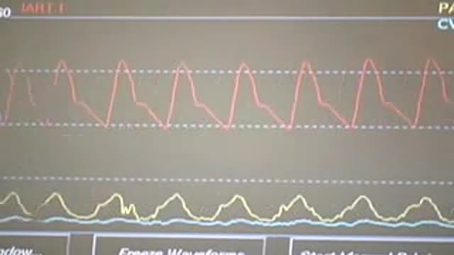
Central Venous Catheter Placement & Pulmonary Artery Catheter Video
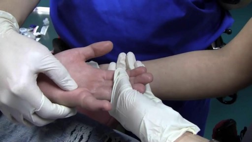
Finger metacarpophalangeal (MCP) joint collateral ligament sprains should not be overtreated. First-degree sprains may require a brief period of protection, usually consisting of buddy taping for 2-3 weeks. Second-degree sprains are immobilized in mid flexion for 3 weeks. Finger MCP joint hyperextension injuries may be treated by gently flexing the proximal phalanx and immobilizing the MCP joint in 30° of flexion for 2-3 weeks. A dorsal extension-block splint protects the healing volar plate while allowing active flexion of the finger. Early protected motion minimizes postinjury stiffness. Thumb MCP joint hyperextension injuries ("locked MCP joint") are immobilized in 20° MCP joint flexion for 3 weeks.
