- Physical Examination
- Surgical Examination
- Ophthalmology
- Clinical Skills
- Orthopedics
- Surgery Videos
- Laparoscopy
- Pediatrics
- Funny Videos
- Cardiothoracic Surgery
- Nursing Videos
- Plastic Surgery
- Otorhinolaryngology
- Histology and Histopathology
- Neurosurgery
- Dermatology
- Pediatric Surgery
- Urology
- Dentistry
- Oncology and Cancers
- Anatomy Videos
- Health and Fitness
- Radiology
- Anaesthesia
- Physical Therapy
- Pharmacology
- Interventional Radiology
- Cardiology
- Endocrinology
- Gynecology
- Emergency Medicine
- Psychiatry and Psychology
- Childbirth Videos
- General Medical Videos
- Nephrology
- Physiology
- Diet and Food Health
- Diabetes Mellitus
- Neurology
- Women Health
- Osteoporosis
- Gastroenterology
- Pulmonology
- Hematology
- Rheumatology
- Toxicology
- Nuclear Medicine
- Infectious Diseases
- Vascular Disease
- Reproductive Health
- Burns and Wound Healing
- Other
Top videos
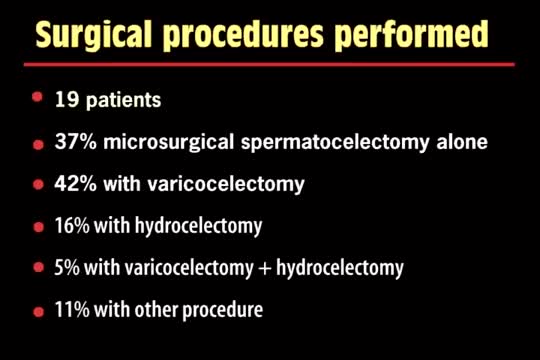
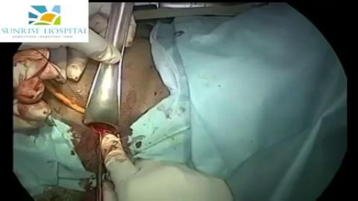
A spermatocelectomy is surgery to remove a spermatocele. A spermatocele is a cyst (sac of fluid) that contains sperm. It forms inside your scrotum on the outside of your testicle. The cyst is most often attached to your epididymis. The epididymis is a tube that stores sperm.

Rectal bleeding can refer to any blood that passes from your anus, although rectal bleeding is usually assumed to refer to bleeding from your lower colon or rectum. Your rectum makes up the last few inches of your large intestine. Rectal bleeding may show up as blood in your stool, on the toilet paper or in the toilet bowl. Blood that results from rectal bleeding can range in color from bright red to dark maroon to a dark, tarry color.

Dont worry sister!
Video of surgical management of cleft lip
A pneumothorax occurs when some of the tiny air sacs (alveoli) in a baby's lung become overinflated and burst. This causes air to leak into the space between the lung and chest wall (pleural space). The most common cause of pneumothorax is respiratory distress syndrome. This is a condition that occurs in babies who are born too early (premature). The baby's lungs lack the slippery substance (surfactant) that helps them stay open. Therefore, the tiny air sacs are not able to expand as easily. If the baby is put on a breathing machine (mechanical ventilator), there is extra pressure on the baby's lungs, which can sometimes burst the air sacs.
Vaginoplasty is any surgical procedure that results in the construction or reconstruction of the vagina. It is a type of genitoplasty. Pelvic organ prolapse is often treated with one or more surgeries to repair the vagina.
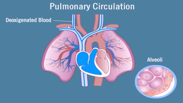
Pulmonary circulation is the portion of the cardiovascular system which carries deoxygenated blood away from the heart, to the lungs, and returns oxygenated (oxygen-rich) blood back to the heart. The function of pulmonary circulation is to exchange carbon dioxide for oxygen in the blood. It is the passage of blood from the heart to the capillaries of the lungs, where the gases are exchanged, and back to the heart to be pumped around the body.

Vaginoplasty is a surgical procedure designed to rejuvenate and tighten a woman’s vagina, by removing excess lining and repairing the surrounding soft tissues. It is designed to decrease the diameter of the vagina, resulting in increased friction during intercourse to make the experience more pleasurable for both partners.
Pancreatitis is inflammation of the pancreas. The pancreas is a gland located behind the stomach. It releases the hormones, insulin and glucagon, as well as digestive enzymes that help you digest and absorb food.
急性坏疽阑尾炎的手术治疗
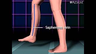
Urogenital neoplasms spreading to the inguinal lymph nodes are penile carcinoma (the most frequent), urethral and scrotum cancers, tumors of the testis with scrotal violation. Penile carcinoma is an uncommon malignant disease and accounts for as many 0.4-0.6% of male cancers. Most patients are elder...ly. It rarely occurs in men under age 60 and its incidence increases progressively until it reaches a peak in the eighth decade 1. The risk of a lymph node invasion is greater with high grade and high stage tumors 2. Some investigators have reported the inaccuracy of the sentinel node biopsy 3, 4, described by Cabanas 5. Patients with metastatic lymph node penis cancer have a very poor prognosis if penectomy only is performed. Ilioinguinal lymphadenectomy is basically carried out as a treatment modality and not only as a staging act. Patients with lymph node invasion have a 30-40% cure rate. Ilioinguinal lymphadenectomy should be also performed in patients with disseminated neoplasms for the local control of the disease. The 5 years survival rate of patients with clinically negative lymph nodes treated with a modified inguinal lymphadenectomy is 88% versus 38% in patients not initially treated with lymphadenectomy 6. This video-tape clearly shows a therapeutic algorithm, the anatomy of the inguinal lymph nodes, according to Rouviere 7 and Daseler 8, the radical ilioinguinal node dissection with transposition of the sartorius muscle and the modified inguinal lymphadenectomy proposed by Catalona 9. References: 1. Lynch D.F. and Schellhammer P: Tumors of the penis. In Campbell’s Urology Seventh Edition, edited by Walsh P.C., Retik A.B., Darracott Vaughan E. and Wein A.J. W.B. Saunders Company, Vol. 3, chapt. 79, p. 2458, 1998. 2. Pizzocaro G., Piva L., Bandieramonte G., Tana S. Up-to-date management of carcinoma of the penis. Eur. Urol. 32: 5-15, 1997 3. Perinetti E., Crane D.B. and Catalona W.J. Unreliability of sentinel lymph node biopsy for staging penile carcinoma. J. Urol. 124: 734, 1980 4. Fowler J.E. Jr. Sentinel lymph node biopsy for staging penile cancer. Urology 23: 352, 1984 5. Cabanas R.M. An approach for the treatment of penile carcinoma. Cancer 39: 456, 1977 6. Russo P. and Gaudin P. Management strategies for carcinoma of the penis. Contemporary Urology;5:48-66, 2000 7. Rouviere H. Anatomy of the human lymphatic system. Edwards Brothers, p. 218, 1938 8. Daseler E.H., Anson B.J., Reimann A.F. Radical excision of the inguinal and iliac lymph glands: a study based on 450 anatomical dissections and upon supportive clinical observations. Surg. Gynecol. Obstet. 87: 679, 1948 9. Catalona W.J. Modified inguinal lymphadenectomy for carcinoma of the penis with preservation of saphenous veins: technique and preliminary results. J. Urol. 140: 306-310, 1988

Revision knee replacement is peformed by Dr.Venkatachalam for lack of mobility. Infection. aseptic loosening are frequent causes requiring a revision. Madras Joint replacement center performs primary and revision knee replacements in a super specialty hospital in Chennai, India. Dr.Venkatachalam, the chief orthopedic surgeon is UK board certified.
Overview
Heart bypass surgery creates a new route, called a bypass, for blood and oxygen to reach the heart.
Heart bypass surgery begins with an incision in the chest, and the breastbone is cut exposing the heart. Next, a portion of the saphenous vein, which is very large, is harvested from the inside of the leg. Pieces of this large vein are used to bypass the blocked coronary arteries, which are arteries that supply blood to the heart. The venous graft is sewn to the aorta, the main artery of the body, and to the affected coronary artery, to bypass the blocked site.
The internal mammary artery from the chest may also be used to bypass a clogged artery.
Several arteries may be bypassed depending on the condition of the heart. After the graft is created, the breastbone and chest are closed.

Watch that video for a Boy Returns from the Beach with a Snail Inside His Knee

-The cremasteric reflex test is considered positive if there is elevation of the testis in response to stroking the upper inner thigh. This reaction is typically absent in testicular torsion and boys under the age of 6 months. Although not completely reliable in older boys and adults, an absent cremasteric reflex is highly suggestive of torsion. Patients with epididymitis usually have a normal cremasteric reflex, with pain and swelling isolated to
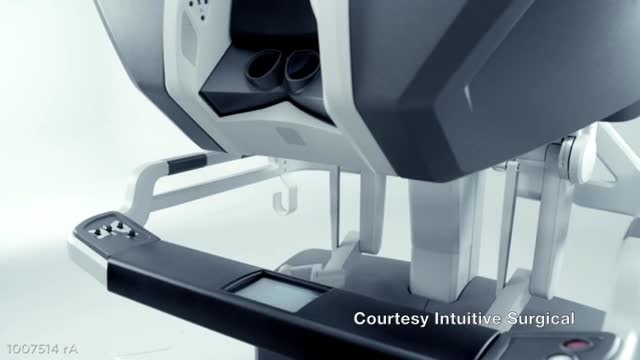
Autonomous Tumor Localization and Extraction: Palpation, Incision, Debridement and Adhesive Closure with the da Vinci Research Kit. This video demonstrates a successful trial of the entire five-step procedure where human input is required only at four points to change tools. We also show failure modes of the current autonomous system and we are working on experiments to characterize the reliability of each step and on incorporating computer vision and new probing algorithms to improve robustness.

LDL (Bad) Cholesterol LDL cholesterol is considered the “bad” cholesterol because it contributes to plaque, a thick, hard deposit that can clog arteries and make them less flexible. This condition is known as atherosclerosis. If a clot forms and blocks a narrowed artery, heart attack or stroke can result. Another condition called peripheral artery disease can develop when plaque buildup narrows an artery supplying blood to the legs. View an animation of cholesterolHDL (Good) Cholesterol HDL cholesterol is considered “good” cholesterol because it helps remove LDL cholesterol from the arteries. Experts believe HDL acts as a scavenger, carrying LDL cholesterol away from the arteries and back to the liver, where it is broken down and passed from the body. One-fourth to one-third of blood cholesterol is carried by HDL. A healthy level of HDL cholesterol may also protect against heart attack and stroke, while low levels of HDL cholesterol have been shown to increase the risk of heart disease.
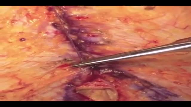
Barbed sutures first received US Food and Drug Administration approval for soft tissue approximation in 2005 and early adopters readily embraced this device to develop new techniques. It has become apparent that the advantages are more than just "skin deep." Superficial and deep fascia, cartilage, tendon, joint capsule, and fibrous periprosthetic capsules can also be manipulated. Barbed sutures have revolutionized our approach to facial rejuvenation and body contouring by enhancing our ability to quilt and powerfully lift tissue. The elimination of surgical drains and shorter surgical times has made this a true boon for plastic surgeons as well as many other surgical specialists. This article summarizes some of the current and evolving applications of this exciting new tool.
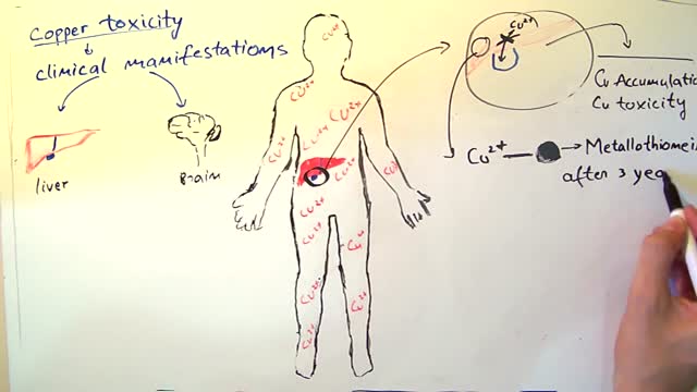
Wilson's disease is a rare inherited disorder that causes too much copper to accumulate in your liver, brain and other vital organs. Symptoms typically begin between the ages of 12 and 23. Copper plays a key role in the development of healthy nerves, bones, collagen and the skin pigment melanin. Normally, copper is absorbed from your food, and any excess is excreted through bile — a substance produced in your liver. But in people with Wilson's disease, copper isn't eliminated properly and instead accumulates, possibly to a life-threatening level. When diagnosed early, Wilson's disease is treatable, and many people with the disorder live normal lives.
