
- Physical Examination
- Surgical Examination
- Ophthalmology
- Clinical Skills
- Orthopedics
- Surgery Videos
- Laparoscopy
- Pediatrics
- Funny Videos
- Cardiothoracic Surgery
- Nursing Videos
- Plastic Surgery
- Otorhinolaryngology
- Histology and Histopathology
- Neurosurgery
- Dermatology
- Pediatric Surgery
- Urology
- Dentistry
- Oncology and Cancers
- Anatomy Videos
- Health and Fitness
- Radiology
- Anaesthesia
- Physical Therapy
- Pharmacology
- Interventional Radiology
- Cardiology
- Endocrinology
- Gynecology
- Emergency Medicine
- Psychiatry and Psychology
- Childbirth Videos
- General Medical Videos
- Nephrology
- Physiology
- Diet and Food Health
- Diabetes Mellitus
- Neurology
- Women Health
- Osteoporosis
- Gastroenterology
- Pulmonology
- Hematology
- Rheumatology
- Toxicology
- Nuclear Medicine
- Infectious Diseases
- Vascular Disease
- Reproductive Health
- Burns and Wound Healing
- Kidney Zone Podcast
- Other





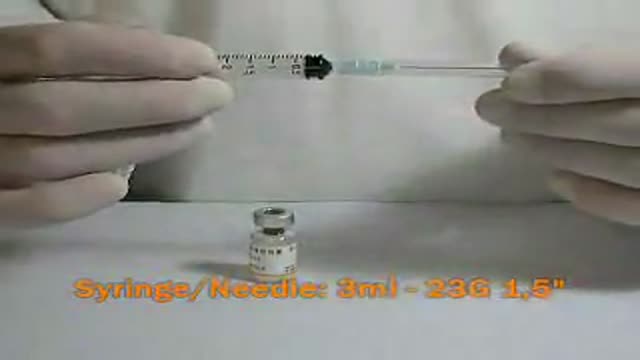


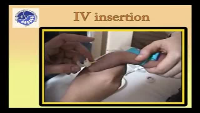
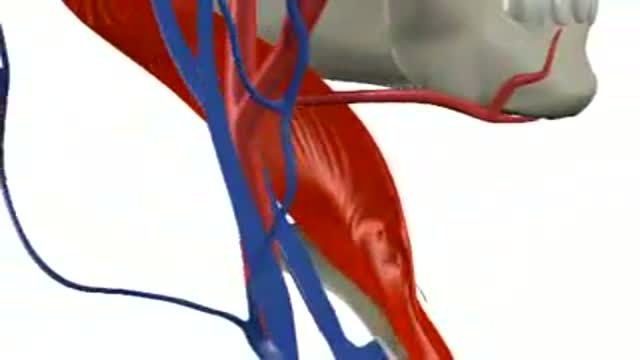

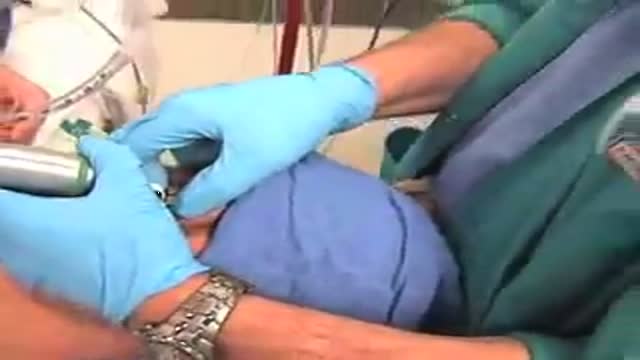









Immunohistochemistry IHC Staining

Educational only: videos on MedicalVideos.com are for general information and are not medical advice. Always consult your own doctor about your health.
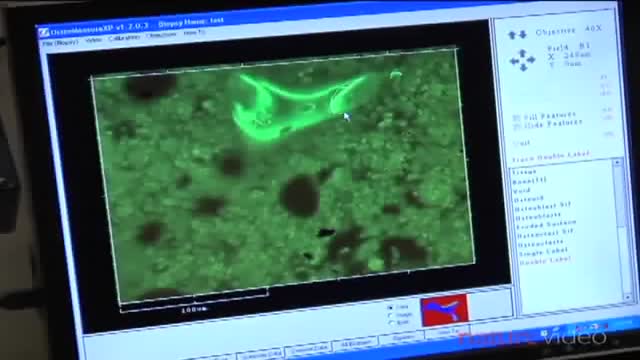
Immunohistochemistry or IHC refers to the process of detecting antigens (e.g., proteins) in cells of a tissue section by exploiting the principle of antibodies binding specifically to antigens in biological tissues.[1] IHC takes its name from the roots "immuno," in reference to antibodies used in the procedure, and "histo," meaning tissue (compare to immunocytochemistry). Immunohistochemical staining is widely used in the diagnosis of abnormal cells such as those found in cancerous tumors. Specific molecular markers are characteristic of particular cellular events such as proliferation or cell death (apoptosis). IHC is also widely used in basic research to understand the distribution and localization of biomarkers and differentially expressed proteins in different parts of a biological tissue. Visualising an antibody-antigen interaction can be accomplished in a number of ways. In the most common instance, an antibody is conjugated to an enzyme, such as peroxidase, that can catalyse a colour-producing reaction. Alternatively, the antibody can also be tagged to a fluorophore, such as fluorescein or rhodamine
This video in 30 seconds
This video explains immunohistochemistry staining, the process of detecting antigens in cells of a tissue section using antibodies that bind specifically to antigens. It describes the use of IHC staining in diagnosing abnormal cells such as cancerous tumors and in research on biomarkers and proteins in tissue.
Key points
- Immunohistochemistry staining is explained
- Antibody binding to antigens in tissue sections is described
- Use in diagnosis of abnormal and cancerous cells is covered
- Enzyme and fluorophore tagging of antibodies is described
Video Overview
This video presents “Immunohistochemistry IHC Staining” — Immunohistochemistry or IHC refers to the process of detecting antigens (e.g., proteins) in cells of a tissue section by exploiting the principle of antibodies binding specifically to antigens in biological tissues.[1] IHC takes its name. Whether you are preparing for a procedure, studying the technique, or learning how a condition is assessed, the recording shows the process as it is performed in practice.
From our Histology and Histopathology collection: Tissue slides under the microscope, biopsy interpretation and pathology basics for medical students. Browse all Histology and Histopathology videos →
Educational content only — not a substitute for professional medical advice.
Medical Videos
Free medical and surgery videos for patients, students and professionals — trusted since 2007.
Founded by Dr. Mohamed Ibrahim, MD, PhD — physician-scientist.
Follow us on Facebook · Instagram
SORT BY-
Top Comments
-
Latest comments