Mga nangungunang video
Watch out which infections could affect your baby or tend to be worse for you during pregnancy period and how to reduce your risk of getting them.

Teeth whitening fit for a beauty queen! Miss. Harris County Teen Angela H. just completed a ZOOM! whitening.
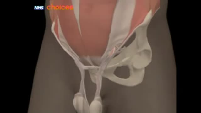
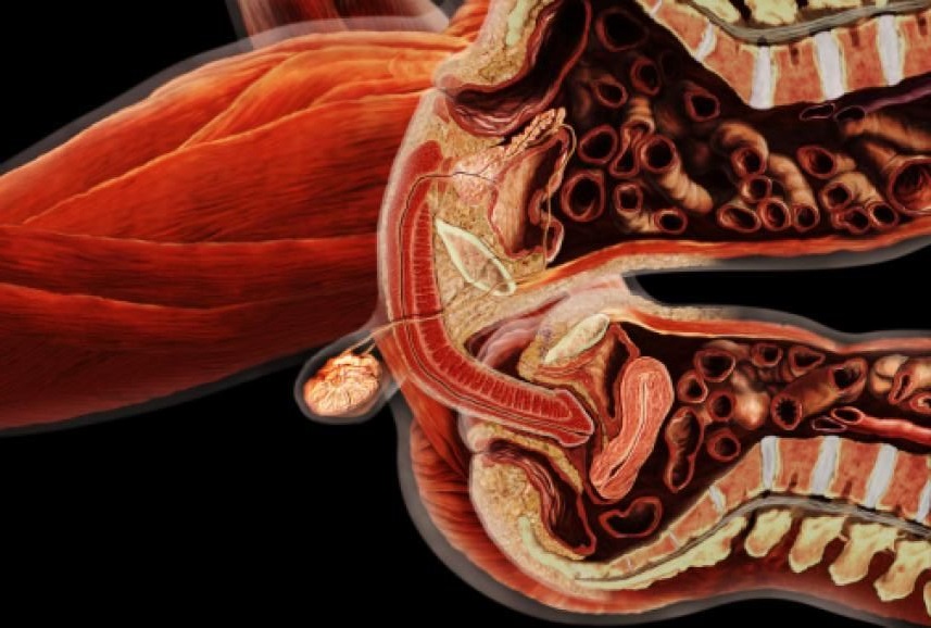
A hernia occurs when an organ or fatty tissue squeezes through a weak spot in a surrounding muscle or connective tissue called fascia. The most common types of hernia are inguinal (inner groin), incisional (resulting from an incision), femoral (outer groin), umbilical (belly button), and hiatal (upper stomach).
Watch that video to know How to Triple Your Chances of Getting Pregnant
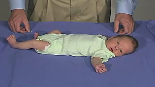
physical exam -Newborn Normal:Behavior

Disordered Eater vs. Eating Disorder - What's the difference?
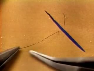
The needle should pass through the tissue at a perpendiculaPlace the tips of the left-hand forceps on the underside of the tissue at the point where the needle will enter, and gently push the edge upward. With the right hand, bring the needle into contact with the tissue, and press downward. These movements create eversion. Pass the needle through. Do not grab the tissue with your left hand forceps since it will damage the intima. If needed, you can pick up adventitia or a nearby suture to help with exposure and eversion. r.The needle must pass through the other side at a perpendicular, too. Bring the tip of the needle to the place where you intend to bring it out on the other side. Put the tip of your left-hand forceps on the upper surface of the tissue at the intended exit point. Press down with the left-hand forceps and push up with the needle to give you the correct eversion. The width of the bite should be about three times the thickness of the needle. The bites on both sides must be equal, and the needle should cross exactly in a straight line (not diagonally). Pull the needle through the tissue following the curve of the needle
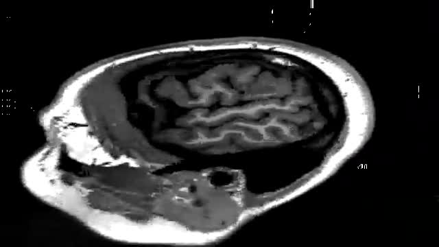
An animated video showing an MRI of the brain

Dr. Shaun Kunisaki is an Associate Professor of Surgery at The Johns Hopkins University and Associate Chief of Strategy and Integration in the Division of General Pediatric Surgery at the Johns Hopkins Children's Center. His clinical practice spans the full breadth of pediatric general surgery, but he is recognized both regionally and nationally for this expertise in complex thoracic surgical problems in the fetus and young child. As Director of Pediatric Esophageal Surgery, he specializes in the management of long-gap esophageal atresia. In this role within the Johns Hopkins Children Center Fetal Program, he helps counsel parents with pregnancies complicated by fetal anomalies.
Learn more about Dr. Kunisaki at https://www.hopkinsmedicine.or....g/profiles/results/d
Cholangitis Email this page to a friend Email this page to a friend Facebook Twitter Google+ Cholangitis is an infection of the bile ducts, the tubes that carry bile from the liver to the gallbladder and intestines. Bile is a liquid made by the liver that helps digest food. Causes Cholangitis is most often caused by bacteria. This can occur when the duct is blocked by something, such as a gallstone or tumor. The infection causing this condition may also spread to the liver. Risk factors include a previous history of gallstones, sclerosing cholangitis, HIV, narrowing of the common bile duct, and rarely, travel to countries where you might catch a worm or parasite infection. Symptoms The following symptoms may occur: Pain on the upper right side or upper middle part of the abdomen. It may also be felt in the back or below the right shoulder blade. The pain may come and go and feel sharp, cramp-like, or dull. Fever and chills. Dark urine and clay-colored stools. Nausea and vomiting. Yellowing of the skin (jaundice), which may come and go.

Neglected elbow dislocations are seen in patients hailing from Africa and Asia. A Nigerian patient with this condition was successfully treated by open reduction and external fixator application
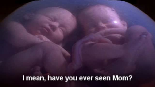
Twins Conversation
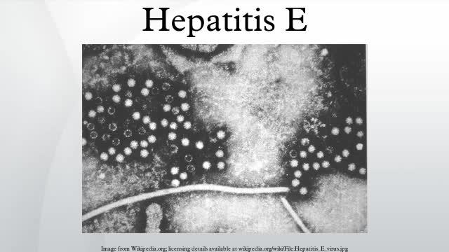
Hepatitis E is a virus that can infect the liver. Unlike other forms of hepatitis, the hepatitis E virus usually doesn't lead to long-term illness or serious liver damage. Most people get well within a few months.

The Human Body in Numbers.
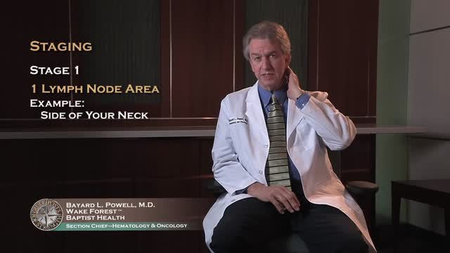
Non-Hodgkin's lymphoma, also called non-Hodgkin lymphoma, is cancer that originates in your lymphatic system, the disease-fighting network spread throughout your body. In non-Hodgkin's lymphoma, tumors develop from lymphocytes — a type of white blood cell. Non-Hodgkin's lymphoma is more common than the other general type of lymphoma — Hodgkin lymphoma. Many different subtypes of non-Hodgkin's lymphoma exist. The most common non-Hodgkin's lymphoma subtypes include diffuse large B-cell lymphoma and follicular lymphoma.

Vocal Cord Surgery HD
Watch that video of Super Model's Butt and Leg Implants Exploded
A Chinese hospital in the process of creating a human ear almost entirely through the human anatomy alone.
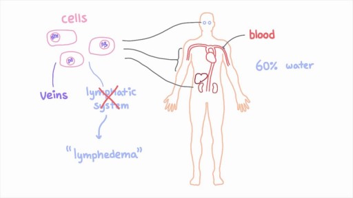
The lymphatic system is a network of specialized vessels (lymph vessels) throughout the body whose purpose is to collect excess lymph fluid with proteins, lipids, and waste products from the tissues. This fluid is then carried to the lymph nodes, which filter waste products and contain infection-fighting cells called lymphocytes. The excess fluid in the lymph vessels is eventually returned to the bloodstream. When the lymph vessels are blocked or unable to carry lymph fluid away from the tissues, localized swelling (lymphedema) is the result.
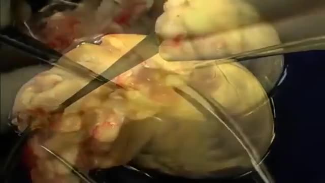
The first operation is harvesting the heart from the donor. The donor is usually an unfortunate person who has suffered irreversible brain injury, called "brain death". Very often these are patients who have had major trauma to the head, for example, in an automobile accident. The victim's organs, other than the brain, are working well with the help of medications and other "life support" that may include a respirator or other devices. A team of physicians, nurses, and technicians goes to the hospital of the donor to remove donated organs once brain death of the donor has been determined. The removed organs are transported on ice to keep them alive until they can be implanted. For the heart, this is optimally less than six hours. So, the organs are often flown by airplane or helicopter to the recipient's hospital.
