Najbolji videi

Aspirin, or acetylsalicylic acid (ASA) is a salicylate drug, and is generally used as an analgesic (something that relieves pain without producing anesthesia or loss of consciousness) for minor aches and pains, to reduce fever (an antipyretic), and also as an anti-inflammatory drug.

Teeth whitening fit for a beauty queen! Miss. Harris County Teen Angela H. just completed a ZOOM! whitening.
Watch that video of Super Model's Butt and Leg Implants Exploded

Dr. Rowe shows how to quickly fix knee popping, clicking, and cracking sounds.
This exercise will focus on lengthening tight muscles and tendons that may be causing a noisy knee. It's especially good for osteoarthritis (wear and tear arthritis) of the knee.
It can be done at home, requires no equipment, and may also give knee pain relief... even within seconds.
Let us know how it works for you!
***************************
Dr. Michael Rowe
St. Joseph, Michigan chiropractor
If you are looking for effective neck, back, or sciatica pain relief, contact us at 269-408-8439 or visit us at https://www.BestSpineCare.com
Facebook: https://www.facebook.com/bestspinecare
Twitter: https://www.twitter.com/stjoechiro
Instagram: https://www.instagram.com/stjoechiro
Your local St. Joseph | Benton Harbor | Stevensville Michigan chiropractor
SpineCare Decompression and Chiropractic Center
3134 Niles Rd
Saint Joseph, MI 49085
**MEDICAL DISCLAIMER**
All information, content, and material of this video or website is for informational and demonstration purposes only. It is not intended to serve as a substitute for the consultation, diagnosis, and/or medical treatment of a qualified physician or healthcare provider.
Don’t use this content as a replacement for treatment and advice given by your doctor or health care provider. Consult with your doctor or healthcare professional before doing anything contained in this content.
By watching this video, you agree to indemnify and hold harmless SpineCare Decompression and Chiropractic Center (and its representatives) for any and all losses, injuries, or damages resulting from any and all claims that arise from your use or misuse of this content. SpineCare Decompression and Chiropractic Center makes no representations about the accuracy or suitability of this content.
USE OF THIS CONTENT IS AT YOUR OWN RISK.
- AFFILIATE DISCLAIMER -
We may receive commissions when you click on this video's links and make purchases. This helps support our channel so we can continue to give you helpful content.
#kneepain #kneepainrelief #kneearthritis
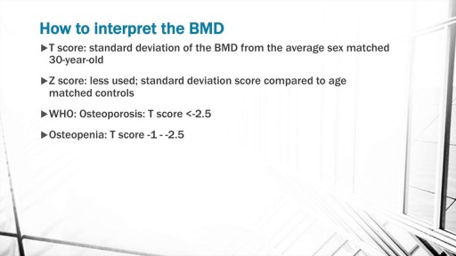
The discussion begins with a basic explanation of Bone biology taking into consideration the osteoblast and osteoclast balance. Concepts of RANK, RANK ligand and Osteoprotegerin are included. Risk factors for Osteoporosis such as Age, alcohol, smoking, sedentary lifestyle are also discussed.
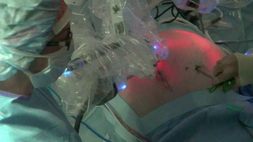
Splenectomy is a surgical procedure to remove your spleen. The spleen is an organ that sits under your rib cage on the upper left side of your abdomen. It helps fight infection and filters unneeded material, such as old or damaged blood cells. With the da Vinci Surgical System, Dr. Olson operates through just a few small incisions. The da Vinci System features a magnified 3D high-definition vision system and tiny wristed instruments that bend and rotate far greater than the human hand. As a result, da Vinci enables surgeons to operate with enhanced vision, precision and control.
Ligation of Aneurysm in ArterioVenous Malformation
Gastrointestinal GI Drug Delivery
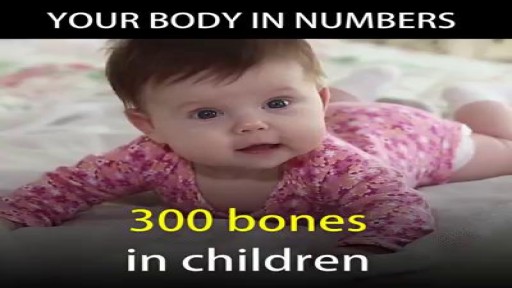
The Human Body in Numbers.

What Is A Body Wrap, Body Wraps Do They Work, Detoxifying Body Wrap, Best Body Wraps For Cellulite -- http://do-body-wraps-work.plus101.com -- Slimming body wraps firm, tighten and detoxify the skin as well as giving instant inch loss whilst removing toxins and from the body. Depending on your body type it is easy to lose up to 15 inches in sixty minutes! All you need to do is start your weight loss program, remain motivated and you will achieve your desired appearance and also reduce stretch marks and cellulite. Slimming body wrap helps achieve an inch loss in every session which can last for approximately 3 months provided you maintain a healthy lifestyle and your current weight. As opposed to other type of treatments body wrap don't need a lot of post treatment. A body wraps helps in detoxification of your body both externally and internally. It cleans blocked body tissues letting your body to firm up as well as aid in holding the newly well cut shape by the firmness body wrap. The slimming body wraps help in getting rid of toxins deposits through detoxification of tissues as well as restraining of lymphatic system. When preparing for a body wrap you should not moisturize, and you should drink plenty of water. During the process women are expected to be only in panties and bra or briefs in the case of men. You are then weighed and measured multiple areas of your body. A solution of citrus and amino nutrient is often applied on your skin to open up your pores. You are the comfortably but firmly wrapped in linen and elastic body wraps for at least half an hour. The main benefits of body wraps include detoxification, skin firming, slimming, body contouring, boosting metabolism, relaxation, redefining your skin texture and stimulating your lymphatic system. Typically spa or salon body wraps costs between 0 and 0 depending on your area, but you can make the same at home for pennies on the dollar! Home body wrap recipes are available at http://do-body-wraps-work.plus101.com
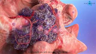
This cancer development medical video is devoted to elaborating the basics of cancer growth. We used advanced medical animation techniques to display such a complicated process.
What is happening in cancer development medical video
The fundamental abnormality described in the cancer development medical video is the nonstop unregulated multiplication of cancer cells. Being uncontrollable by body’s signals that regulate normal cell behavior; cancerous cells divide and grow populating neighboring normal tissues or even spread throughout the body. The overall lack of growth control acquired by cancer cells is due to the accumulated abnormalities in numerous cell regulatory mechanisms and is considered in some aspects of cell behavior that differs them from their healthy counterparts. The interaction of these cells is shown in our previous medical animation video.
Read full article on our webpage http://bit.ly/2LQj9ln
Follow us on Facebook https://www.facebook.com/Nanob....ot.Medical.Animation
Follow us on LinkedIn https://www.linkedin.com/compa....ny/nanobotmodels-med
Follow us on Twitter https://twitter.com/Nanobot_Studio
Follow us on Instagram https://www.instagram.com/nano....bot_medical_animatio
Follow us on Clutch https://clutch.co/profile/nano....bot-medical-animatio
Follow us on Behance https://www.behance.net/NanobotStudio
#cancer #tumor #oncology #metatastic #nanobot #visualscience #scientificcommunication #medicalanimation #animationvideo #animationdesign #animationstudio #animationmovie #nanotechnology #medicine #health #science #education #medschool #medicaleducation #animation_studio #animationstudio
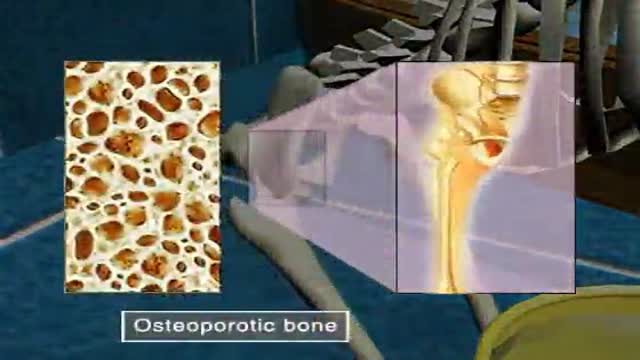
Osteoporosis
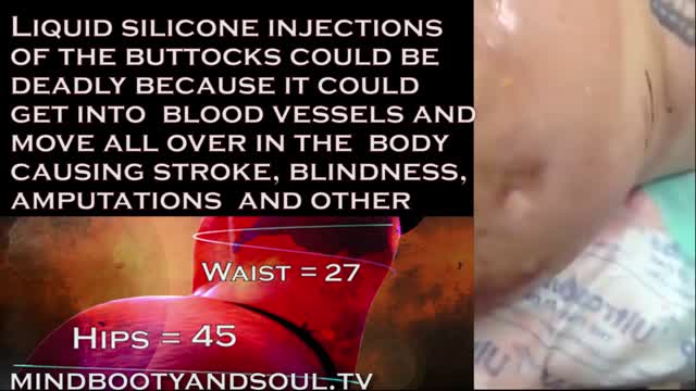
2016 marks 10 years when illegal injections started to gain momentum and become a popular alternative to butt implants. The Brazilian butt lift wasn't well know at the time but the goal of finding an unlicensed person to inject a foreign substance into the body was in high demand.
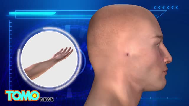
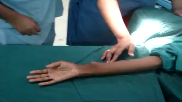
A Chinese hospital in the process of creating a human ear almost entirely through the human anatomy alone.
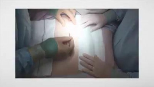
Laparotomy (opening and closing)
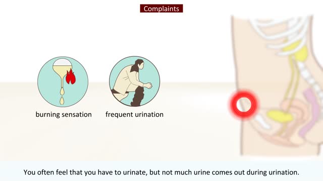
Most women are put on a 3 to 5 day antibiotic. Men might be put on an antibiotic for 7 to 14 days. While symptoms usually clear up around three days after antibiotic treatment, it can take up to five days for all the bacteria in your urinary tract to die off. It may take even longer for men.

Sialadenitis is an infection of the salivary glands. It is usually caused by a virus or bacteria . The parotid (in front of the ear) and submandibular (under the chin) glands are most commonly affected. Sialadenitis may be associated with pain, tenderness, redness, and gradual, localized swelling of the affected area.
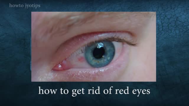
Red eyes usually are caused by allergy, eye fatigue, over-wearing contact lenses or common eye infections such as pink eye (conjunctivitis). However, redness of the eye sometimes can signal a more serious eye condition or disease, such as uveitis or glaucoma.
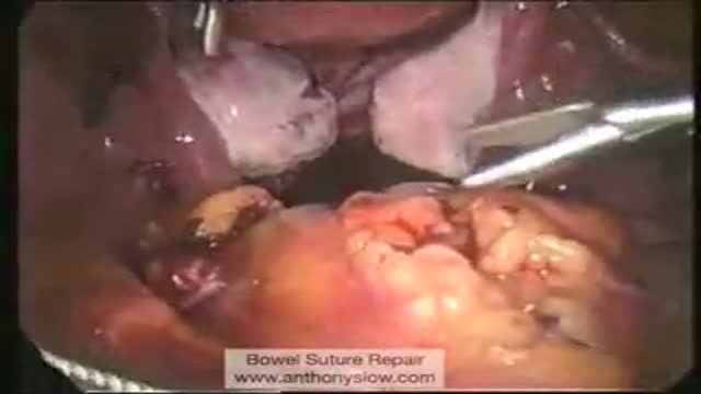
Laparoscopic Suture Repair of Bowel

Demonstration of simple interrupted suturing technique for laceration repair.
