- Physical Examination
- Surgical Examination
- Ophthalmology
- Clinical Skills
- Orthopedics
- Surgery Videos
- Laparoscopy
- Pediatrics
- Funny Videos
- Cardiothoracic Surgery
- Nursing Videos
- Plastic Surgery
- Otorhinolaryngology
- Histology and Histopathology
- Neurosurgery
- Dermatology
- Pediatric Surgery
- Urology
- Dentistry
- Oncology and Cancers
- Anatomy Videos
- Health and Fitness
- Radiology
- Anaesthesia
- Physical Therapy
- Pharmacology
- Interventional Radiology
- Cardiology
- Endocrinology
- Gynecology
- Emergency Medicine
- Psychiatry and Psychology
- Childbirth Videos
- General Medical Videos
- Nephrology
- Physiology
- Diet and Food Health
- Diabetes Mellitus
- Neurology
- Women Health
- Osteoporosis
- Gastroenterology
- Pulmonology
- Hematology
- Rheumatology
- Toxicology
- Nuclear Medicine
- Infectious Diseases
- Vascular Disease
- Reproductive Health
- Burns and Wound Healing
- Other
Top videos
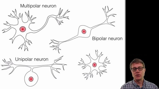
A neuron, also known as a neurone (British spelling) and nerve cell, is an electrically excitable cell that receives, processes, and transmits information through electrical and chemical signals. These signals between neurons occur via specialized connections called synapses.
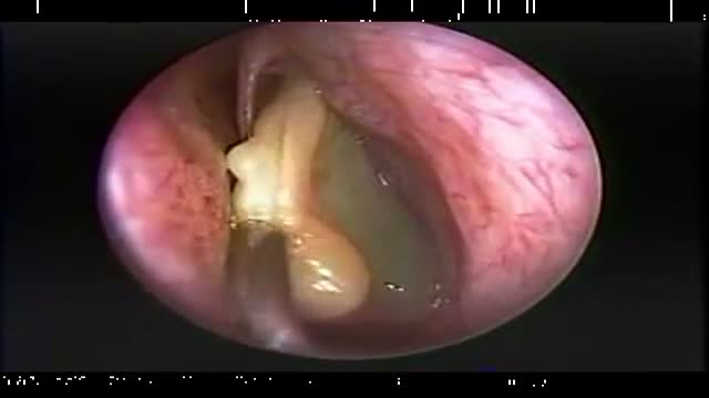
Endoscopic Nasal Polypectomy
A detailed description of the Hepato-pulmonary syndrome including its definition, pathophysiology, diagnosis and treatment. The pathophysiology includes nitric oxide in the pulmonary vasculature which results in intrapulmonary vasodilatation. This causes the classical and unique symptom of platypnea and orthodeoxia.
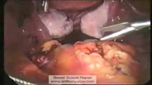
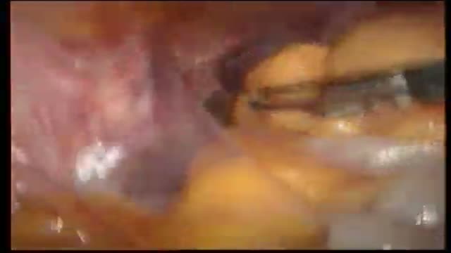
Laparoscopic Suture Repair of Bowel

Demonstration of simple interrupted suturing technique for laceration repair.
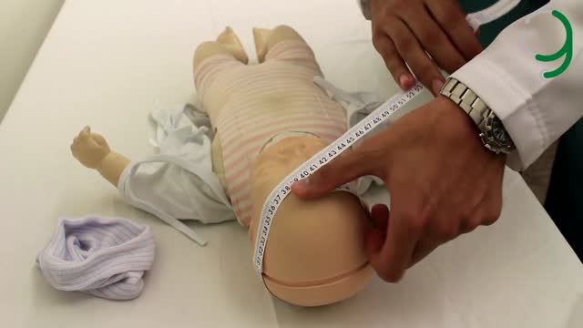
A brief screening examination should be conducted checking the face, eyes, mouth, chest, abdomen, spine and limbs to exclude major abnormalities. A strong cry and a widespread pink blush over the face and body are good signs that all is well. Some children may be born with ambiguous genitalia. Ambiguous genitalia is a medical emergency and requires urgent assessment by a paediatrician. If you have sufficient clinical experience, an orogastric tube should be passed when the neonate's mother has suffered polyhydramnios. This excludes oesophageal atresia.
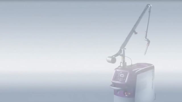
New Minimally Invasive Procedure with No Pain or Downtime… From Dr. Michael Goodman, Caring For Women Wellness Center Laser Vaginal Tightening for Improved Sexual Pleasure and Relief from Minimal Urinary Incontinence Laser Vaginal Therapy for reversing Vaginal Atrophy (Good also for Breast Cancer Survivors with Vaginal Atrophy)
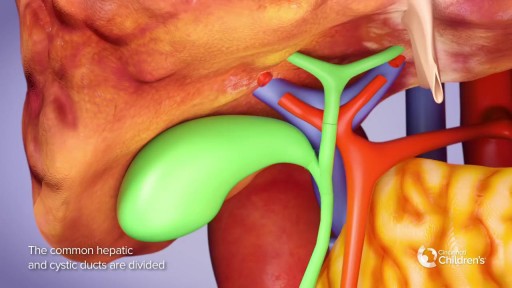
Liver Transplant Surgery Explained
Sialadenitis is an infection of the salivary glands. It is usually caused by a virus or bacteria . The parotid (in front of the ear) and submandibular (under the chin) glands are most commonly affected. Sialadenitis may be associated with pain, tenderness, redness, and gradual, localized swelling of the affected area.
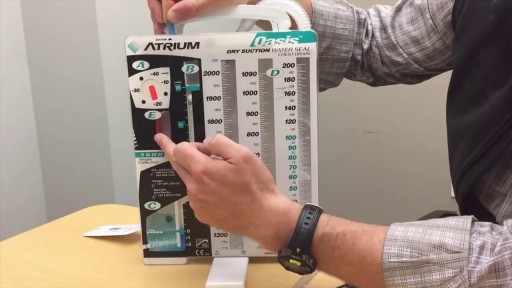
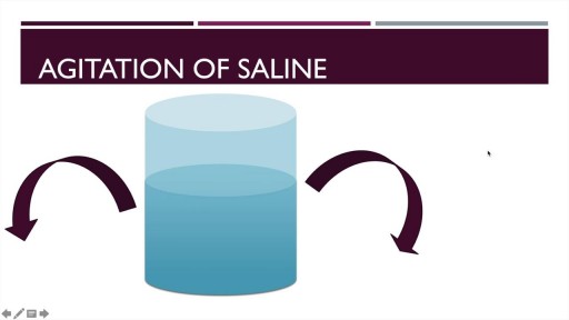
If you’re like me, you probably hook your chest tube up to a Pleur-Evac, put it on the ground, then back away slowly. Who knows what goes on in that mysterious bubbling white box? Hopefully this will post shed some light. Isn’t this just a container for stuff that comes out of the chest? Why does it look so complicated? It’s complicated because the detection/collection of air and fluid require different setups. Most commercial models also allow you to hook the drainage system to wall suction, so you can quickly evacuate the pleural space. This requires its own setup. Because of the need to juggle air, fluid and suction, the most common commercial system includes 3 distinct chambers. If you were to simplify the device, or build one out of spare bottles and tubes, it might look like this:
Laparoscopic anterior resection for cancer colon in Qatar by Dr. Al-Emadi
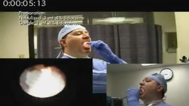
ROTIGS medical device by Honolulu inventor Dr. Brad NaPier makes difficult airway intubations easier for medical professionals.

► Sign up here and try our FREE content: http://lectur.io/freecontentyt
► If you’re an medical educator or faculty member, visit: http://lectur.io/medytb2u
This video “Connective Tissue” is part of the Lecturio course “Histology” ► WATCH the complete course on http://lectur.io/connectivetissue
► LEARN ABOUT:
- Cells and Basic Tissue
- Nerve Tissues
- Muscle Tissues
- Epithelial Tissues
- Connective Tissues
► THE PROF: Your lecturer is Professor Geoff Meyer. He is currently teaching at the School of Anatomy, Physiology and Human Biology at the University of Western Australia (UWA). As a leading anatomy and histology expert he is also coordinating the Federative International Program for Anatomical Terminologies (FIPAT) of the International Federation of Associations of Anatomists (IFAA). Besides medical research on the ovarian function, steroidogenesis, corpus luteum, angiogenesis, and microcirculation, Geoff Meyer’s research activities also focus on developing innovative, computer-aided learning and teaching tools. For his inventiveness, Geoff Meyer has received a number of awards, including the Australian University Teaching Award.
► LECTURIO is your single-point resource for medical school:
Study for your classes, USMLE Step 1, USMLE Step 2, MCAT or MBBS with video lectures by world-class professors, recall & USMLE-style questions and textbook articles. Create your free account now: http://lectur.io/connectivetissue
► INSTALL our free Lecturio app
iTunes Store: https://app.adjust.com/z21zrf
Play Store: https://app.adjust.com/b01fak
► READ TEXTBOOK ARTICLES related to this video:
Types of Tissue: Connective Tissue, Muscle Tissue, Epithelial Tissue, and Nervous Tissue
http://lectur.io/connectivetissuearticle
► SUBSCRIBE to our YouTube channel: http://lectur.io/subscribe
► WATCH MORE ON YOUTUBE: http://lectur.io/playlists
► LET’S CONNECT:
• Facebook: https://www.facebook.com/lectu....rio.medical.educatio
• Instagram: https://www.instagram.com/lecturio_medical_videos
• Pinterest: https://www.pinterest.de/lecturiomedical
• LinkedIn: https://www.linkedin.com/company/lecturio-medical/
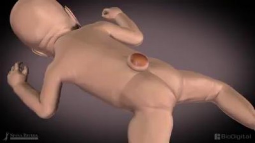
Spina bifida is a type of birth defect called a neural tube defect. It occurs when the bones of the spine (vertebrae) don't form properly around part of the baby's spinal cord. Spina bifida can be mild or severe. The mild form is the most common.
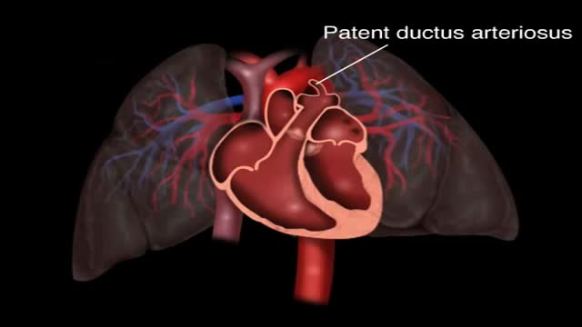
Patent ductus arteriosus (PDA) is a persistent opening between two major blood vessels leading from the heart. The opening, called the ductus arteriosus, is a normal part of a baby's circulatory system before birth that usually closes shortly after birth. If it remains open, however, it's called a patent ductus arteriosus.
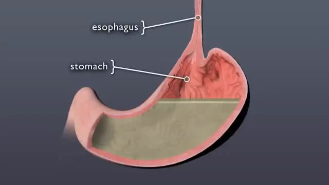
Discover what happens to pill when it swallowed
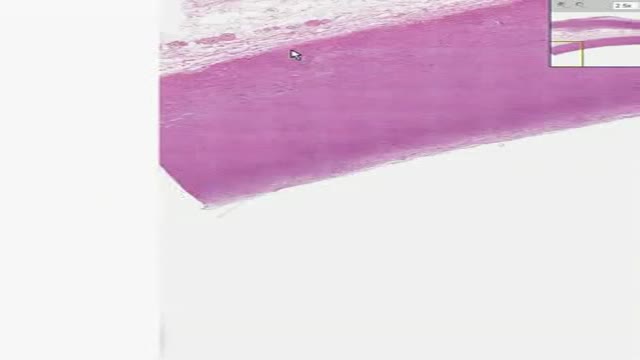
Histology of Aorta
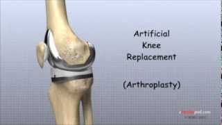
In this animated episode of eOrthopodTV, orthopedic surgeon Randale C. Sechrest, MD narrates the procedure to replace an arthritic knee with an artificial joint.
Yannas had been studying collagen, a protein found in human skin. Teaming up during the 1970s, the two made a polymer (a chemical compound made of multiple repeating units). Using collagen fibers and a long sugar molecule, they formed a porous (full of small holes) material resembling skin.
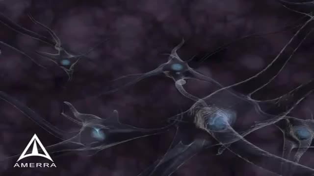
Ca2+ binds with the membrane of the synaptic vesicles, which causes the vesicles to break and release the neurotransmitter into the synaptic cleft. After the neurotransmitters are released, they diffuse across the synaptic cleft and interact with receptors on the postsynaptic membrane. When the action potential reaches the presynaptic terminal, it provokes the release of a small quantity of neurotransmitter molecules, which bind to chemical receptor molecules located in the membrane of another neuron, the postsynaptic neuron, on the opposite side of the synaptic cleft.
