
- Physical Examination
- Surgical Examination
- Ophthalmology
- Clinical Skills
- Orthopedics
- Surgery Videos
- Laparoscopy
- Pediatrics
- Funny Videos
- Cardiothoracic Surgery
- Nursing Videos
- Plastic Surgery
- Otorhinolaryngology
- Histology and Histopathology
- Neurosurgery
- Dermatology
- Pediatric Surgery
- Urology
- Dentistry
- Oncology and Cancers
- Anatomy Videos
- Health and Fitness
- Radiology
- Anaesthesia
- Physical Therapy
- Pharmacology
- Interventional Radiology
- Cardiology
- Endocrinology
- Gynecology
- Emergency Medicine
- Psychiatry and Psychology
- Childbirth Videos
- General Medical Videos
- Nephrology
- Physiology
- Diet and Food Health
- Diabetes Mellitus
- Neurology
- Women Health
- Osteoporosis
- Gastroenterology
- Pulmonology
- Hematology
- Rheumatology
- Toxicology
- Nuclear Medicine
- Infectious Diseases
- Vascular Disease
- Reproductive Health
- Burns and Wound Healing
- Kidney Zone Podcast
- Other
Top videos
The most-watched medical videos on MedicalVideos.com — ranked by views from our community of medical students, healthcare professionals, and patients learning how the body works.
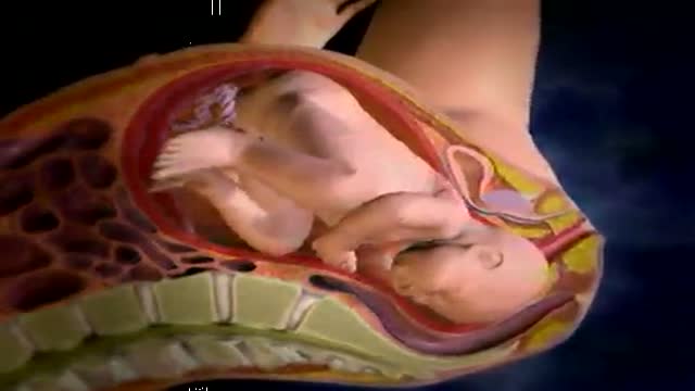

Vacuum Extraction Childbirth Video with ventouse
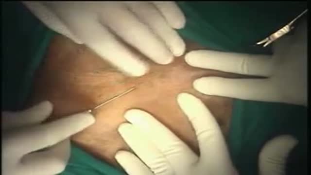
Hernia Repair with Prolene Hernia System
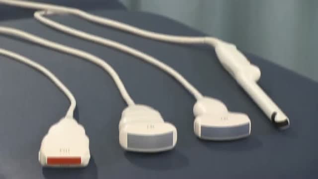
Identify the anatomy and explain the physiology of the scrotum on diagrams and sonograms.
Describe and demonstrate the protocol for sonographic scanning of the scrotum.
Identify and describe sonographic images of congenital abnormalities of the scrotum.
Identify and describe sonographic images of pathologies of the scrotum.
Identify and describe sonographic images of extratesticular disease processes.
Identify the anatomy and explain the physiology of the prostate on diagrams and sonograms.
Describe and demonstrate the protocol for transabdominal and endorectal sonographic scanning of the prostate.
Identify and describe sonographic images of benign and malignant pathologies of the prostate, including benign hyperplasia, prostatitis, carcinoma, and calculi.
Explain the technique for prostate biopsy.
Define the criteria for an ultrasound appearance of prostate tumor staging.
Explain the technique for radiation seed implantation.
Explain the Patient Privacy Rule (HIPAA) and Patient Safety Act (see reference).

Most intact aortic aneurysms do not produce symptoms. As they enlarge, symptoms such as abdominal pain and back pain may develop. Compression of nerve roots may cause leg pain or numbness. Untreated, aneurysms tend to become progressively larger, although the rate of enlargement is unpredictable for any individual. Rarely, clotted blood which lines most aortic aneurysms can break off and result in an embolus. They may be found on physical examination. Medical imaging is necessary to confirm the diagnosis. Symptoms may include: anxiety or feeling of stress; nausea and vomiting; clammy skin; rapid heart rate. In patients presenting with aneurysm of the arch of the aorta, a common symptom is a hoarse voice as the left recurrent laryngeal nerve (a branch of the vagus nerve) is stretched. This is due to the recurrent laryngeal nerve winding around the arch of the aorta. If an aneurysm occurs in this location, the arch of the aorta will swell, hence stretching the left recurrent laryngeal nerve. The patient therefore has a hoarse voice as the recurrent laryngeal nerve allows function and sensation in the voicebox. Abdominal aortic aneurysms, hereafter referred to as AAAs, are the most common type of aortic aneurysm. One reason for this is that elastin, the principal load-bearing protein present in the wall of the aorta, is reduced in the abdominal aorta as compared to the thoracic aorta (nearer the heart). Another is that the abdominal aorta does not possess vasa vasorum, hindering repair. Most are true aneurysms that involve all three layers (tunica intima, tunica media and tunica adventitia), and are generally asymptomatic before rupture. The most common sign for the aortic aneuysm is the Erythema nodosum also known as leg lesions typically found near the ankle area. The prevalence of AAAs increases with age, with an average age of 65–70 at the time of diagnosis. AAAs have been attributed to atherosclerosis, though other factors are involved in their formation. An AAA may remain asymptomatic indefinitely. There is a large risk of rupture once the size has reached 5 cm, though some AAAs may swell to over 15 cm in diameter before rupturing. Before rupture, an AAA may present as a large, pulsatile mass above the umbilicus. A bruit may be heard from the turbulent flow in a severe atherosclerotic aneurysm or if thrombosis occurs. Unfortunately, however, rupture is usually the first hint of AAA. Once an aneurysm has ruptured, it presents with a classic pain-hypotension-mass triad. The pain is classically reported in the abdomen, back or flank. It is usually acute, severe and constant, and may radiate through the abdomen to the back. The diagnosis of an abdominal aortic aneurysm can be confirmed at the bedside by the use of ultrasound. Rupture could be indicated by the presence of free fluid in potential abdominal spaces, such as Morison's pouch, the splenorenal space (between the spleen and left kidney), subdiaphragmatic spaces (underneath the diaphragm) and peri-vesical spaces. A contrast-enhanced abdominal CT scan is needed for confirmation. Only 10–25% of patients survive rupture due to large pre- and post-operative mortality. Annual mortality from ruptured abdominal aneurysms in the United States alone is about 15,000. Another important complication of AAA is formation of a thrombus in the aneurysm.
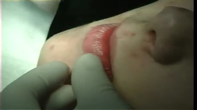
Surgical removal of mucocele from lower lip
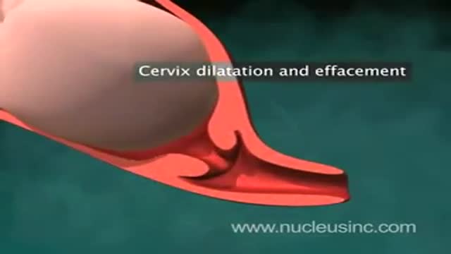

An animation showing vaginal childbirth (delivery)
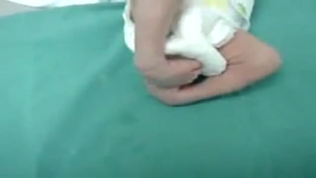
A club foot, or congenital talipes equinovarus (CTEV), is a congenital deformity involving one foot or both. The affected foot appears rotated internally at the ankle. TEV is classified into 2 groups: Postural TEV or Structural TEV.
Without treatment, persons afflicted often appear to walk on their ankles, or on the sides of their feet. It is a common birth defect, occurring in about one in every 1,000 live births. Approximately 50% of cases of clubfoot are bilateral. In most cases it is an isolated dysmelia. This occurs in males more often than in females by a ratio of 2:1. A condition of the same name appears in animals, particularly horses.

The only sure way to prevent genital warts is to not have sex. But everyone wants sex, so here is how to have safe sex if you are living with Genital Warts.

The World Health Organization's official handwashing demonstration: the full soap-and-water technique, surface by surface. The Key Steps checklist on this page follows the WHO sequence in order. General hygiene education — follow your institution's hand-hygiene policy.
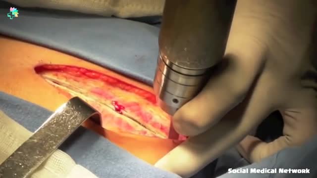
Heart Surgery | 8 Years Old Girl Open Heart Surgery Repair
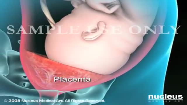
A Cesarean section (C-section) is surgery to deliver a baby. The baby is taken out through the mother's abdomen. In the United States, almost one in three women has their babies this way. Some C-sections are planned, but many are done when unexpected problems happen during delivery. Reasons for a C-section may include
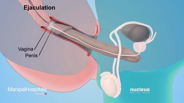
How To Use Male Condom Correctly - Manipal Hospitals

Best Sex Position to Get Pregnant Fast
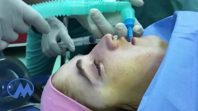
If a patient comes to you with a painful, throbbing, swollen, red face (a ''fat face'), perhaps with fever, trismus and lymphadenitis, he is probably suffering from an acute dental or oral infection, most probably an alveolar abscess. He may have: (1) An alveolar abscess begins as an infection in the bone around a non-vital infected tooth. He has severe pain, which becomes less as pus is released into more superficial tissues and his face starts to swell. After 36 hours of cellulitis he usually has a fluctuant abscess which needs draining. If drainage is delayed, the pus in his abscess discharges spontaneously through a sinus (26-8) in his gum or face, which may become chronic. First, control infection with antibiotics, and then drain the abscess, either by incising it where it is pointing, or by removing the infected tooth, which acts as a cork to prevent the pus escaping, or by doing both these things. If you remove a tooth before you have controlled the infection with antibiotics, and while his face is still severely swollen, you may spread the infection; your task will also be more difficult. (2) A periodontal abscess at the side of a tooth, caused by spread from an infected gum. (3) A pericoronal abscess caused by infection of the gum over the crown of an unerupted and impacted tooth, usually a lower third molar (''an infected wisdom tooth'). Often, an abscess does not form, and the gum round the tooth is merely inflamed.
A Pap smear (also called a Pap test) is a screening procedure for cervical cancer. It tests for the presence of precancerous or cancerous cells on the cervix, the opening of the uterus. It's named after the doctor who determined that this was a useful way to detect signs of cervical cancer.
Dysentery is an infection of the intestines causing diarrhoea that contains blood or mucus. There are two main types of dysentery: Shigellosis, or bacillary dysentery, is the most common type experienced in the UK, caused by the shigella bacteria. Amoebic dysentery, also called amoebiasis, is caused by a single-celled parasite called Entamoeba histolytica. This form of dysentery is more common abroad in tropical countries. This article focuses on amoebic dysentery, This is usually caused by poor hygiene or contaminated food or water. Amoebic dysentery is a notifiable disease, so your GP must let the local health authority know if you have contracted it. Causes of amoebic dysentery Once inside the body, amoeba clump together to form a cyst that is protected by the stomach’s digestive acid. When the cyst passes through the intestines it breaks open infecting the body. The amoebae burrow into the intestinal wall and cause small ulcers or abscesses. Cysts exit the body via faeces but are still able to live outside, which is how many people become infected. Severe dysentery is more common in developing countries due to compromised hygiene. You can get sick in a number of ways including: Eating contaminated food Drinking contaminated water Contracting dysentery from another infected person. Symptoms of amoebic dysentery Symptoms can appear as many as 10 days after exposure and infection by the parasite. Signs of infection include: Watery diarrhoea with blood or pus in it Nausea or vomiting Stomach pain High temperature Chills Bleeding from back passage (rectum) Weight loss Loss of appetite. Complications of amoebic dysentery If the parasite gets into your bloodstream it can spread to other parts of your body, including the liver. When this happens you run the risk of developing a liver abscess. Symptoms include: High temperature Weakness Cough Jaundice Nausea Loss of appetite Weight loss
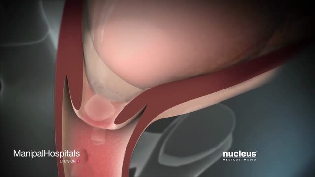
Labor And Delivery During Vaginal Child Birth
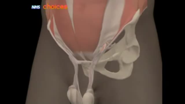
A hernia occurs when an organ or fatty tissue squeezes through a weak spot in a surrounding muscle or connective tissue called fascia. The most common types of hernia are inguinal (inner groin), incisional (resulting from an incision), femoral (outer groin), umbilical (belly button), and hiatal (upper stomach).
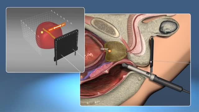
A prostate gland biopsy is a test to remove small samples of prostate tissue to be looked at under a microscope. ... For a prostate biopsy, a thin needle is inserted through the rectum (transrectal biopsy), through the urethra, or through the area between the anus and scrotum (perineum).
When is endoscopy used? Endoscopes were first developed to look at parts of the body that couldn’t be seen any other way. This is still a common reason to use them, but endoscopy now has many other uses too. It’s often used in the prevention, early detection, diagnosis, staging, and treatment of cancer. To prevent and screen for cancer Some types of endoscopes are used to look for cancer in people who have no symptoms. For example, colonoscopy (KO-lun-AH-skuh-pee) and sigmoidoscopy (SIG-moid-AH-skuh-pee) are used to screen for colon and rectal cancer. These procedures can also help prevent cancer because they let doctors find and remove polyps (growths) that might become cancer if left alone. To find cancer early Endoscopy can sometimes be used to find cancer early, before it has had a chance to grow or spread. Looking for causes of symptoms When people go to the doctor with certain symptoms, endoscopy can sometimes be used to help find a cause. For instance: Laryngoscopy to look at the vocal cords in people with long-term hoarseness Upper endoscopy in people having trouble swallowing Colonoscopy in people with anemia (low red blood cell counts) with an unknown cause Colonoscopy in people with blood in their stool Looking at problems found on imaging tests Imaging tests such as x-rays and CT scans can sometimes show physical changes within the body. But these tests may only give information about the size, shape, and location of the problem. Doctors use endoscopes to see more details, like color and surface texture, when trying to find out what’s going on. Newer methods of endoscopy that include high magnification are being tested to find out whether they are more useful in detecting cancer and other abnormal cells on the inner surfaces of the body. To diagnose and find out the stage (extent) of cancer To get a tissue sample Going one step further, most types of endoscopes have tools on the end that the doctor can use to take out small tissue samples. This procedure is called a biopsy (BY-op-see). Samples can be taken from suspicious areas and then looked at under a microscope or tested in other ways to see if cancer is there. A biopsy is usually the best way to find out if a growth or change is cancer or something else. Getting a closer look In some cases endoscopes are used to help find out how far a cancer has spread. Thoracoscopy (THOR-uh-KAHS -kuh-pee) and laparoscopy (LAP-uh-RAHS-kuh-pee) can be very useful in finding out if cancer has spread into the thorax (chest) or abdomen (belly). The surgeon can look into these places making only a small incision (cut) in the skin.
Medical Videos
Free medical and surgery videos for patients, students and professionals — trusted since 2007.
Founded by Dr. Mohamed Ibrahim, MD, PhD — physician-scientist.
Follow us on Facebook · Instagram