- Physical Examination
- Surgical Examination
- Ophthalmology
- Clinical Skills
- Orthopedics
- Surgery Videos
- Laparoscopy
- Pediatrics
- Funny Videos
- Cardiothoracic Surgery
- Nursing Videos
- Plastic Surgery
- Otorhinolaryngology
- Histology and Histopathology
- Neurosurgery
- Dermatology
- Pediatric Surgery
- Urology
- Dentistry
- Oncology and Cancers
- Anatomy Videos
- Health and Fitness
- Radiology
- Anaesthesia
- Physical Therapy
- Pharmacology
- Interventional Radiology
- Cardiology
- Endocrinology
- Gynecology
- Emergency Medicine
- Psychiatry and Psychology
- Childbirth Videos
- General Medical Videos
- Nephrology
- Physiology
- Diet and Food Health
- Diabetes Mellitus
- Neurology
- Women Health
- Osteoporosis
- Gastroenterology
- Pulmonology
- Hematology
- Rheumatology
- Toxicology
- Nuclear Medicine
- Infectious Diseases
- Vascular Disease
- Reproductive Health
- Burns and Wound Healing
- Other
Top videos
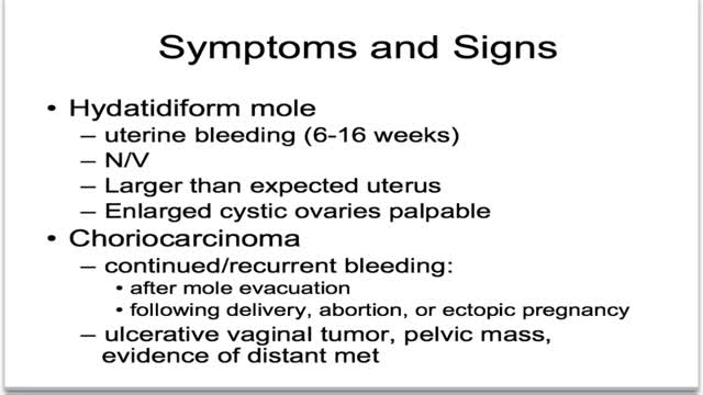
What is gestational trophoblastic disease? Cancer starts when cells in the body begin to grow out of control. Cells in nearly any part of the body can become cancer, and can spread to other areas of the body. To learn more about how cancers start and spread, see What Is Cancer? Gestational trophoblastic (jeh-STAY-shuh-nul troh-fuh-BLAS-tik) disease (GTD) is a group of rare tumors that involve abnormal growth of cells inside a woman's uterus. GTD does not develop from cells of the uterus like cervical cancer or endometrial (uterine lining) cancer do. Instead, these tumors start in the cells that would normally develop into the placenta during pregnancy. (The term gestational refers to pregnancy.) GTD begins in the layer of cells called the trophoblast (troh-fuh-BLAST) that normally surrounds an embryo. (Tropho- means nutrition, and -blast means bud or early developmental cell.) Early in normal development, the cells of the trophoblast form tiny, finger-like projections known as villi. The villi grow into the lining of the uterus. In time, the trophoblast layer develops into the placenta, the organ that protects and nourishes the growing fetus.
The usual reason given for people getting fat is that they eat too much and/or exercise too little. That reflects one of the basic laws of thermodynamics—I forget which one. The amount of energy you put into a system minus the energy you take out has to be stored somewhere i.e. FAT! This formulation—true though it is—does not entirely explain obesity since some people seem to eat more than fat people and exercise no more than these same fat people, and yet they are not fat! Chalking this fact up to the general perversity of the universe is not sufficient explanation. Other factors must come into play. I mention below some of the ideas thoughtful people have proposed to explain why fat people become fat:
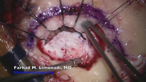
Brain Surgery: Microvascular Decompression of facial nerve for hemifacial spasm
Cytomegalovirus is a genus of viruses in the order Herpesvirales, in the family Herpesviridae, in the subfamily Betaherpesvirinae. Humans and monkeys serve as natural hosts.
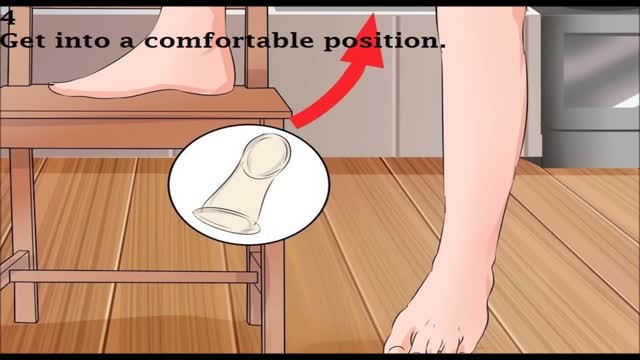
female condom
Pyogenic liver abscess Email this page to a friend Email this page to a friend Facebook Twitter Google+ Pyogenic liver abscess is a pus-filled area in the liver. Causes There are many potential causes of liver abscesses, including: Abdominal infection, such as appendicitis, diverticulitis, or a perforated bowel Infection in the blood Infection of the bile draining tubes Recent endoscopy of the bile draining tubes Trauma that damages the liver The most common bacteria that cause liver abscesses are: Escherichia coli Bacteroides Enterococcus Klebsiella pneumoniae Staphylococcus aureus Streptococcus In most cases, more than one type of bacteria is found.

Callus Peel is a luxury, spa foot treatment that removes hard, callused skin leaving your feet feeling soft and revitalised. The treatment is a 15 minute...
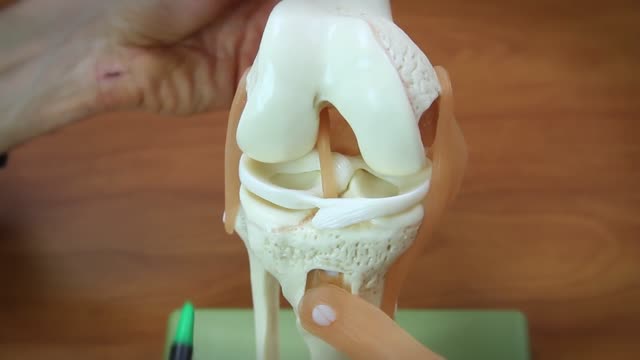
A torn meniscus is one of the most common knee injuries. Any activity that causes you to forcefully twist or rotate your knee, especially when putting your full weight on it, can lead to a torn meniscus. Each of your knees has two menisci — C-shaped pieces of cartilage that act like a cushion between your shinbone and your thighbone. A torn meniscus causes pain, swelling and stiffness. You also might feel a block to knee motion and have trouble extending your knee fully. Conservative treatment — such as rest, ice and medication — is sometimes enough to relieve the pain of a torn meniscus and give the injury time to heal on its own. In other cases, however, a torn meniscus requires surgical repair.
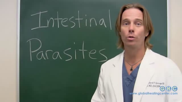
One of the most common parasites to infect human beings is the yeast-like Blastocystis hominis, a single-celled parasitic organism that causes abdominal cramping, bloating, gas, and sometimes anal itching. Other common parasites are: Tapeworms, which can grow as long as 60 feet while living in the human intestines.
Indications for intervention in patients with a renal artery aneurysm (RAA) include the following [20, 8, 13, 14] : Rupture Symptomatic RAA - Hypertension (from associated renal artery stenosis, refractory to medical management), pain, renal ischemia or infarction secondary to embolization from the aneurysm sac RAAs in females who are pregnant or are contemplating pregnancy Diameter greater than 2 cm Enlarging RAA RAA associated with acute dissection Currently, there is no consensus regarding the size at which an RAA should be repaired in an asymptomatic patient. Experts have recommended RAA repair at diameters ranging from 1.5 to 3 cm, [8] though most suggest 2 cm. Some reports have even suggest that larger asymptomatic saccular aneurysms may be managed expectantly. Note that aneurysm rupture at a diameter of 1.5 cm has been reported. Complete calcification of the wall of the aneurysm sac manifests in about 40% of patients. This was once believed to confer protection against rupture [21] ; however, this belief has since been questioned. [30] Asymptomatic, small (<2 cm in diameter) RAAs do not usually require treatment. One notable exception is an RAA in a woman who is pregnant or contemplating pregnancy. In view of the increased risk of rupture in such cases, even small asymptomatic aneurysms should be repaired in this population. For diagnosis and preinterventional planning, gadolinium-enhanced magnetic resonance angiography (MRA) and computed tomography (CT) angiography (CTA) with three-dimensional (3D) reconstruction have essentially replaced conventional arteriography. Regular follow-up examination with ultrasonography (US) or CT) is recommended in patients who are treated expectantly. Spontaneous cure by thrombosis of small aneurysms has been described. Further refinements in endovascular techniques may allow more RAAs to be treated in this manner. So far, excellent short- and intermediate-term results have been described in the literature [40] ; however, there remains a need for further long-term outcome data.

Find Out More at https://medwayhealthcare.com/

Male babies leave their DNA in the mother
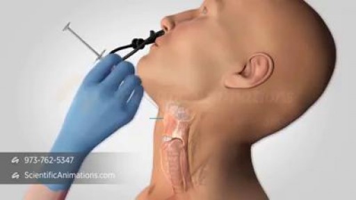
The voice box, or larynx, has three important functions. It is necessary for breathing, voice and swallowing. The vocal folds have two positions, open (apart) for breathing (picture I) and closed (together) for making sound, coughing and sealing off the lungs when swallowing (picture II). When one of the vocal folds are paralyzed, it usually rests in an in-between position (picture III), and neither opens for breathing, nor closes for voicing, coughing, or swallowing. Usually, the effects on the voice are the most dramatic. The voice becomes weak and breathy. People can only say a few words per breath, and are frequently out-of-breath, or physically tired when trying to speak for more than a few minutes straight. The voice may also get somewhat high and squeaky, with a diminished range. Swallowing may be affected as well, where you may notice some choking or coughing with certain liquids. Your cough is frequently different and very weak. This is a serious problem for patients with with vocal fold paralysis because one of the most important functions of the larynx is to keep liquids out of the lungs, and to be able to cough up mucus. When this does not happen, you are at risk for getting an "aspiration" pneumonia. The surgical procedure to restore these important functions is called "medialization laryngoplasty"
Cardioversion takes minutes. The patient is sedated (for a few minutes) and then a shock is delivered. The heart nearly always goes back to regular sinus rhythm. ... Patients without prior ablation or heart surgery rarely develop non-right atrial flutter.
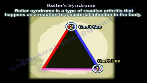
Reiter syndrome is a type of reactive arthritis that happens as a reaction to a bacterial infection in the body. The infection usually happens in the intestines, genitals, or urinary tract. Reiter syndrome includes redness, joint swelling and pain, often in knees, ankles, and feet, along with inflammation of the eyes and urinary tract. It is not contagious. But the bacteria that trigger it can be passed from one person to another. There is no cure for Reiter syndrome, but you can control the symptoms. For most people, symptoms go away in 2 to 6 months.
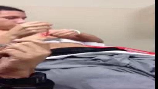
A foot of gauze out of nipple abscess
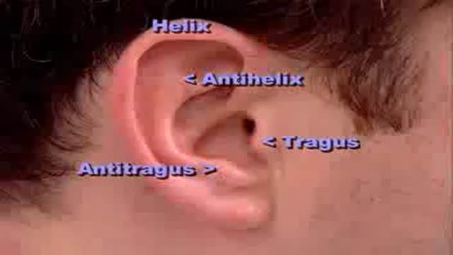
Complete clinical examination of the ears with all the associated tests
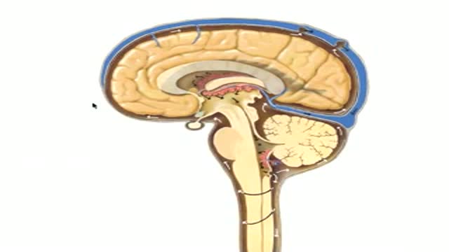
Neuroanatomy of CSF Flow
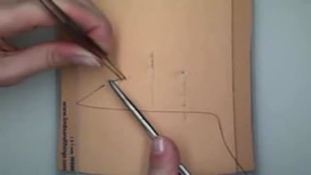
Demonstration of subcuticular or intradermal suturing technique for wound closure in the operating room.

Video shows a Hip resurfacing operation done using the Durom hip from Zimmer.
The patient is a young active male. Hip resurfacing is emerging as the surgical procedure of choice in young and active patients for pain relief from Hip arthritis.
