
- Physical Examination
- Surgical Examination
- Ophthalmology
- Clinical Skills
- Orthopedics
- Surgery Videos
- Laparoscopy
- Pediatrics
- Funny Videos
- Cardiothoracic Surgery
- Nursing Videos
- Plastic Surgery
- Otorhinolaryngology
- Histology and Histopathology
- Neurosurgery
- Dermatology
- Pediatric Surgery
- Urology
- Dentistry
- Oncology and Cancers
- Anatomy Videos
- Health and Fitness
- Radiology
- Anaesthesia
- Physical Therapy
- Pharmacology
- Interventional Radiology
- Cardiology
- Endocrinology
- Gynecology
- Emergency Medicine
- Psychiatry and Psychology
- Childbirth Videos
- General Medical Videos
- Nephrology
- Physiology
- Diet and Food Health
- Diabetes Mellitus
- Neurology
- Women Health
- Osteoporosis
- Gastroenterology
- Pulmonology
- Hematology
- Rheumatology
- Toxicology
- Nuclear Medicine
- Infectious Diseases
- Vascular Disease
- Reproductive Health
- Burns and Wound Healing
- Other
Top videos
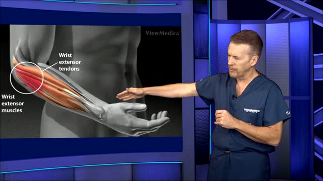

This video: Tennis elbow or lateral epicondylitis is a condition in which the outer part of the elbow becomes sore and tender. The forearm muscles and tendons become damaged from overuse — repeating the same strenuous motions again and again.

Laparoscopy in acute bowel obstruction following previous surgery is a difficult procedure and avoided by most of the surgeons due to the difficulty in obtaining pneumoperitoneum, port placement, lack of working space, adhesions and risk of bowel injury.
Here is a patient who had a previous laparotomy for trauma with a midline incision from xyphysternum to pubis; after unsuccessful conservative management he underwent a laparoscopy; a prior CT scan showed adhesions in the left side and a distal-mid small bowel obstruction. The pneumoperitoneum was obtained with the Visiport placed in the right lower quadrant; although the abdomen was grossly distended, under significant tension and distended loops of small bowel were occupying most the peritoneal cavity, with muscle relaxation there is usually enough space to perform a thorough inspection of the abdominal cavity. Port placement has to be done with special care as there is no room to push and usually a blunt trocar directed away from the bowel is employed in my practice. The collapsed loops of small bowel point quickly to the site of obstruction -- it is better to avoid manipulating the distended bowel as it is heavy, oedematous and prone to be lacerated with the instruments; once the pathology is identified, in this case the obstructive band, light packing is performed in order to expose the working space and protect the bowel from instruments like scissors or diathermy. In this case the band adhesion was slightly more difficult to separate from the bowel and required a combination of sharp and gentle blunt dissection.
Once the obstruction is release and the transit of contents is confirmed in the collapsed bowel the procedure is terminated. No abdominal drainage is usually necessary.

TV interview with Adina Nack, Ph.D. about her own cervical HPV experiences, STD research, her new book (Damaged Goods? Women Living with Incurable Sexually Transmitted Diseases), and women's lives after genital warts, HPV and herpes infections. More info is available on STDdatings.com, which is the official STD dating & support site.

Periodontal Disease and Respiratory Infections
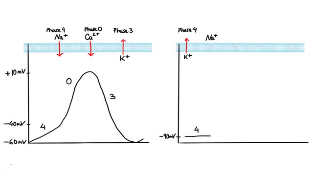
Antiarrhythmics are drugs that are used to treat abnormal heart rhythms resulting from irregular electrical activity of the heart. There are many different types of antiarrhythmic drugs. Examples include: Amiodarone (Cordarone) Flecainide (Tambocor) Procainamide (Procanbid) Sotalol (Betapace) In addition, there are other types of heart drugs that can be used to treat arrhythmias, including: Beta-blockers such as metoprolol or Toprol XL, which reduce the heart's workload and heart rate. Calcium channel blockers such as verapamil or Calan, which also reduces the heart rate.

bad breath odor
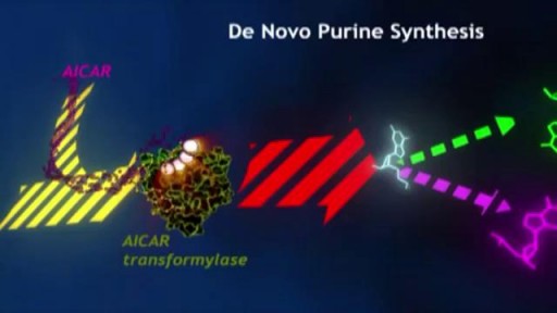
Methotrexate anti-tumor activity is a result of the inhibition of folic acid reductase, leading to inhibition of DNA synthesis and inhibition of cellular replication. The mechanism involved in its activity against rheumatoid arthritis is not known.
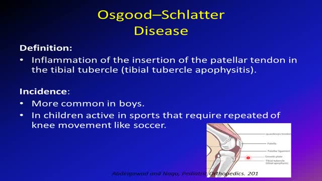
Osgood-Schlatter disease can cause a painful lump below the kneecap in children and adolescents experiencing growth spurts during puberty. Osgood-Schlatter disease occurs most often in children who participate in sports that involve running, jumping and swift changes of direction — such as soccer, basketball, figure skating and ballet. While Osgood-Schlatter disease is more common in boys, the gender gap is narrowing as more girls become involved with sports. Age ranges differ by sex because girls experience puberty earlier than do boys. Osgood-Schlatter disease typically occurs in boys ages 13 to 14 and girls ages 11 to 12. The condition usually resolves on its own, once the child's bones stop growing.

A Medical Video showing an overview of the endocrine and gland system of the human body
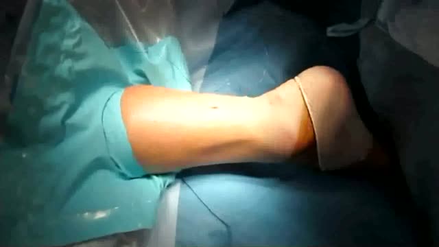
an incision made on the back of the lower leg starting just above the heel bone. After the surgeon finds the two ends of the ruptured tendon, these ends are sewn together with sutures. The incision is then closed. Another repair method makes a small incision on the back of the lower leg at the site of the rupture.

Conjoined twins Alex and Angel Mendoza from Phoenix, Ariz. were successfully separated after more than 12 hours in surgery, reports Dr. Debbye Turner Bell.
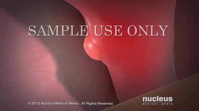
What is an Aneurysm? A cerebral or intracranial aneurysm is an abnormal focal dilation of an artery in the brain that results from a weakening of the inner muscular layer (the intima) of a blood vessel wall. The vessel develops a "blister-like" dilation that can become thin and rupture without warning. The resultant bleeding into the space around the brain is called a subarachnoid hemorrhage (SAH). This kind of hemorrhage can lead to a stroke, coma, and/or death. Aneurysms are usually found at the base of the brain just inside the skull, in an area called the subarachnoid space. In fact, 90 percent of SAHs are attributed to ruptured cerebral aneurysms and the two terms are often used synonymously.

UW Health orthopedic surgeon Richard Illgen has pioneered robotic-assisted knee replacement and serves as a regional and national expert in these techniques. Learn more: http://www.uwhealth.org/49421
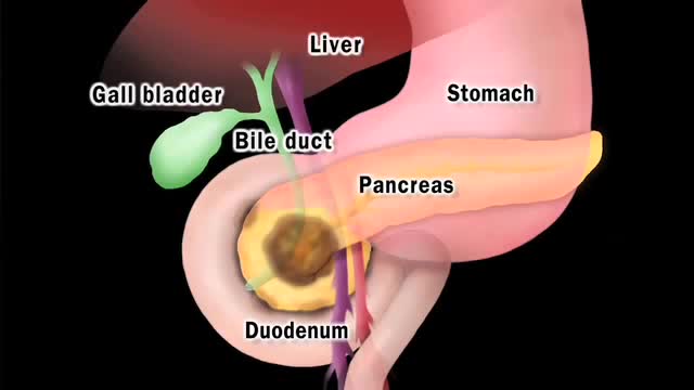
Pancreatic cancer begins in the tissues of your pancreas — an organ in your abdomen that lies horizontally behind the lower part of your stomach. Your pancreas secretes enzymes that aid digestion and hormones that help regulate the metabolism of sugars. Pancreatic cancer often has a poor prognosis, even when diagnosed early. Pancreatic cancer typically spreads rapidly and is seldom detected in its early stages, which is a major reason why it's a leading cause of cancer death. Signs and symptoms may not appear until pancreatic cancer is quite advanced and complete surgical removal isn't possible.

Wet dreams occur when you ejaculate during your sleep. The medical term for a wet dream is “nocturnal emission.“ Most wet dreams are reported in teenage boys and young men, and sometimes they occur well into adulthood.

An U/S showing first trimesteric scan
LASER SURGERY Pilonidal Cyst removal
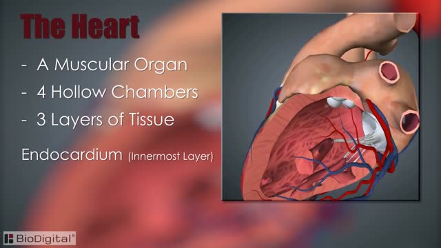
The heart, blood vessels, and blood are the parts that make up the circulatory system, which is defined as a closed system of blood vessels for the transport of gasses and nutrients. The heart is the key organ in the circulatory system. As a hollow, muscular pump, its main function is to propel blood throughout the body.
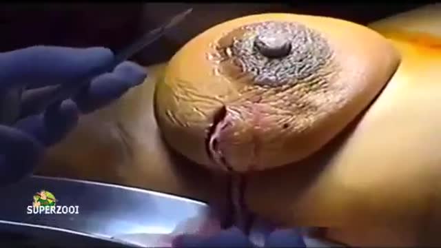
Breast abscesses are often linked to mastitis – a condition that causes breast pain and swelling (inflammation), and usually affects women who are breastfeeding. Infections can occur during breastfeeding if bacteria enter your breast tissue, or if the milk ducts (tiny tubes) become blocked. This can cause mastitis which, if not treated, can result in an abscess forming. Women who aren't breastfeeding can also develop mastitis if bacteria enter the milk ducts through a sore or cracked nipple, or a nipple piercing. White blood cells are sent to attack the infection, which causes tissue at the site of the infection to die. This creates a small, hollow area that fills with pus (an abscess).
