Top Videos
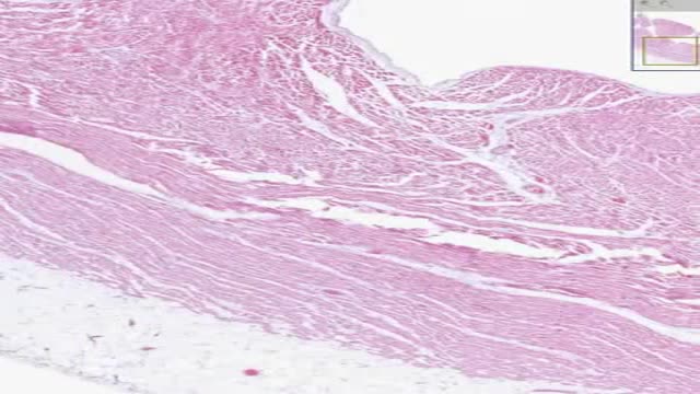
Histology of Heart Cardiac Muscle
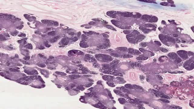
Histology of Thymus
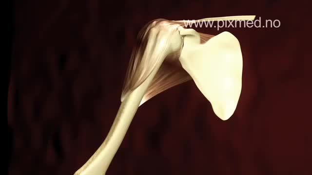
The shoulder is one of the largest and most complex joints in the body. The shoulder joint is formed where the humerus (upper arm bone) fits into the scapula (shoulder blade), like a ball and socket. Other important bones in the shoulder include: The acromion is a bony projection off the scapula. The clavicle (collarbone) meets the acromion in the acromioclavicular joint. The coracoid process is a hook-like bony projection from the scapula.

This could be caused by an infection, food poisoning, parasites, Crohn's disease, or reduced blood flow in the colon. Hemorrhoids are another common cause of GI or rectal bleeding. A hemorrhoid is an enlarged vein in your rectum or anus. These enlarged veins can rupture and bleed, causing rectal bleeding.

Colorectal cancer (also known as colon cancer, rectal cancer or bowel cancer) is the development of cancer in the colon or rectum (parts of the large intestine). It is due to the abnormal growth of cells that have the ability to invade or spread to other parts of the body. People with HNPCC tend to develop colon cancer before age 50. Familial adenomatous polyposis (FAP). FAP is a rare disorder that causes you to develop thousands of polyps in the lining of your colon and rectum. People with untreated FAP have a greatly increased risk of developing colon cancer before age 40.

Hodgkin's lymphoma — formerly known as Hodgkin's disease — is a cancer of the lymphatic system, which is part of your immune system. In Hodgkin's lymphoma, cells in the lymphatic system grow abnormally and may spread beyond the lymphatic system. As Hodgkin's lymphoma progresses, it compromises your body's ability to fight infection. Hodgkin's lymphoma is one of two common types of cancers of the lymphatic system. The other type, non-Hodgkin's lymphoma, is far more common. Advances in diagnosis and treatment of Hodgkin's lymphoma have helped give people with this diagnosis the chance for a full recovery. The prognosis continues to improve for people with Hodgkin's lymphoma.
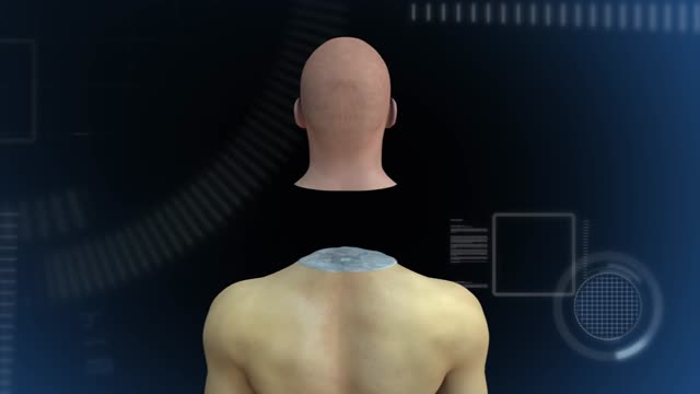
Head transplant successfully performed on monkey,

How to memorize more in pharma: Drug names, dental implications, numbers

Hepatitis A signs and symptoms, which typically don't appear until you've had the virus for a few weeks, may include: Fatigue Nausea and vomiting Abdominal pain or discomfort, especially in the area of your liver on your right side beneath your lower ribs Clay-colored bowel movements Loss of appetite Low-grade fever Dark urine Joint pain Yellowing of the skin and eyes (jaundice) If you have hepatitis A, you may have a mild illness that lasts a few weeks or a severe illness that lasts several months. Not everyone with hepatitis A develops signs or symptoms.
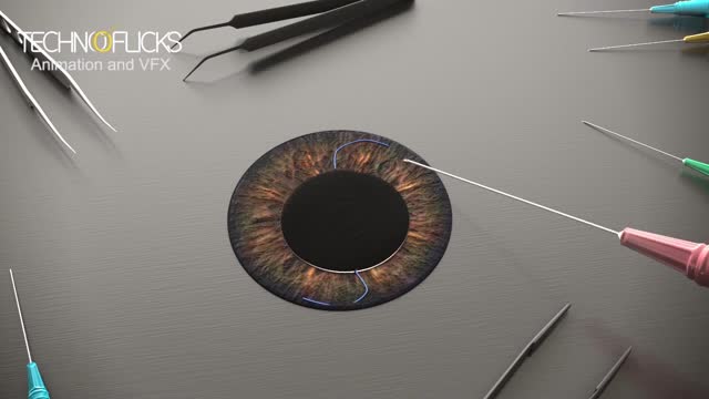
Cosmetic iris implants have not been evaluated by any U.S. regulatory agency or tested for safety in clinical trials. While the implants are not approved by the U.S. Food and Drug Administration, it has been reported in the media this month that the surgery is being performed overseas. During iris implant surgery, an artificial iris made of silicone is folded and inserted into a slit that has been cut into the cornea. Then the iris is unfolded and adjusted to cover the natural iris. Local anesthesia is used.

ESCLEROTERAPIA
Femoroacetabular impingement (FAI) is a condition in which extra bone grows along one or both of the bones that form the hip joint — giving the bones an irregular shape. Because they do not fit together perfectly, the bones rub against each other during movement. Over time this friction can damage the joint, causing pain and limiting activity.
Watch that video of A man with one inch-wide hole in his face
Presence of abdominal pain and distension. Presence of urinary symptoms - Such as dysuria, oliguria, flank pain, and hematuria. Occurrence of any symptoms of hypocalcemia - Such as anorexia, vomiting, cramps, seizures, spasms, altered mental status, and tetany. Symptoms of hyperkalemia - Such as weakness and paralysis.
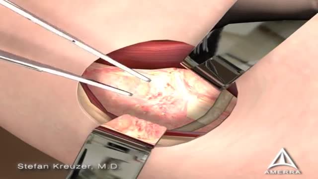
Dacryocystorhinostomy (DCR) is a procedure performed for the treatment of tearing (epiphora) due to blockage of the nasolacrimal duct. Tears originate in the lacrimal gland, located at the upper outer margin of the eye. As tears cross the eye with each blink, they are directed into small openings in the eyelids called puncta. From this point, tears travel through a pathway known as the canalicular system into the lacrimal sac. The lacrimal sac is located between the eye and the nose, and funnels tears into the nasal cavity through the nasolacrimal duct (Figure 1). As this is quite a long path for tears to travel, there can be many causes of excessive tearing. Blockage of the nasolacrimal duct is one common cause, and can be treated by creating a direct opening from the lacrimal sac into the nasal cavity in a procedure known as DCR. The evaluation and management of tearing may involve both an ophthalmologist and an otolaryngologist.
The following guidelines are an interpretation of the evidence presented in the 2010 International Consensus on Cardiopulmonary Resuscitation and Emergency Cardiovascular Care Science With Treatment Recommendations1). They apply primarily to newly born infants undergoing transition from intrauterine to extrauterine life, but the recommendations are also applicable to neonates who have completed perinatal transition and require resuscitation during the first few weeks to months following birth. Practitioners who resuscitate infants at birth or at any time during the initial hospital admission should consider following these guidelines. For the purposes of these guidelines, the terms newborn and neonate are intended to apply to any infant during the initial hospitalization. The term newly born is intended to apply specifically to an infant at the time of birth.
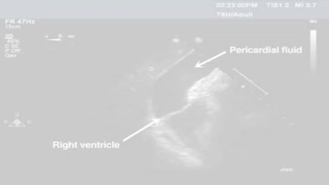
Pericardiocentesis is the aspiration of fluid from the pericardial space that surrounds the heart. This procedure can be life saving in patients with cardiac tamponade, even when it complicates acute type A aortic dissection and when cardiothoracic surgery is not available. [1] Cardiac tamponade is a time sensitive, life-threatening condition that requires prompt diagnosis and management. Historically, the diagnosis of cardiac tamponade has been based on clinical findings. Claude Beck, a cardiovascular surgeon, described 2 triads of clinical findings that he found associated with acute and chronic cardiac tamponade. The first of these triads consisted of hypotension, an increased venous pressure, and a quiet heart. It has come to be recognized as Beck's triad, a collection of findings most commonly produced by acute intrapericardial hemorrhage. Subsequent studies have shown that these classic findings are observed in only a minority of patients with cardiac tamponade. [2] The detection of pericardial fluid has been facilitated by the development and continued improvement of echocardiography. [3] Cardiac ultrasound is now accepted as the criterion standard imaging modality for the assessment of pericardial effusions and the dynamic findings consistent with cardiac tamponade. With echocardiography, the location of the effusion can be identified, the size can be estimated (small, medium, or large), and the hemodynamic effects can be examined by assessing for abnormal septal motion, right atrial or right ventricular inversion, and decreased respiratory variation of the diameter of the inferior vena cava.
Wilson's disease is a rare inherited disorder that causes too much copper to accumulate in your liver, brain and other vital organs. Symptoms typically begin between the ages of 12 and 23. Copper plays a key role in the development of healthy nerves, bones, collagen and the skin pigment melanin. Normally, copper is absorbed from your food, and any excess is excreted through bile — a substance produced in your liver.
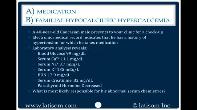
There are 3 genetic types of FHH based on chromosome location. FHH type 1 accounts for 65% of cases and is due to inactivating mutations in the CASR gene, localized to 3q21.1. This gene encodes the calcium-sensing receptor (CaSR). Loss of CaSR function results in a reduction in the sensitivity of parathyroid and renal cells to calcium levels so hypercalcemia is perceived as normal. The other 35% have either a mutation GNA11 (19p13.3) seen in FHH type 2 or AP2S1 (19q13.2-q13.3) seen in FHH type 3 (see these terms) or in genes not yet discovered. FHH is rarely caused by auto-antibodies against CaSR in those without a mutation.

Indian Boy has 232 teeth removed from his mouth !
