- Physical Examination
- Surgical Examination
- Ophthalmology
- Clinical Skills
- Orthopedics
- Surgery Videos
- Laparoscopy
- Pediatrics
- Funny Videos
- Cardiothoracic Surgery
- Nursing Videos
- Plastic Surgery
- Otorhinolaryngology
- Histology and Histopathology
- Neurosurgery
- Dermatology
- Pediatric Surgery
- Urology
- Dentistry
- Oncology and Cancers
- Anatomy Videos
- Health and Fitness
- Radiology
- Anaesthesia
- Physical Therapy
- Pharmacology
- Interventional Radiology
- Cardiology
- Endocrinology
- Gynecology
- Emergency Medicine
- Psychiatry and Psychology
- Childbirth Videos
- General Medical Videos
- Nephrology
- Physiology
- Diet and Food Health
- Diabetes Mellitus
- Neurology
- Women Health
- Osteoporosis
- Gastroenterology
- Pulmonology
- Hematology
- Rheumatology
- Toxicology
- Nuclear Medicine
- Infectious Diseases
- Vascular Disease
- Reproductive Health
- Burns and Wound Healing
- Other
Top videos
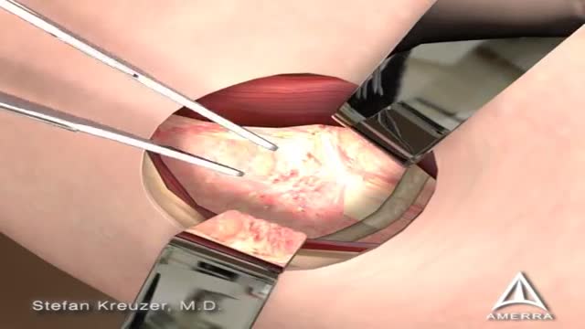
Femoroacetabular impingement (FAI) is a condition in which extra bone grows along one or both of the bones that form the hip joint — giving the bones an irregular shape. Because they do not fit together perfectly, the bones rub against each other during movement. Over time this friction can damage the joint, causing pain and limiting activity.
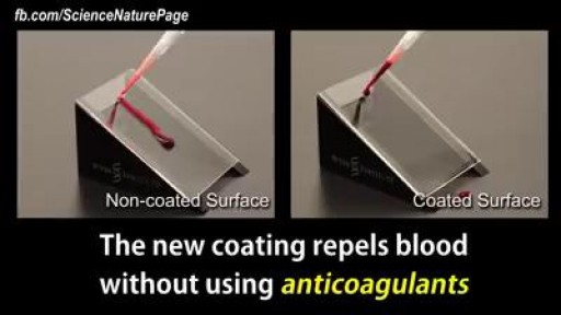
This coating prevents blood from sticking on medical devices
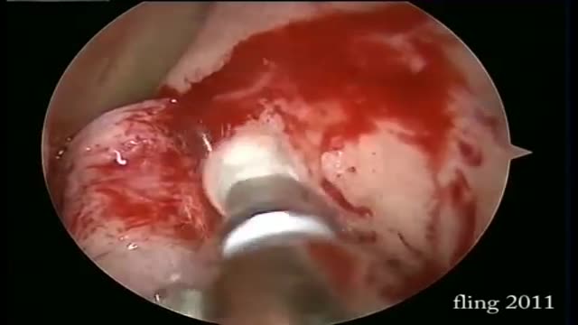
Dacryocystorhinostomy (DCR) is a procedure performed for the treatment of tearing (epiphora) due to blockage of the nasolacrimal duct. Tears originate in the lacrimal gland, located at the upper outer margin of the eye. As tears cross the eye with each blink, they are directed into small openings in the eyelids called puncta. From this point, tears travel through a pathway known as the canalicular system into the lacrimal sac. The lacrimal sac is located between the eye and the nose, and funnels tears into the nasal cavity through the nasolacrimal duct (Figure 1). As this is quite a long path for tears to travel, there can be many causes of excessive tearing. Blockage of the nasolacrimal duct is one common cause, and can be treated by creating a direct opening from the lacrimal sac into the nasal cavity in a procedure known as DCR. The evaluation and management of tearing may involve both an ophthalmologist and an otolaryngologist.
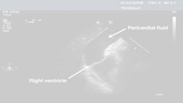
Pericardiocentesis is the aspiration of fluid from the pericardial space that surrounds the heart. This procedure can be life saving in patients with cardiac tamponade, even when it complicates acute type A aortic dissection and when cardiothoracic surgery is not available. [1] Cardiac tamponade is a time sensitive, life-threatening condition that requires prompt diagnosis and management. Historically, the diagnosis of cardiac tamponade has been based on clinical findings. Claude Beck, a cardiovascular surgeon, described 2 triads of clinical findings that he found associated with acute and chronic cardiac tamponade. The first of these triads consisted of hypotension, an increased venous pressure, and a quiet heart. It has come to be recognized as Beck's triad, a collection of findings most commonly produced by acute intrapericardial hemorrhage. Subsequent studies have shown that these classic findings are observed in only a minority of patients with cardiac tamponade. [2] The detection of pericardial fluid has been facilitated by the development and continued improvement of echocardiography. [3] Cardiac ultrasound is now accepted as the criterion standard imaging modality for the assessment of pericardial effusions and the dynamic findings consistent with cardiac tamponade. With echocardiography, the location of the effusion can be identified, the size can be estimated (small, medium, or large), and the hemodynamic effects can be examined by assessing for abnormal septal motion, right atrial or right ventricular inversion, and decreased respiratory variation of the diameter of the inferior vena cava.
Wilson's disease is a rare inherited disorder that causes too much copper to accumulate in your liver, brain and other vital organs. Symptoms typically begin between the ages of 12 and 23. Copper plays a key role in the development of healthy nerves, bones, collagen and the skin pigment melanin. Normally, copper is absorbed from your food, and any excess is excreted through bile — a substance produced in your liver.

The CSICU rounds are an opportunity for residents to come together with attendings and review all the patients in the ICU.
Cedars-Sinai is committed to educating exceptional cardiothoracic surgeons through outstanding personal mentorship, operative training and research leadership. Residents of the Thoracic Surgery—Integrated Residency at Cedars-Sinai will be part of an incredibly rich, academic environment—each year our research and thought leadership features in hundreds of publications in journals including Nature, New England Journal of Medicine, JAMA, Lancet and leading specialty journals.
Learn more about the Cedars-Sinai Thoracic Surgery—Integrated Residency: https://ceda.rs/3UDrZFL
Connect with us:
https://twitter.com/CedarsSinai
https://www.facebook.com/CedarsSinai
https://www.instagram.com/CedarsSinai
Cedars-Sinai is a leader in providing high-quality healthcare encompassing primary care, specialized medicine and research. Since 1902, Cedars-Sinai has evolved to meet the needs of one of the most diverse regions in the nation, setting standards in quality and innovative patient care, research, teaching and community service. Today, Cedars- Sinai is known for its national leadership in transforming healthcare for the benefit of patients. Cedars-Sinai impacts the future of healthcare by developing new approaches to treatment and educating tomorrow’s health professionals. Additionally, Cedars-Sinai demonstrates a commitment to the community through programs that improve the health of its most vulnerable residents.
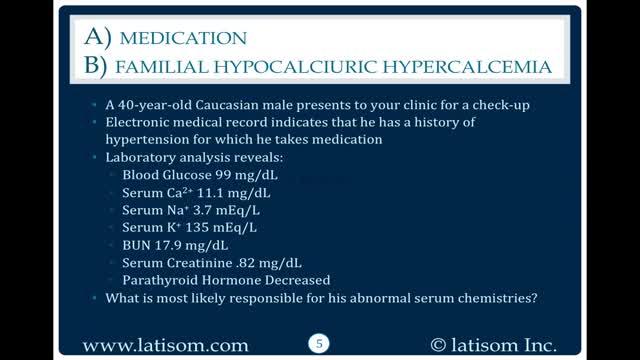
There are 3 genetic types of FHH based on chromosome location. FHH type 1 accounts for 65% of cases and is due to inactivating mutations in the CASR gene, localized to 3q21.1. This gene encodes the calcium-sensing receptor (CaSR). Loss of CaSR function results in a reduction in the sensitivity of parathyroid and renal cells to calcium levels so hypercalcemia is perceived as normal. The other 35% have either a mutation GNA11 (19p13.3) seen in FHH type 2 or AP2S1 (19q13.2-q13.3) seen in FHH type 3 (see these terms) or in genes not yet discovered. FHH is rarely caused by auto-antibodies against CaSR in those without a mutation.
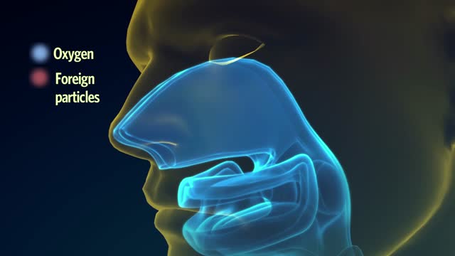
The lungs and respiratory system allow oxygen in the air to be taken into the body, while also enabling the body to get rid of carbon dioxide in the air breathed out. Respiration is the term for the exchange of oxygen from the environment for carbon dioxide from the body's cells.
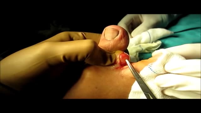
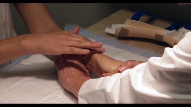
Lipoma From Foot (Inter Digital Web Space) Removal Technique
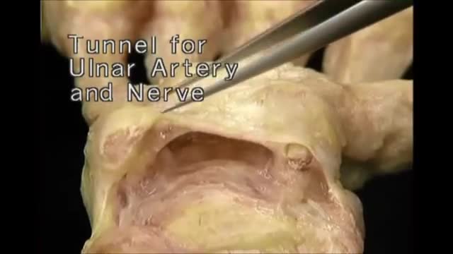
Hand Anatomy
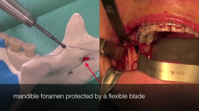
The bilateral sagittal split osteotomy is an indispensable tool in the correction of dentofacial abnormalities. The technique has been in practice since the late 1800s, but did not reach widespread acceptance and use until several modifications were described in the 1960s and 1970s. Those modifications came from a desire to make the procedure safer, more reliable, and more predictable with less relapse. Those goals continue to stimulate innovation in the field today and have helped the procedure evolve to be a very dependable, consistent method of correction of many types of malocclusion. The operative surgeon should be well versed in the history, anatomy, technical aspects, and complications of the bilateral sagittal split osteotomy to fully understand the procedure and to counsel the patient.
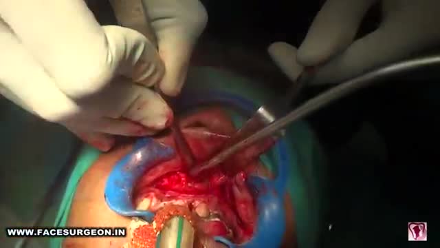
Anterior maxillary distraction for cleft retruded maxilla
Simple blood test could locate gene defects associated with cancer
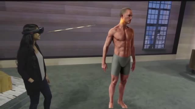
Microsoft HoloLens. Medical Education
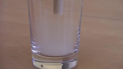
We are aware that the "official" way to use an ear candle is small end down into the ear, but for this video, we have elected to use it the way most "lay" public would (small end up). Ear candling is an alternative medicine practice that is thought to remove earwax. However, this video illustrates how ineffective this practice is in removing earwax... and can potentially be even harmful. And yes... It is still frequently practiced.
De Quervain's tenosynovitis (dih-kwer-VAINS ten-oh-sine-oh-VIE-tis) is a painful condition affecting the tendons on the thumb side of your wrist. If you have de Quervain's tenosynovitis, it will probably hurt when you turn your wrist, grasp anything or make a fist. Although the exact cause of de Quervain's tenosynovitis isn't known, any activity that relies on repetitive hand or wrist movement — such as working in the garden, playing golf or racket sports, or lifting your baby — can make it worse. Symptoms ShareTweet June 13, 2015 References Products and Services Mayo Clinic Sports Medicine Newsletter: Mayo Clinic Health Letter See also Prednisone risks, benefits Prednisone withdrawal: Why taper down slowly? Integrative approaches to treating pain Lifestyle strategies for pain management Nutrition and pain Pain rehabilitation Self-care approaches to treating pain Show more Advertisement Mayo Clinic does not endorse companies or products. Advertising revenue supports our not-for-profit mission. Advertising & Sponsorship PolicyOpportunitiesAd Choices Mayo Clinic Store Check out these best-sellers and special offers on books and newsletters from Mayo Clinic. NEW! – The Mayo Clinic Diet, Second Edition Healthy Heart for Life! Mayo Clinic on Better Hearing and Balance Treatment Strategies for Arthritis The Mayo Clinic Diet Online

How to memorize more in pharma: Drug names, dental implications, numbers
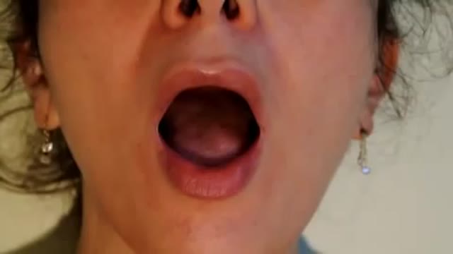
Tongue fassiculations

Phencyclidine (PCP) was developed in the 1950s as an intravenous anesthetic but, due to the side effects of confusion and delirium, its development for human medical use was discontinued. In its pure form, it is a white crystalline powder that readily dissolves in water or alcohol and has a distinctive bitter chemical taste. On the illicit drug market, Phencyclidine contains a number of contaminants as a result of makeshift manufacturing, causing the color to range from tan to brown, and the consistency to range from powder to a gummy mass. It is available in a variety of tablets, capsules, and colored powders, which are either taken orally or snorted. The liquid form of phencyclidine is actually phencyclidine base dissolved most often in ether, a highly flammable solvent. For smoking, phencyclidine is typically sprayed onto leafy material such as mint, parsley, oregano, or marijuana.
