- Physical Examination
- Surgical Examination
- Ophthalmology
- Clinical Skills
- Orthopedics
- Surgery Videos
- Laparoscopy
- Pediatrics
- Funny Videos
- Cardiothoracic Surgery
- Nursing Videos
- Plastic Surgery
- Otorhinolaryngology
- Histology and Histopathology
- Neurosurgery
- Dermatology
- Pediatric Surgery
- Urology
- Dentistry
- Oncology and Cancers
- Anatomy Videos
- Health and Fitness
- Radiology
- Anaesthesia
- Physical Therapy
- Pharmacology
- Interventional Radiology
- Cardiology
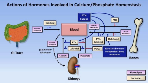
- Endocrinology
- Gynecology
- Emergency Medicine
- Psychiatry and Psychology
- Childbirth Videos
- General Medical Videos
- Nephrology
- Physiology
- Diet and Food Health
- Diabetes Mellitus
- Neurology
- Women Health
- Osteoporosis
- Gastroenterology
- Pulmonology
- Hematology
- Rheumatology
- Toxicology
- Nuclear Medicine
- Infectious Diseases
- Vascular Disease
- Reproductive Health
- Burns and Wound Healing
- Other
Top videos
Cinematic Rendering of the Aorta Plus
Treatment of a stroke interventionaly

Most minor cuts you can treat yourself -- but know when to see a doctor:

You have a good hearing again baby

The inflatable penile prosthesis consists of two attached cylinders -- a reservoir and a pump -- which are placed surgically in the body. The two cylinders are inserted in the penis and connected by tubing to a separate reservoir of saline. The reservoir is implanted under the rectus muscles in the lower abdomen. A pump is also connected to the system and sits under the loose skin of the scrotal sac, between the testicles. This penile prosthesis is referred to as a 3-piece inflatable penile prosthesis, due to the three different components. A 2-piece inflatable penile prosthesis consists of only two components: the attached cylinders and the combined reservoir and pump unit. Instead of the reservoir being placed behind the groin, it is combined with the pump into one housing unit that fits comfortably within the scrotum. The advantage of a 2-piece prosthesis in that the surgery is shorter and less complicated and there is no device parts in the abdomen. The disadvantage of the 2-piece prosthesis is that the smaller reservoir may not result in adequate erections in some men. To inflate the prosthesis, the man presses on the pump. The pump transfers saline from the reservoir to the cylinders in the penis, inflating them and causing an erection. Pressing on a deflation valve at the base of the pump returns the fluid to the reservoir, deflating the penis and returning it to the normal flaccid state.

Child CPR
Anatomy of The Orbit

Histology of Fibrocartilage

What's helping me become a better doctor
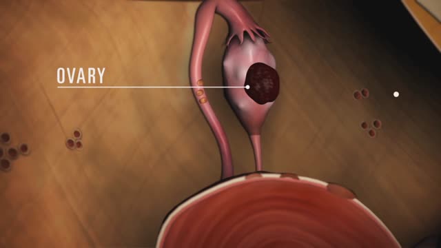
Endometriosis (en-doe-me-tree-O-sis) is an often painful disorder in which tissue that normally lines the inside of your uterus — the endometrium — grows outside your uterus. Endometriosis most commonly involves your ovaries, fallopian tubes and the tissue lining your pelvis. Rarely, endometrial tissue may spread beyond pelvic organs.
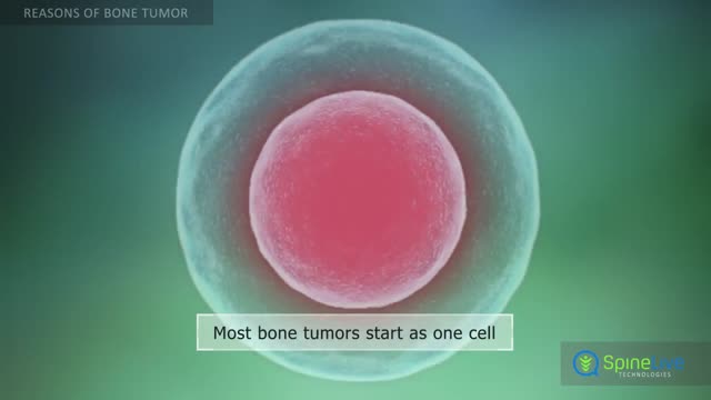
Bone tumors develop when cells in the bone divide without control, forming a mass of tissue. Most bone tumors are benign, which means they are not cancer and cannot spread. However, they may still weaken bone and lead to fractures or cause other problems. Bone cancer destroys normal bone tissue and may spread to other parts of the body (called metastasis). Benign Bone Tumors Benign tumors are more common than malignant tumors of the bones. These are a few common types of benign bone tumors: Osteochondroma is the most common benign bone tumor. It is more common in people under age 20. Giant cell tumor is a benign tumor, typically affecting the leg (malignant types of this tumor are uncommon). Osteoid osteoma is a bone tumor, often occurring in long bones, that occurs commonly in the early 20s. Osteoblastoma is a single tumor that occurs in the spine and long bones, mostly in young adults. Enchondroma usually appears in bones of the hand and feet. It often has no symptoms. It is the most common type of hand tumor.
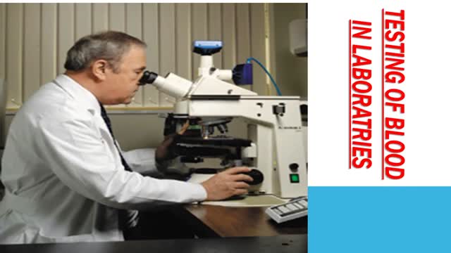
COMMON BLOOD DISORDERS
Myelodysplastic syndromes are a group of cancers in which immature blood cells in the bone marrow do not mature or become healthy blood cells. In a healthy person, the bone marrow makes blood stem cells (immature cells) that become mature blood cells over time.Aug 12, 2015

The cell cycle or cell-division cycle is the series of events that take place in a cell leading to its division and duplication of its DNA (DNA replication) to produce two daughter cells.

Laser-assisted in situ keratomileusis (LASIK) eye surgery can correct or improve your sight by using a laser to change the shape of the cornea. Find out more here: https://www.bupa.co.uk/health-....information/eyes-sig and https://www.bupa.co.uk/health-....information/eyes-sig/laser-eye-surgery
The content is intended for general information only and does not replace the need for personal advice from a qualified health professional.

You may have heard that some positions, such as your partner on top (missionary position), are better than others for getting pregnant. In fact, there's no evidence to back these theories up. Experts just haven't done the research yet. What experts have done, though, is use scanning to show what's going on inside when you're doing the deed. The research looked at two positions: the missionary position and doggy style. (Doggy style being when you're on all fours, and your partner enters you from behind). Common sense tells us that these positions allow for deep penetration. This means that they're more likely to place sperm right next to your cervix (the opening of your uterus). The scans confirm that the tip of the penis reaches the areas between the cervix and vaginal walls in both of these positions. The missionary position allows the penis to reach the area at the front of the cervix. The rear entry position reaches the area at back of the cervix. It's amazing what some experts spend their time doing, isn't it! Other positions, such as standing up, or woman on top, may be just as good for getting sperm right next to the cervix. We just don't know yet. http://www.babycentre.co.uk/sex-for-getting-pregnant#ixzz4XKnPLbxL
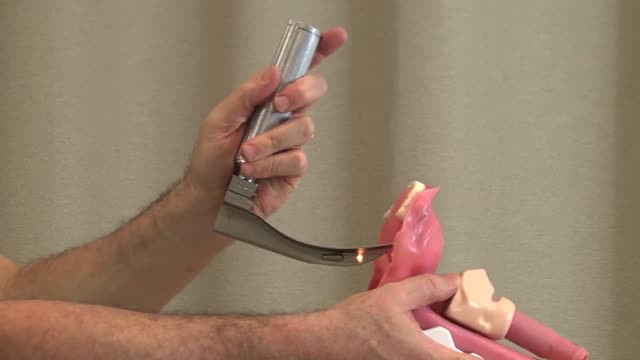
Direct Laryngoscopy: MICU Fellows Airway Course

How to place an NG tube in a baby,
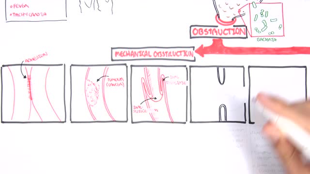
A small-bowel obstruction (SBO) is caused by a variety of pathologic processes. The leading cause of SBO in industrialized countries is postoperative adhesions (60%), followed by malignancy, Crohn disease, and hernias, although some studies have reported Crohn disease as a greater etiologic factor than neoplasia.
