
- Physical Examination
- Surgical Examination
- Ophthalmology
- Clinical Skills
- Orthopedics
- Surgery Videos
- Laparoscopy
- Pediatrics
- Funny Videos
- Cardiothoracic Surgery
- Nursing Videos
- Plastic Surgery
- Otorhinolaryngology
- Histology and Histopathology
- Neurosurgery
- Dermatology
- Pediatric Surgery
- Urology
- Dentistry
- Oncology and Cancers
- Anatomy Videos
- Health and Fitness
- Radiology
- Anaesthesia
- Physical Therapy
- Pharmacology
- Interventional Radiology
- Cardiology
- Endocrinology
- Gynecology
- Emergency Medicine
- Psychiatry and Psychology
- Childbirth Videos
- General Medical Videos
- Nephrology
- Physiology
- Diet and Food Health
- Diabetes Mellitus
- Neurology
- Women Health
- Osteoporosis
- Gastroenterology
- Pulmonology
- Hematology
- Rheumatology
- Toxicology
- Nuclear Medicine
- Infectious Diseases
- Vascular Disease
- Reproductive Health
- Burns and Wound Healing
- Other
Top videos


Mr. Dan underwent 5 suregries in a single go-Both sided hernias+both sided varicocoele and one hydrocoele.Our web site: http://www.desarda.com “Complete cure from groin hernia is now possible with Dr.Desarda's repair technique.......” Mesh is a foreign body, a simple piece of cloth prepared from the synthetic threads. Therefore, its use in inguinal hernia repairs is known to cause all sorts of complications like pain, recurrence, infection etc. We have developed an innovative new technique of inguinal hernia repair without mesh. It uses your own body muscle for repair and gives virtually complete cure from inguinal hernia problem. An undetached strip of the external oblique aponeurosis is stitched on the weak area between the muscle arch and the inguinal ligament to form a new, strong and physiologically dynamic posterior wall that gives protection and prevents re-herniation. Normally patient goes home in a day after surgery and can drive car and go to office in 3-4 days time. This "Dr.Desarda's hernia repair" is now followed in many countries all over the world. We are surprised to see the enquiries from many patients in the developed countries asking for this repair in their country. This is because this operation does not use any foreign body like mesh for repair and therefore there are no complications that are seen in mesh repairs. A visit to Topix or other hernia forums show thousands of posts showing sufferings of many patients due to mesh repairs. But still why surgeons from developed countries are interested in mesh repairs is a big question for us. Please visit our website for more details: http://www.desarda.com or http://herniasurgery.tripod.com Our cell number: +91 9373322178
腹腔镜联合胆囊+阑尾切除术——普外寰潮网,汕头市第二人民医院
Pregnancy All Weeks HD Animation: From www.MedicalVideos.us Showing the progress and mechanisms of pregnancy in each week.

The Role and Function of Nursing

Venepuncture Tutorial HD Drawing Blood

Congestive Heart Failure Animation
Eye Phacoemulsification

Immunization Mechanism Animation
RLE and Alcon Restor IOL Implantation
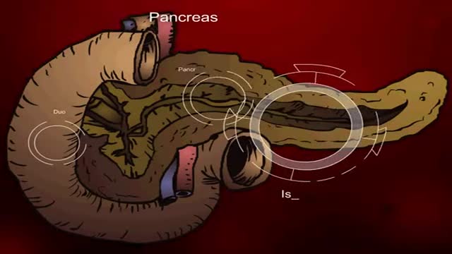
Insulin Processes Mechanism Animation 3D

Coconut Oil Provides Thyroid Gland Benefits

Considering having an Austin plastic surgery procedure like Smartlipo? Then you’ll want to watch this quick video where staff members of renowned Austin plastic surgeon Dr. William Davis give you an overview of what you can expect.
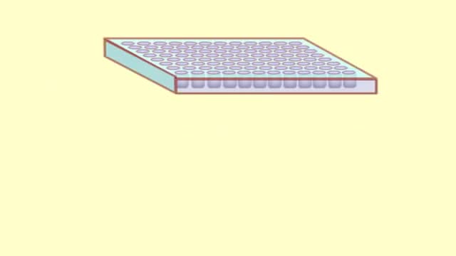
The purpose of an ELISA is to determine if a particular protein is present in a sample and if so, how much. There are two main variations on this method: you can determine how much antibody is in a sample, or you can determine how much protein is bound by an antibody. The distinction is whether you are trying to quantify an antibody or some other protein. In this example, we will use an ELISA to determine how much of a particular antibody is present in an individuals blood.
ELISAs are performed in 96-well plates which permits high throughput results. The bottom of each well is coated with a protein to which will bind the antibody you want to measure. Whole blood is allowed to clot and the cells are centrifuged out to obtain the clear serum with antibodies (called primary antibodies). The serum is incubated in a well, and each well contains a different serum (see figure below). A positive control serum and a negative control serum would be included among the 96 samples being tested.

Mitral Valve Repair video

Quran describes the stages of human creation accurately
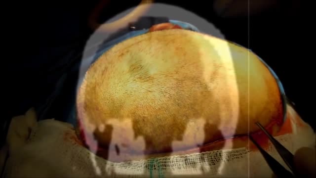
A subdural hematoma (SDH) is a collection of blood below the inner layer of the dura but external to the brain and arachnoid membrane (see the images below). Subdural hematoma is the most common type of traumatic intracranial mass lesion. Subdural hematoma occurs not only in patients with severe head injury but also in patients with less severe head injuries, particularly those who are elderly or who are receiving anticoagulants. Subdural hematoma may also be spontaneous or caused by a procedure, such as a lumbar puncture (see Etiology). Rates of mortality and morbidity can be high, even with the best medical and neurosurgical care (see Prognosis). Subdural hematomas are usually characterized on the basis of their size and location and the amount of time elapsed since the inciting event age (ie, whether they are acute, subacute, or chronic). When the inciting event is unknown, the appearance of the hematoma on neuroimaging studies can help determine when the hematoma occurred. These factors, as well as the neurologic and medical condition of the patient, determine the course of treatment and may also influence the outcome. Generally, acute subdural hematomas are less than 72 hours old and are hyperdense compared with the brain on computed tomography scans. The subacute phase begins 3-7 days after acute injury. Chronic subdural hematomas develop over the course of weeks and are hypodense compared with the brain. However, subdural hematomas may be mixed in nature, such as when acute bleeding has occurred into a chronic subdural hematoma. Presentation varies widely in acute subdural hematoma (see Clinical). Many of these patients are comatose on admission. However, approximately 50% of patients with head injuries who require emergency neurosurgery present with head injuries that are classified as moderate or mild (Glasgow Coma Scale scores 9-13 and 14-15, respectively). Many of these patients harbor intracranial mass lesions. In a large series of patients who developed intracranial hematomas requiring emergent decompression, more than half had lucid intervals and were able to make conversation between the time of their injury and subsequent deterioration. In a more comprehensive review of the literature on the surgical treatment of acute subdural hematomas, lucid intervals were noted in up to 38% of cases. These patients may be more likely to benefit from medical and surgical intervention when instituted in a timely fashion (ie, before further neurological deterioration).
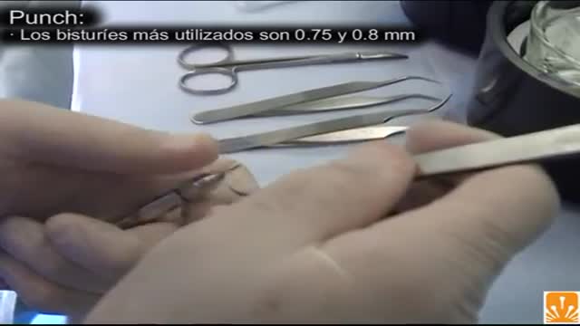
FUE (follicular unit extraction) is a minimally invasive method in hair transplantation. Unlike strip harvesting, the traditional technique in which a strip of skin is removed from a donor site and cut into individual units, FUE uses an instrument to remove multiple groups of one to four hairs. The great thing about FUE is that there’s no linear scar. The downside is that a fewer number of hair grafts can be gathered per session.

10 Diseases That Will Kill You In A Day
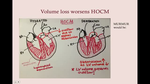
Dilated cardiomyopathy is a disease of the heart muscle, usually starting in your heart's main pumping chamber (left ventricle). The ventricle stretches and thins (dilates) and can't pump blood as well as a healthy heart can. The term "cardiomyopathy" is a general term that refers to the abnormality of the heart muscle itself. Dilated cardiomyopathy might not cause symptoms, but for some people it can be life-threatening. A common cause of heart failure — the heart's inability to supply the body with enough blood — dilated cardiomyopathy can also contribute to irregular heartbeats (arrhythmias), blood clots or sudden death. The condition affects people of all ages, including infants and children, but is most common in men ages 20 to 60.
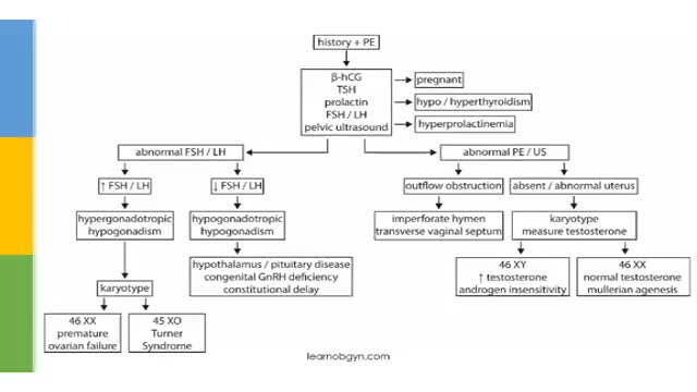
Amenorrhea is the absence of menstrual bleeding.[1] Amenorrhea is a normal feature in prepubertal, pregnant, and postmenopausal females. In females of reproductive age, diagnosing amenorrhea is a matter of first determining whether pregnancy is the etiology. In the absence of pregnancy, the challenge is to determine the exact cause of absent menses.[2] Primary amenorrhea is the failure of menses to occur by age 16 years, in the presence of normal growth and secondary sexual characteristics. If by age 13 menses has not occurred and the onset of puberty, such as breast development, is absent, a workup for primary amenorrhea should start. Secondary amenorrhea is defined as the cessation of menses sometime after menarche has occurred. Oligomenorrhea is defined as menses occurring at intervals longer than 35 days apart. No consensus has been reached regarding the point at which oligomenorrhea becomes amenorrhea. Some authors suggest the absence of menses for 6 months constitutes amenorrhea, but the basis for this recommendation is unclear. For a post-menarchal girl or a reproductive-aged woman to experience a menstrual cycle interval of more than 90 days is statistically unusual. Practically speaking, this should be an indication for an evaluation to seek the cause.
